Last Updated on November 27, 2025 by Bilal Hasdemir

At Liv Hospital, we are dedicated to top-notch healthcare for all patients. It’s key to know the differences between aortic and mitral stenosis for better care. Valvular stenosis happens when heart valves narrow. This makes blood flow harder and puts more work on the heart.
The aortic and mitral valves are usually affected. Aortic stenosis often comes from age-related buildup and birth defects. Mitral stenosis is usually linked to rheumatic heart disease.
Heart valve stenosis happens when the valves narrow, blocking blood flow. This can lead to serious problems. It makes the heart work harder, which is why knowing about it is key.
Valvular stenosis means the heart valves get narrower. This blocks blood flow between the heart’s chambers or to the rest of the body. Aortic stenosis and mitral stenosis are two main types, each with its own causes and effects.
Many things can cause the valves to narrow. These include getting older, being born with it, or having rheumatic heart disease. Knowing the causes helps doctors diagnose and treat it better.
Normal heart valves let blood flow in one direction. They open and close with each heartbeat. This ensures blood flows well and doesn’t go back.
Stenosed valves block blood flow, making the heart work harder. This can cause symptoms like shortness of breath, chest pain, and tiredness.
| Valve Condition | Effect on Blood Flow | Common Symptoms |
|---|---|---|
| Aortic Stenosis | Obstruction to outflow from the left ventricle | Exertional dyspnea, angina, syncope |
| Mitral Stenosis | Impediment to inflow into the left ventricle | Dyspnea, atrial fibrillation, fatigue |
Knowing how stenosed valves affect blood flow is important. It helps us see the differences between aortic stenosis and mitral stenosis. It also guides how to treat them.
It’s important to know about heart valve stenosis to treat it well. This condition happens when heart valves narrow. This narrowing can block blood flow and cause heart problems.
The heart has four valves: aortic, mitral, pulmonary, and tricuspid. Each valve is special and helps blood flow right. The valves open and close with each heartbeat, letting blood move forward and stopping it from going back.
Stenosis happens when valve leaflets get thick or damaged. This makes it hard for them to open fully. As a result, the heart has to work harder to push blood through. This can damage the heart over time.
The aortic and mitral valves are most often affected by stenosis. Aortic stenosis can come from age or birth defects. Mitral stenosis is often linked to rheumatic heart disease. Pulmonary stenosis is less common but can be due to birth defects.
| Valve | Causes of Stenosis | Effects on Blood Flow |
|---|---|---|
| Aortic Valve | Age-related calcification, Congenital bicuspid valve | Obstruction to left ventricular outflow |
| Mitral Valve | Rheumatic heart disease, Calcification | Impediment to left ventricular inflow |
| Pulmonary Valve | Congenital defects | Obstruction to right ventricular outflow |
It’s important to know what causes aortic stenosis to understand how common it is. This heart disease makes the aortic valve narrow. It mainly affects older people.
Calcification is a big reason for aortic stenosis. As we get older, calcium builds up on the aortic valve. This makes the valve stiff and narrow. It’s like atherosclerosis but in the valve.
A congenital bicuspid aortic valve is another cause. This means the valve has only two leaflets instead of three. People born with this are more likely to get aortic stenosis early in life.
Aortic stenosis is very common in older people. Studies say 2% to 7% of those 65 and up have it. If not treated, it can cause serious health problems and even death.
| Age Group | Prevalence of Aortic Stenosis |
|---|---|
| 65-75 years | 2% |
| 75-85 years | 4% |
| 85 years and above | 7% |
The risk of aortic stenosis goes up with age. This shows why older adults need regular check-ups and quick treatment if needed.
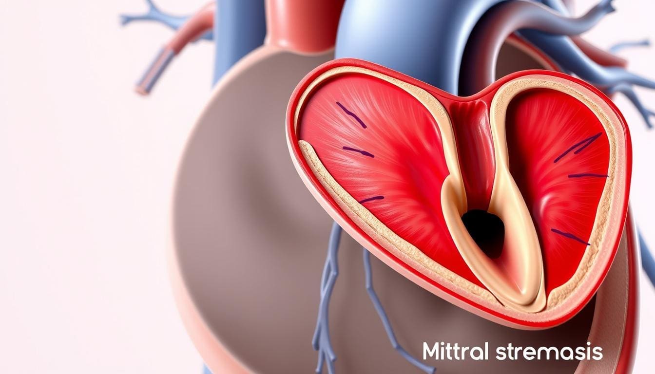
To understand mitral stenosis, we must look at its causes. Rheumatic heart disease is the main reason. It makes the mitral valve opening narrow. This blocks blood flow from the left atrium to the left ventricle.
Rheumatic heart disease, caused by rheumatic fever, is the top reason for mitral stenosis globally. Rheumatic fever can damage the mitral valve, causing it to narrow over time. It’s important to know how rheumatic fever leads to mitral stenosis.
While rheumatic heart disease is the main cause, other factors can also lead to mitral stenosis. These include:
These causes are less common but are important in diagnosing and treating mitral stenosis.
In developed countries, the number of people with mitral stenosis has gone down. This is thanks to better healthcare, like better treatment for rheumatic fever. But, mitral stenosis is a big problem in developing countries.
The global impact of mitral stenosis is not the same everywhere. This shows we need to keep working on public health to fight rheumatic heart disease.
The heart works differently in aortic and mitral stenosis. Aortic stenosis blocks blood flow from the left ventricle to the aorta. Mitral stenosis blocks blood flow from the left atrium to the left ventricle.
The aortic valve gets narrower in aortic stenosis. This blocks blood flow from the left ventricle. The left ventricle then works harder, leading to left ventricular hypertrophy.
Over time, this can cause the heart to pump less efficiently. If not treated, it may lead to heart failure.
Mitral stenosis blocks blood flow from the left atrium to the left ventricle. This raises pressure in the left atrium. It can cause left atrial enlargement and atrial fibrillation.
The heart may not fill with enough blood. This can lower cardiac output, making it harder to work during exercise.
Aortic stenosis makes the left ventricle work harder to pump blood. Mitral stenosis may make the left ventricle work less, but it doesn’t pump as much blood.
| Hemodynamic Effect | Aortic Stenosis | Mitral Stenosis |
|---|---|---|
| Primary Impact | Obstruction to outflow | Impediment to inflow |
| Chamber Affected | Left ventricle | Left atrium |
| Compensatory Mechanism | Left ventricular hypertrophy | Left atrial enlargement |
It’s important to understand these differences for treating stenosis heart conditions. Knowing how aortic and mitral stenosis affect the heart helps doctors choose the right treatment for each patient.
Cardiac remodeling is key in both aortic and mitral stenosis. Each condition affects the heart differently. The heart changes in unique ways to handle the blockage caused by these conditions.
In aortic stenosis, the heart thickens the left ventricle walls. This thickening, or left ventricular hypertrophy (LVH), helps the heart pump blood through the narrowed valve. It’s a way the heart tries to keep up despite the blockage.
But, over time, LVH can cause problems. The ventricle might get smaller and stiffer. This makes it harder for the ventricle to relax and fill with blood during diastole.
Mitral stenosis causes the left atrium to grow. The blockage at the mitral valve raises pressure in the left atrium. This can lead to atrial fibrillation, a common complication.
The increased pressure and volume in the left atrium can also cause structural changes. These changes can lead to atrial fibrillation and other arrhythmias.
Both aortic and mitral stenosis can cause lasting changes in the heart. In aortic stenosis, long-term LVH can lead to scarring and fibrosis in the ventricular walls. This can lead to heart failure.
In mitral stenosis, chronic enlargement of the left atrium can cause persistent atrial fibrillation. It also increases the risk of blood clots and stroke.
| Characteristics | Aortic Stenosis | Mitral Stenosis |
|---|---|---|
| Primary Cardiac Change | Left Ventricular Hypertrophy | Left Atrial Enlargement |
| Main Complication | Heart Failure, Sudden Death | Atrial Fibrillation, Pulmonary Hypertension |
| Long-term Impact | Ventricular Fibrosis, Reduced Compliance | Atrial Fibrosis, Thromboembolic Risk |
Understanding these differences in cardiac remodeling is key for treating aortic and mitral stenosis. By knowing how each condition affects the heart, doctors can tailor treatments to meet each patient’s needs.
Aortic and mitral stenosis show different symptoms. Knowing these differences helps doctors treat patients better.
Aortic stenosis often causes three main symptoms: exertional dyspnea, angina, and syncope. Exertional dyspnea happens when the heart works harder during exercise. This leads to lung problems. Angina is when the heart needs more oxygen but can’t get it. Syncope is when the heart can’t pump enough blood, often when exercising.
“The presence of symptoms in aortic stenosis is a critical turning point in the disease’s natural history,” as emphasized by cardiology guidelines. Symptoms appearing means the disease is getting worse, making it urgent to act.
Mitral stenosis mainly causes dyspnea because of high pressure in the left atrium. This leads to lung problems. Atrial fibrillation also happens, caused by a big left atrium. This irregular heartbeat can make the heart work worse and raise the risk of blood clots.
Physical checks also show differences. Aortic stenosis has a loud sound when the heart pumps, heard in the carotids. Mitral stenosis has a diastolic rumbling murmur at the heart’s tip, with an opening snap sometimes.
These symptoms and physical signs are key to telling aortic and mitral stenosis apart. Accurate diagnosis is vital for the right treatment and better patient care.
When it comes to aortic stenosis and mitral stenosis, the diagnostic findings are different. These differences help doctors diagnose and treat these heart conditions accurately.
Echocardiography is key in spotting heart valve stenosis. For aortic stenosis, it shows a narrowed valve and thickened leaflets. Mitral stenosis, on the other hand, has thickened leaflets and a smaller valve area.
Echocardiographic assessment is vital. It tells us about the valve’s shape, how severe the stenosis is, and its impact on blood flow.
We use echocardiography to check the valve’s structure and function. This includes measuring the valve area, mean gradient, and peak velocity. These measurements help us understand how severe the stenosis is and decide on treatment.
Electrocardiograms (ECGs) offer more clues for diagnosing aortic and mitral stenosis. Aortic stenosis often shows signs of left ventricular hypertrophy. Mitral stenosis might show signs of left atrial enlargement.
While ECGs aren’t enough to diagnose on their own, they add important information. This is when combined with echocardiographic and clinical findings.
Other imaging methods are also used in diagnosing and managing aortic and mitral stenosis. Cardiac catheterization gives us hemodynamic data. Cardiac MRI and CT scans provide detailed anatomy.
Cardiac MRI is great for checking ventricular function and regurgitant volumes. Stress testing helps us see how well patients can function and if there’s ischemia.
These diagnostic findings help us plan treatment and predict outcomes. They are all important for managing stenosis heart conditions.
The fifth key difference between aortic and mitral stenosis is in their complications and how well patients do. Knowing these differences helps doctors manage and treat patients better.
Aortic stenosis and mitral stenosis have different problems that affect how well patients do. We’ll look at the specific issues each condition can cause.
Aortic stenosis can lead to heart failure and sudden death. The stenosis makes the left ventricle work harder, leading to heart failure. The risk of sudden death is high because of arrhythmias or not enough blood flow to the heart.
Heart failure in aortic stenosis is a serious problem that needs quick action. We need to find good treatment options for stenosis heart conditions to deal with these issues.
Mitral stenosis causes problems like high blood pressure in the lungs and blood clots. The narrowed valve makes it hard for blood to flow from the left atrium to the left ventricle. This leads to high pressure in the left atrium and lungs.
High blood pressure in the lungs can cause right ventricular failure. Blood clots, often from irregular heartbeat, can cause strokes. It’s important to know the causes of heart valve stenosis to prevent these problems.
Aortic and mitral stenosis have different outlooks. Aortic stenosis tends to progress in a predictable way, with a high risk of sudden death even when patients don’t show symptoms. Mitral stenosis can lead to severe high blood pressure in the lungs but its course can vary a lot.
Both conditions need close monitoring and timely treatment to improve patient outcomes. The outlook for patients with valvular stenosis depends on how bad their symptoms are, how well their left ventricle is working, and if they have other health problems.
Managing valvular stenosis requires a mix of medical and interventional strategies. We look at many factors to find the best treatment for each patient.
Medical care for valvular stenosis changes based on the valve and how severe it is. For example, those with aortic stenosis might just need to watch for symptoms. But, people with mitral stenosis might need blood thinners to avoid blood clots.
We adjust treatment plans based on each patient’s health and other conditions. For instance, those with severe symptoms or high risk of problems might get more aggressive treatment.
New surgical and interventional methods have changed how we treat valvular stenosis. For aortic stenosis, TAVR is now a good option for high-risk patients. This is thanks to advances like the partnership between Medtronic and DASI Simulations to improve TAVR through predictive modeling and personalized planning.
For mitral stenosis, PMBC is often chosen for patients who can handle it. It’s a less invasive option compared to surgery.
| Treatment Option | Aortic Stenosis | Mitral Stenosis |
|---|---|---|
| Medical Management | Monitoring, symptom management | Anticoagulation, symptom management |
| Surgical/Interventional | TAVR, SAVR | PMBC, Surgical Mitral Valve Replacement |
Valve replacement is needed when stenosis is severe and causing symptoms, or when the heart is not working well. Choosing to replace the valve involves looking at the patient’s health, symptoms, and the risks and benefits of the surgery.
We help patients decide when to replace the valve. We balance the need for surgery with the risks it carries.
Understanding aortic and mitral stenosis is key to managing heart conditions. At Liv Hospital, we’ve explained the main differences. This includes their causes, symptoms, and treatment options.
Getting a correct diagnosis is essential for choosing the right treatment. This could be medication or surgery. We focus on personalized care for each patient’s needs.
Healthcare providers need to understand the complexities of stenosis heart. This knowledge helps in providing better and more caring treatment. At Liv Hospital, we aim to offer top-notch care for our patients.
Heart valve stenosis is when the heart valves narrow. This blocks blood flow. It can happen in valves like the aortic and mitral valves.
Symptoms include shortness of breath when exerting, chest pain, and fainting. These happen because blood can’t flow well from the left ventricle to the aorta.
Mitral stenosis often comes from rheumatic heart disease. But other things can also narrow the mitral valve.
Aortic stenosis blocks blood flow from the left ventricle to the aorta. Mitral stenosis blocks blood flow from the left atrium to the left ventricle. This affects the heart’s workload differently.
Echocardiography, electrocardiograms, and other imaging help tell the two apart. These tools show key differences in the conditions.
Treatments include medicine, surgery like valve replacement, and interventional procedures. The right choice depends on the stenosis’s severity and type.
Aortic stenosis can lead to heart failure and sudden death. Mitral stenosis can cause high blood pressure in the lungs and blood clots. Both need timely treatment.
In developed countries, mitral stenosis is less common thanks to better healthcare. But, it’s a big problem in places with less medical access.
Echocardiography is key for checking how severe valve stenosis is. It helps decide treatment and track how the disease changes over time.
Some mild cases might be treated with medicine. But severe stenosis usually needs surgery or interventional treatments like valve replacement. This helps relieve symptoms and prevent worse problems.
American Heart Association (AHA): Problem: Heart Valve Stenosis
National Heart, Lung, and Blood Institute (NHLBI): Heart Valve Disease Types
PubMed Central (NCBI): Heart Valve Stenosis Pathogenesis and Treatment (Specific PMC ID)
Subscribe to our e-newsletter to stay informed about the latest innovations in the world of health and exclusive offers!