At Liv Hospital, we know how tough it is to treat arteriovenous malformations (AVMs) in the leg and skin. AVMs are weird links between arteries and veins found in different body parts.
Our team is all about giving top-notch care and custom plans for each AVM. We look at the size, where it is, and how it makes you feel. We have many treatment options like watching it closely, using embolization, or surgery.
Knowing about the different treatment modalities helps patients make smart choices about their health. This way, they get the best care for their AVM.
Key Takeaways
- AVMs are abnormal connections between arteries and veins that can occur in the leg and skin.
- Liv Hospital offers advanced care and expert protocols for AVM treatment.
- Treatment options range from conservative management to embolization and surgery.
- Personalized care is key for managing AVMs well.
- Understanding treatment modalities helps patients make informed decisions about their health.
What Are Arteriovenous Malformations?

Arteriovenous malformations, or AVMs, are abnormal connections between arteries and veins. They can happen in different parts of the body. These vascular anomalies can cause serious health problems, depending on where they are and how big they are.
Definition and Pathophysiology
AVMs are a tangled network of blood vessels that connect arteries to veins. They skip the capillary system. This can cause hemodynamic changes, leading to symptoms and complications.
The growth of AVMs involves genetics and environment. Studies show that genetic mutations can affect how blood vessels develop.
Common Locations: Skin, Leg, and Arm AVMs
AVMs can appear anywhere in the body, but they often show up in the skin, leg, and arm.
- Skin AVMs can cause visible lesions and cosmetic concerns.
- Leg AVMs may lead to pain, swelling, and mobility issues.
- Arm AVMs can cause similar symptoms, affecting limb function.
Are AVMs Genetic? Understanding the Causes
The exact causes of arteriovenous malformations are not fully understood. But, there’s evidence that genetics play a role. Some AVMs are linked to genetic syndromes, like hereditary hemorrhagic telangiectasia (HHT).
| Genetic Factor | Description | Association with AVMs |
| HHT | Hereditary hemorrhagic telangiectasia | Strong association with AVMs in various organs |
| CAP2B | CAPillary malformation-Arteriovenous Malformation syndrome | Linked to AVMs and capillary malformations |
| RASA1 | RASA1 gene mutations | Associated with AVMs and vascular anomalies |
Understanding the genetic factors behind AVMs helps in diagnosing and managing them. More research is needed to understand how genetics and environment interact in AVM development.
Recognizing AVM Symptoms and Complications

It’s important to know the signs and risks of arteriovenous malformations (AVMs). AVMs can show up in different ways, based on where they are, how big they are, and how serious they are.
Clinical Presentation of AV Malformation in Leg
AVMs in the leg can cause pain, swelling, and limited mobility. The pain from leg AVMs can get worse over time if not treated. Swelling happens because of the abnormal blood flow and high blood pressure in veins.
Skin AVM Manifestations and Warning Signs
Skin AVMs can show up as visible lesions, discoloration, or bleeding. These spots might look like a red or purple patchon the skin and feel warm because of the extra blood flow. Warning signs include the spots getting bigger, changing color, or hurting.
Potential Complications: Bleeding AVM and Rupture Risk
One big risk of AVMs is bleeding or rupture. AVMs near the skin or that are big are more likely to bleed. If an AVM ruptures, it can cause serious bleeding, which can be deadly.
It’s key to watch for signs of a possible rupture, like sudden pain or swelling. If you see these signs, get medical help right away.
Diagnosis and Assessment of Arteriovenous Malformations
Diagnosing AVMs starts with a detailed physical check-up and advanced imaging. Getting the diagnosis right is key to finding the best treatment and improving patient care.
Physical Examination Findings
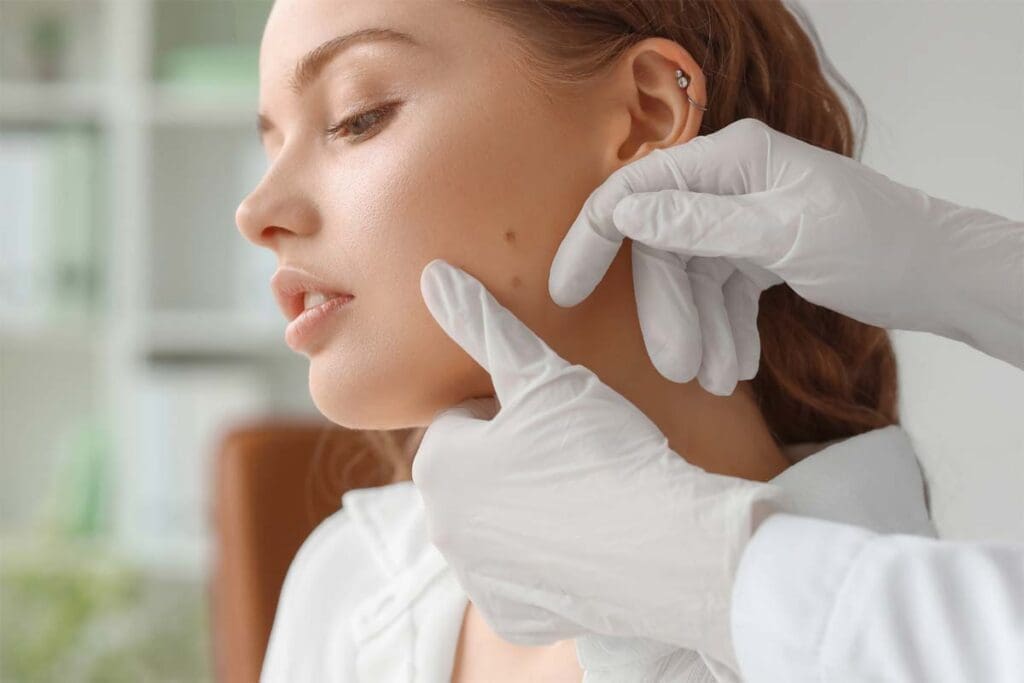
The first step in finding AVMs is a thorough physical check-up. Doctors look for signs like visible swelling, skin discoloration, or a palpable mass. If the AVM is in a limb, patients might feel pain, heaviness, or have trouble moving it.
We also check the size and where the AVM is, and look for any bleeding or ulcers. This info helps us decide what tests to do next and plan treatment.
MRI AVM Imaging: The Gold Standard
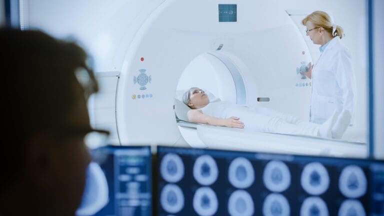
Magnetic Resonance Imaging (MRI) is the top choice for finding AVMs. It gives high-resolution images of the AVM’s structure, including its feeding arteries and draining veins. This info is key for understanding the AVM’s complexity and planning treatment.
We use MRI to see how big the AVM is, its relation to nearby tissues, and any complications. The detailed images from MRI help us make a treatment plan that fits each patient’s needs.
Other Diagnostic Modalities
While MRI is the main tool for AVMs, other imaging methods can help too. These include:
- Ultrasound: Good for first checks and watching AVMs, mainly in easy-to-reach spots.
- Computed Tomography (CT) scans: Give detailed views of the AVM and nearby areas, useful in bleeding cases.
- Angiography: Shows the blood vessels inside the AVM and its vascular layout.
By using info from these tests, we get a full picture of the AVM. This helps us make a treatment plan that works well.
Arteriovenous Malformation Treatment: A Holistic Approach
Treating arteriovenous malformations (AVMs) requires a detailed plan. Every patient is different, so treatments must fit their specific needs.
Factors Influencing Treatment Selection
Choosing the right treatment for AVMs involves several important factors. Size, location, and symptoms play a big role. For example, big AVMs or those in sensitive spots might need stronger treatments. Smaller, symptom-free AVMs might be treated more gently.
A leading medical expert says, “The choice of treatment depends on many things, like size, location, and symptoms of the AVM”
This shows how complex AVM treatment is and why each patient needs a custom plan.
Treatment Goals and Expected Outcomes
The main goals of AVM treatment are to reduce symptoms, prevent problems, and improve life quality. We tailor treatments to meet these goals based on each patient’s needs and situation.
- Lessen symptoms and boost function
- Stop future issues like bleeding or rupture
- Improve life quality
Risk-Benefit Assessment
It’s key to weigh the risks and benefits of each treatment. This helps us pick the best option for each patient. We look at their health and medical history.
When looking at treatments like embolization, surgery, and watchful waiting, we must think about the risks and benefits of each. This helps us choose the safest and most effective treatment.
Conservative Management for Mild AVMs
For those with mild arteriovenous malformations (AVMs), a conservative approach can work well. We understand that not every AVM needs immediate treatment. This method helps manage symptoms and avoid complications.
Monitoring and Observation Protocols
Keeping an eye on mild AVMs is key. Regular check-ups with doctors are important. Imaging like MRI or ultrasound helps track the AVM’s size and changes. It’s also vital for patients to know the signs of worsening symptoms, like more pain or swelling.
Compression Therapy for Extremity AVMs
Compression therapy is great for AVMs in the legs or arms. It uses special garments or bandages to reduce swelling and ease symptoms. This method improves blood flow and lowers the chance of problems.
Pain Management and Symptom Control
Managing pain is critical for AVM patients. We use various methods, from medicines to lifestyle changes, to control pain and symptoms.
“Pain management is a critical component of AVM care, requiring a tailored approach to meet each patient’s needs.”
Our team creates a custom pain management plan for each patient.
Embolization of AVM: Minimally Invasive Intervention
Embolization is a top choice for treating arteriovenous malformations (AVMs). It’s a less invasive option compared to surgery. This method blocks the blood vessels that feed the AVM, cutting down blood flow to it.
Procedure Steps
The steps for embolizing AVMs are straightforward:
- Start with a small incision in the groin to access the blood vessels.
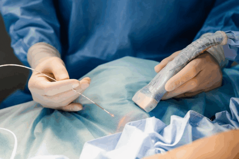
- Use imaging like fluoroscopy to guide a catheter to the AVM.
- Inject embolic agents through the catheter to block the feeding vessels.
- Watch the procedure in real-time to place the embolic material correctly.
Embolization Agents and Their Selection
The right embolization agent depends on the AVM’s size, location, and the patient’s health. Common agents include:
- N-butyl cyanoacrylate (NBCA): A liquid that hardens in blood.
- Ethylene vinyl alcohol copolymer (Onyx): A liquid that doesn’t stick to blood vessels.
- Coils: Devices that cause blood clots in the vessel.
Studies show that picking the right embolic agent is key to success. Research highlights how the right material can lead to better outcomes in AVM treatment.
Recovery Timeline and Success Rates
After embolization, patients need time to recover, which can take days to weeks. The procedure’s success is measured by how much the AVM is reduced and symptoms improved. Many studies show that embolization can significantly shrink AVMs and improve symptoms for many patients.
The recovery timeline can differ based on the AVM’s complexity and the patient’s health. Most patients can get back to normal in a few weeks. The success rate of embolization is high, with many patients seeing a big improvement in their symptoms.
Surgical Resection for Complex Arteriovenous Malformations
Dealing with complex AVMs needs careful planning and precise methods. Surgery is a key treatment for these malformations. It can offer a cure for some patients.
Preoperative Planning and Considerations
Good planning before surgery is key for complex AVMs. We look at the AVM’s size, location, and how it drains blood. MRI and angiography help us understand its structure and how it affects nearby tissues.
Key considerations for preoperative planning include:
- Detailed imaging to assess AVM characteristics
- Evaluation of the patient’s overall health and comorbidities
- Multidisciplinary team discussion to determine the best treatment approach
Surgical Techniques for Different AVM Locations
How we operate on AVMs changes based on where they are. For leg AVMs, we use tourniquets and careful dissection to reduce bleeding. Skin AVMs need a shallower approach, keeping in mind how it will look after.
| AVM Location | Surgical Considerations | Techniques Used |
| Leg | Tourniquet control, risk of significant blood loss | Microsurgical dissection, tourniquet application |
| Skin | Cosmetic considerations, superficial dissection | Laser-assisted excision, suture techniques |
| Arm | Similar to leg AVMs, with attention to functional preservation | Microsurgical techniques, nerve-sparing approaches |
Postoperative Care and Rehabilitation
After surgery, we focus on wound care, pain management, and watching for complications. For bigger AVMs or those in sensitive areas, we may need to help with recovery. This helps patients regain function and improve their life quality.
Postoperative care includes:
- Close monitoring for signs of bleeding or hematoma
- Pain management using multimodal analgesia
- Early mobilization and rehabilitation as needed
Laser and Sclerotherapy for Superficial AVMs
Superficial AVMs can be treated with laser therapy and sclerotherapy. These methods are less invasive and have shown to be effective. They help reduce the size and symptoms of superficial AVMs.
Modalities for Laser Treatment
Laser treatment for superficial AVMs uses different laser types. Each type is chosen based on the AVM’s size and the patient’s skin. The main lasers include:
- Nd:YAG Laser: Good for deeper lesions because it penetrates deeper.
- Alexandrite Laser: Best for AVMs closer to the surface.
- Pulse Dye Laser: Used for smaller, surface-level AVMs or after treatment.
Sclerotherapy Techniques and Agents
Sclerotherapy uses agents to close off the AVM. This leads to its reduction. The agents used are:
| Sclerosing Agent | Concentration | Use in AVMs |
| Sodium Tetradecyl Sulfate | 1-3% | Works for different AVM sizes |
| Polidocanol | 0.5-3% | Less risk of skin damage |
| Ethanol | Absolute or diluted | Best for big or complex AVMs |
We choose the agent based on the AVM’s size, location, and flow. We also consider the patient’s health and preferences.
Combining Laser and Sclerotherapy for Enhanced Results
Using both laser and sclerotherapy can improve results for superficial AVMs. This approach treats different parts of the AVM at once. It may reduce the need for more treatments and make patients happier.
Studies show that laser and sclerotherapy together work well for superficial AVMs. This method treats both the looks and symptoms of these malformations.
Emergency Management of Bleeding AVMs
An AVM rupture needs quick and effective action. It can cause a lot of blood loss, pain, and serious health risks. We will cover the key steps in handling bleeding AVMs, from first aid to hospital care and ways to stop future bleeding.
First Aid for AVM Rupture
Act fast when an AVM ruptures. Apply direct pressure to the wound if you can. Elevate the affected limb to slow blood flow. Call emergency services right away for professional help.
Keep an eye on the person’s pulse and breathing while waiting for help. If they’re awake, try to keep them calm and comfortable.
Hospital-Based Interventions for Acute Bleeding
At the hospital, a team will figure out the best plan. They might use imaging studies like angiography or MRI to see how bad the bleeding is.
They might try embolization to stop the bleeding or surgery to remove the AVM. The choice depends on the AVM’s size, location, and the patient’s health.
| Treatment Option | Description | Indications |
| Embolization | Minimally invasive procedure to block blood flow to the AVM | Bleeding AVMs, high-risk AVMs |
| Surgical Resection | Surgical removal of the AVM | Accessible AVMs, failed embolization |
| Conservative Management | Monitoring and supportive care | Small, asymptomatic AVMs |
Prevention of Recurrent Hemorrhage
Stopping future bleeding is key. This might mean regular check-ups and imaging studies to watch the AVM. Sometimes, more treatments are needed.
People who’ve had AVM rupture should know the signs of another bleed. They should get medical help right away. Making lifestyle changes and managing risks can also help avoid future problems.
Conclusion: Navigating AVM Treatment Decisions
Understanding the different ways to treat arteriovenous malformations (AVMs) is key. We’ve looked at various methods, from watching and waiting to more active treatments. Each method is chosen based on the AVM’s details and the patient’s health.
Every AVM is different, so a custom care plan is vital. This plan considers the AVM’s size, location, and how it affects the patient. This way, we can pick the best treatment and reduce risks.
It’s important for patients and doctors to work together when deciding on treatment. By sharing information and using the latest treatments, we can give patients the best care for their AVM.
The main aim of AVM treatment is to improve patients’ lives. By tailoring care and keeping up with new treatments, we can help patients manage their AVMs well.
FAQ
What does AVM stand for in medical terms?
AVM stands for Arteriovenous Malformation. It’s a condition where arteries and veins are abnormally connected.
What is an arteriovenous malformation?
An arteriovenous malformation (AVM) is a mix of blood vessels in the body. It can happen in places like the skin or legs. It disrupts normal blood flow.
Are AVMs genetic?
The exact cause of AVMs is not fully known. But, research suggests genetics might play a part in their development.
What are the symptoms of an AVM in the leg?
Symptoms of an AVM in the leg include swelling and pain. You might also see varicose veins or a mass. Sometimes, it can cause skin discoloration or ulcers.
How is an AVM diagnosed?
Doctors first do a physical exam. Then, they use imaging tests like MRI. MRI is the best way to find AVMs.
What is the treatment for an arteriovenous malformation?
Treatment for AVMs depends on the malformation’s size, location, and severity. It can include watching it, embolization, surgery, laser therapy, or sclerotherapy.
What is embolization of AVM?
Embolization of AVM is a procedure to block blood flow to the malformation. It uses agents to reduce symptoms and prevent complications.
Can AVMs bleed or rupture?
Yes, AVMs can bleed or rupture. This can lead to serious problems. It’s important to recognize warning signs and get medical help quickly.
How are bleeding AVMs managed?
Bleeding AVMs need immediate care. This includes first aid and hospital treatments to stop the bleeding and prevent more.
Is it possible to prevent recurrent hemorrhage from an AVM?
Yes, you can prevent bleeding from an AVM with the right treatment. This might include embolization, surgery, or other treatments based on your case.
References:
- Steiger, H.-J. (2021). Recent progress understanding pathophysiology and genesis of brain AVM — a narrative review. Neurosurgical Review, 44(6), 3165–3175. https://doi.org/10.1007/s10143-021-01526-0 Retrieved from https://pmc.ncbi.nlm.nih.gov/articles/PMC8592945/ PMC
- Mansur, A., & Radovanovic, I. (2024). Defining the role of oral pathway inhibitors as targeted therapeutics in arteriovenous malformation care. Biomedicines, 12(6), 1289. https://doi.org/10.3390/biomedicines12061289Retrieved from https://pmc.ncbi.nlm.nih.gov/articles/PMC11201820/PMC
- Clinical Presentations and Treatment Approaches in a Retrospective Analysis of 128 Intracranial Arteriovenous Malformation Cases. (n.d.). Retrieved from https://pmc.ncbi.nlm.nih.gov/articles/PMC11591554/ PMC
- Extracranial arteriovenous malformations: Towards etiology‑based therapeutic management. (n.d.). Retrieved from https://pmc.ncbi.nlm.nih.gov/articles/PMC11910209/ PMC
- Seattle Children’s Hospital. (n.d.). Arteriovenous malformation (AVM). Retrieved from https://www.seattlechildrens.org/conditions/avm
- Services/Neurology‑Neurosurgery/AVM‑Arteriovenous‑Malformation/AVM‑Treatment
- Foundation for Urologic & Spinal FUS. (n.d.). Diseases and conditions: Arteriovenous malformations (AVMs). Retrieved from https://www.fusfoundation.org/diseases-and-conditions/arteriovenous-malformations-avms


