Last Updated on October 21, 2025 by mcelik

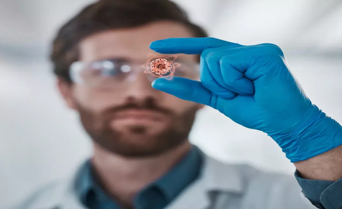
At Liv Hospital, we’re changing how we diagnose blood diseases. We use bone marrow images and leukemia microscopy to see cells up close. This helps us spot diseases like leukemia early.
We use high-quality bone marrow pictures to help diagnose and study blood diseases. This gives our patients the best care and new treatment options.
Key Takeaways
- Advanced bone marrow images enhance diagnostic accuracy for leukemia.
- Leukemia microscopy plays a critical role in understanding disease progression.
- Synthetic augment images support research and treatment development.
- Liv Hospital combines the latest technology with patient-focused care.
- High-quality bone marrow pictures help identify abnormal cells.
The Critical Role of Bone Marrow Imaging in Hematology

Bone marrow imaging is key in hematology. It helps us understand bone marrow’s role in health and disease. This imaging is vital for diagnosing and treating blood-related conditions.
Cellular Composition of Healthy Bone Marrow
Healthy bone marrow has many cell types, like stem cells and red and white blood cells. The balance of these cells is important for blood production. Any imbalance can cause blood disorders.
Healthy bone marrow’s cells can be divided into several parts:
| Cell Type | Function | Percentage of Total Cells |
|---|---|---|
| Stem Cells | Self-renewing cells that give rise to all blood cell types | 1-2% |
| Erythroid Cells | Precursor cells that mature into red blood cells | 20-30% |
| Myeloid Cells | Precursor cells that mature into white blood cells and platelets | 40-50% |
| Lymphoid Cells | Cells that play a key role in the immune system | 10-20% |
Visualization Techniques in Bone Marrow Analysis
Many techniques are used to study bone marrow cells, like microscopy and advanced imaging. These methods help us see cell details, aiding in diagnosing blood disorders.
Some main techniques for studying bone marrow include:
- Light microscopy: Examines cell shape
- Fluorescence microscopy: Finds specific cell markers
- Flow cytometry: Analyzes cells based on surface antigens
By using these techniques and knowing about healthy bone marrow, we can accurately diagnose and treat blood diseases, including leukemia.
Understanding Leukemia Microscopy Fundamentals

Learning about leukemia microscopy is key for diagnosing and treating leukemia. Leukemia is marked by abnormal cells in the bone marrow. Microscopy helps spot these cells.
Looking at bone marrow samples under a microscope is vital. It helps tell healthy cells from cancerous ones. This is important for planning treatment.
Identifying Leukemic Blasts Under the Microscope
Leukemic blasts are young, abnormal cells found in leukemia. They can be seen under a microscope because of their unique look. Spotting these cells is key for making a diagnosis.
Here’s a table showing the differences between healthy and cancerous cells:
| Cell Characteristics | Healthy Cells | Leukemic Cells |
|---|---|---|
| Morphology | Normal size and shape | Abnormal size and shape |
| Nuclear Characteristics | Normal nuclear structure | Abnormal nuclear structure |
| Cell Distribution | Normal distribution | Abnormal distribution |
A leading hematologist says, “Spotting leukemic blasts is essential for diagnosing leukemia.”
“Microscopy is the It lets us see the details of cells that are vital for diagnosis.”
Distinguishing Between Healthy and Leukemic Marrow
Telling healthy marrow from cancerous one is critical. Cancerous marrow has abnormal cells that can be seen under a microscope.
Using synthetic pictures helps in teaching doctors to recognize cancer cells. Mixing real and fake images helps us learn more about leukemia. This improves how well we can diagnose it.
In summary, knowing about leukemia microscopy is vital for diagnosing and treating it. By spotting cancer cells and telling healthy from cancerous marrow, we can give better care to patients.
Bone Marrow Real Pictures: Clinical Applications and Sources
High-quality bone marrow pictures are key in clinical diagnosis. They help doctors spot problems accurately. These images help us grasp the complexities of blood disorders and plan treatments.
Diagnostic Value of High-Quality Bone Marrow Images
The importance of bone marrow images in diagnosis is huge. They show the detailed structure of cells. This helps identify conditions like leukemia. Accurate diagnosis is key for the right treatment, and clear images are vital.
We use top-notch imaging to see bone marrow’s fine details. This helps in diagnosing, tracking disease progress, and checking treatment success.
Medical Image Banks and Educational Resources
Having access to big medical image banks is essential for learning and research. They offer a lot of info, including bone marrow pictures for both diagnosis and teaching.
| Resource | Description | Access |
|---|---|---|
| National Cancer Institute | Provides images and data related to various cancers, including leukemia. | Public |
| American Society of Hematology | Offers educational resources, including images and case studies. | Members and subscribers |
| Radiological Society of North America | Features a collection of medical images, including bone marrow images. | Public and members |
These resources are priceless for doctors and researchers. They give the tools needed to better understand bone marrow and related issues.
7 Essential Bone Marrow Real Pictures for Clinical Reference
We have seven key bone marrow real pictures for doctors to use. These images help doctors understand different blood diseases, like leukemia. They can use these pictures to get better at diagnosing and caring for patients.
Picture 1: Normal Bone Marrow Aspirate
A normal bone marrow aspirate image shows what healthy marrow looks like. It has cells at different stages of growth, fat cells, and other parts. Seeing trilineage hematopoiesis means the marrow is working right.
Picture 2: Acute Myeloid Leukemia (AML) Sample
An AML sample has myeloid blasts, which are young cells that didn’t grow right. These cells fill up the marrow and mess with blood making. Knowing the AML type is key for the right treatment.
Picture 3: Acute Lymphoblastic Leukemia (ALL) Presentation
ALL is when lymphoblasts grow too much in the marrow. The image shows many lymphoblasts with special shapes. Finding ALL right is important because it needs its own treatment plan.
Picture 4: Chronic Myeloid Leukemia (CML) Features
CML has the Philadelphia chromosome, from a chromosome swap. The marrow looks busy with many myeloid cells. Doctors check for the BCR-ABL gene to confirm CML.
These images are part of a big collection important for learning and diagnosing. By looking at these bona fide bone marrow samples, doctors can get better at understanding blood diseases.
The Evolution of Synthetic Bone Marrow Pictures
The field of medical imaging has seen big changes with synthetic bone marrow pictures. These changes come from new algorithms, like Generative Adversarial Networks (GANs).
Role of GANs in Medical Imaging
GANs have changed how we make synthetic medical images, like bone marrow pictures. They have a generator and a discriminator. The generator makes images, and the discriminator checks if they look real.
This back-and-forth process makes GANs create images that look very real. These images can look just like real bone marrow samples.
We can now augment our datasets with these synthetic pictures. This makes our data better for training AI and for teaching.
Quality Assessment of Synthetic Marrow Images
It’s important to check how good synthetic bone marrow images are. Experts use many ways to judge these images. They look at how real they look, their quality, and if they show different health issues well.
By using GANs and careful checks, we make sure these images are not just good-looking. They also have to be useful in real medical situations. This makes them better for learning and for helping doctors.
Augmenting Clinical Datasets with Synthetic Images
We can make clinical datasets better by mixing real and synthetic images. This is very helpful in bone marrow imaging. High-quality images are key for accurate diagnoses.
Using synthetic images has many benefits. It makes AI models better by giving them more images to learn from. It also keeps patient privacy safe by needing less real patient data.
Benefits of Combined Real and Synthetic Data
Real and synthetic bone marrow images together improve AI model training. Real images give the needed detail and accuracy. Synthetic images fill gaps, like rare conditions or hard-to-capture variations.
| Dataset Type | Benefits | Limitations |
|---|---|---|
| Real Images | High accuracy, detailed information | Limited availability for rare conditions |
| Synthetic Images | Diverse, can fill gaps in dataset, enhance AI training | May lack the fine detail of real images |
| Combined Dataset | Balances accuracy with diversity, improves AI performance | Requires careful integration to maintain data quality |
Experts say, “Adding synthetic data to clinical datasets is a big change for medical imaging. It’s a way to deal with the problem of not having enough data.”
This mix not only makes AI models stronger. It also opens new doors for research and development in medical imaging.
Addressing Patient Privacy Through Synthetic Alternatives
One big plus of synthetic images is they help protect patient privacy. By using them, researchers need less real patient data. This lowers the chance of sensitive info getting out.
- Enhanced patient privacy
- Increased dataset diversity
- Improved AI model performance
As we look into synthetic images in medical diagnostics, it’s clear they’re key. They’re not just helpful; they’re essential for moving healthcare tech forward.
Practical Applications of Augmented Bone Marrow Images
Augmented bone marrow images are changing hematology. They combine real and synthetic data. This helps us better diagnose and treat blood disorders.
We’re seeing big changes in how bone marrow images are used. Synthetic bone marrow pictures are being added to datasets. This makes training data for AI better and diagnostic tools more accurate.
AI Training and Diagnostic Algorithm Development
AI tools need good training data. Augmented bone marrow images help by adding synthetic images to real ones. This makes diagnostic algorithms stronger.
For example, leukemia microscopy images get better with synthetic data. This boosts diagnosis accuracy and helps catch diseases early.
Educational Resources for Medical Professionals
Augmented bone marrow images are great for teaching doctors. They offer a wide range of images, including rare cases. This helps doctors learn to spot and diagnose blood disorders better.
Using bone marrow pictures in class helps doctors understand bone marrow better. This makes them better at making diagnoses and treatment plans.
With augmented bone marrow images, we can make better teaching tools. This improves care for patients with blood disorders.
Future Innovations in Bone Marrow Imaging Technology
The use of AI and advanced imaging is changing bone marrow diagnostics. We’re seeing big steps forward in how we take and use bone marrow images in medicine.
AI-enhanced diagnostic capabilities are a big area of innovation. AI helps doctors spot early signs of diseases like leukemia in bone marrow images.
AI-Enhanced Diagnostic Capabilities
AI is making diagnosis better and faster. It can learn from lots of images to find patterns linked to leukemia. This helps doctors give better care and treatment plans.
AI also makes diagnoses more consistent. It helps doctors across different places agree on what they see in images. This makes care more standard.
| Diagnostic Feature | Traditional Method | AI-Enhanced Method |
|---|---|---|
| Pattern Recognition | Manual analysis by clinicians | Automated pattern recognition using machine learning algorithms |
| Diagnostic Accuracy | Dependent on clinician expertise | Enhanced accuracy through AI-driven analysis |
| Diagnostic Speed | Time-consuming manual analysis | Rapid analysis using AI algorithms |
Emerging Visualization Techniques
New ways to see bone marrow images are also important. 3D reconstruction and virtual microscopy give doctors new tools. They help in making diagnoses and teaching.
These new methods improve how we see and understand bone marrow. For example, virtual microscopy lets doctors share and study images together. This helps everyone learn and grow.
Looking ahead, AI and new imaging will keep changing bone marrow tech. These advances will make diagnosis better, care better, and save lives.
Conclusion
We’ve looked into how bone marrow pictures are key in finding and studying leukemia. Using real and synthetic images has changed hematology. It helps doctors make better diagnoses and treatments.
High-quality bone marrow images are vital for learning and teaching. They help train AI and improve diagnostic tools. This leads to better care for patients.
As we keep improving bone marrow imaging, we’ll see big changes in diagnosis and treatment. The future of fighting leukemia looks promising. We’re on track to help more patients worldwide.
FAQ
What is the significance of bone marrow real pictures in leukemia diagnosis?
Bone marrow real pictures are key in diagnosing leukemia. They help doctors see the cells and spot any odd ones. This is vital for the right diagnosis and treatment.
How do visualization techniques aid in bone marrow analysis?
Visualization tools, like microscopy, help in studying bone marrow. They let doctors see the cells and find any that don’t look right. This helps in diagnosing blood diseases.
What are the characteristics of a normal bone marrow aspirate image?
A normal bone marrow image has a mix of cell types. You’ll see hematopoietic cells, fat cells, and more. The cells look right and there’s the right amount of them.
How are synthetic bone marrow pictures generated?
Synthetic bone marrow images are made with Generative Adversarial Networks (GANs). These AI tools create images that look real, based on what they’ve learned from other images.
What are the benefits of using synthetic images to augment clinical datasets?
Using synthetic images can grow your dataset and add variety. It also keeps patient info safe. This helps AI models get better and makes diagnosis more accurate.
How can augmented bone marrow images be used in medical education?
Augmented bone marrow images are great for teaching. They help students learn about leukemia and other blood diseases. They also improve students’ skills in making diagnoses.
What is the role of AI in bone marrow imaging technology?
AI is very important in bone marrow imaging. It makes diagnosis better, improves how images are analyzed, and helps create more accurate algorithms.
How can high-quality bone marrow images be accessed?
You can find top-notch bone marrow images in medical image banks and educational sites. These places have lots of images for learning, research, and clinical use.
What are the synthetic bone marrow pictures in research?
Synthetic bone marrow pictures are useful in research. They help in making new diagnostic tools, improving AI, and exploring new ways to see images.
Reference
Nature. Research Article. https://www.nature.com/articles/s41746-025-01563-9
American Society of Hematology (ASH). Image Bank. https://imagebank.hematology.org
Adobe Stock. Search Results (acute myeloid leukemia). https://stock.adobe.com/search?k=%22acute+myeloid+leukemia%22


