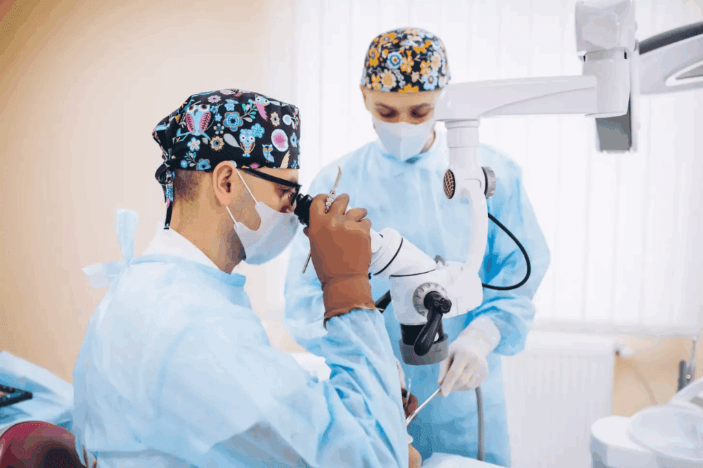
We are seeing big steps forward in brain surgery, thanks to new tech like intraoperative MRI and awake neurosurgery. Real brain surgery has changed, giving patients new treatment options that were once impossible.
These brain operation videos give us a close look at neurosurgery’s details. They show us how complex things like removing tumors and treating movement disorders work. Watching these neurosurgery videos helps us appreciate the skill and care in today’s neurosurgery.
Key Takeaways
- Real brain surgery has become more sophisticated with advanced technologies.
- Brain operation videos offer a detailed look into neurosurgical procedures.
- Modern neurosurgery involves complex treatments like tumor removal and deep brain stimulation.
- Intraoperative MRI and awake neurosurgery are now standard in top centers.
- These advancements improve patient outcomes and expand treatment possibilities.
The Remarkable World of Modern Neurosurgery
Neurosurgery is changing fast, thanks to new tech and methods. Now, brain surgeries are more precise and safer for patients.
Technological Advancements in Brain Surgery
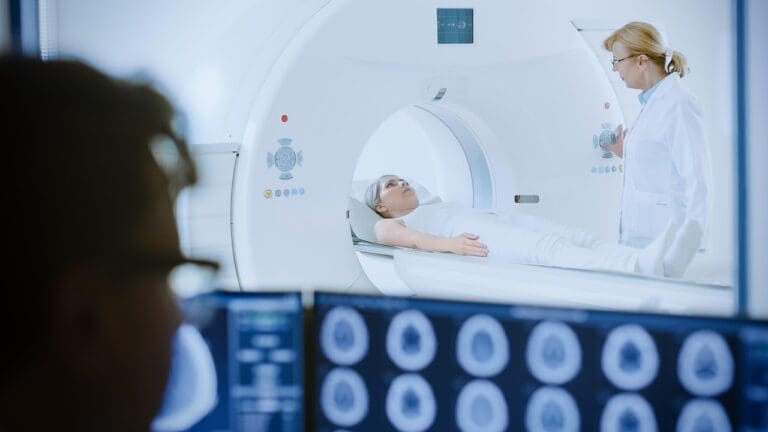
New tech is key in modern neurosurgery. Tools like intraoperative MRI and awake neurosurgery make surgeries safer and more precise. These tools let surgeons see the brain live, helping them remove tumors better while keeping important brain functions.
Some major tech advances include:
- Intraoperative MRI: It lets surgeons see the brain during surgery, helping remove tumors better.
- Awake Neurosurgery: This method lets surgeons work while patients are awake. It helps keep important brain functions safe.
- Advanced Surgical Tools: New tools and methods make neurosurgery safer and more accurate.
Why Surgical Videos Transform Medical Education
Surgical videos change medical education by showing complex procedures clearly. By watching brain surgery videos, doctors can learn from real surgeries. This helps them improve their skills and know neurosurgery better.
Using surgical videos in education has many benefits:
- Enhanced Visual Learning: Videos show complex surgeries clearly.
- Improved Skill Development: Doctors can learn by watching real surgeries.
- Better Patient Outcomes: Educated doctors can give better care, leading to better patient results.
The Value of Real Brain Operation Videos in Medicine
Real brain operation videos have changed medical education and patient care for the better. They have shifted how doctors are trained and how patients learn about their health.
Professional Training Applications
These videos are key for teaching neurosurgeons. They show detailed steps of complex surgeries. This helps trainees learn advanced surgical techniques better.
Watching these videos also boosts critical thinking skills. Trainees learn to make decisions during surgery. This prepares them for tough cases.
Patient Education and Informed Consent
Brain operation videos help patients understand their health and surgery options. They see what the surgery involves. This leads to better informed consent.
Using these videos can lower patient anxiety. It helps them know what to expect during and after surgery. This builds trust between patients and doctors.
Breaking Down Surgical Complexity
Real brain operation videos simplify complex surgeries. They show the steps of neurosurgery clearly. This helps both doctors and patients grasp the surgery’s details.
These videos also help in preoperative planning. Surgeons can plan their approach. Patients know what to expect, preparing them for surgery.
Video 1: Open Brain Surgery for Glioblastoma Removal
Removing a glioblastoma through open brain surgery is a detailed process. It’s key for a patient’s recovery. A skilled neurosurgical team performs a craniotomy to remove the tumor while keeping brain function intact.
Craniotomy Technique and Surgical Approach
The craniotomy technique is vital in open brain surgery. It means temporarily removing a part of the skull to reach the brain. Our team uses advanced imaging to plan the best approach, reducing risks to brain tissue.
Recent studies show that precise craniotomy techniques greatly improve patient outcomes.
Tumor Visualization and Resection Methods
Clear tumor visualization is key for removing glioblastomas. We use top-notch imaging and navigation systems to see the tumor’s edges. This helps our surgeons use precise resection methods to remove the tumor safely.
Post-Operative Care and Recovery Timeline
Post-operative care is essential for recovery after open brain surgery. Our team offers full care, including pain management and rehabilitation. The recovery timeline varies, but with our care, many patients see big improvements.
Watching brain tumor surgery videos helps patients and doctors understand glioblastoma removal better. These videos show the complexity of modern neurosurgery and our team’s commitment to the best results.
Video 2: Awake Neurosurgery with Real-Time Language Mapping
Awake neurosurgery with real-time language mapping gives us a deep look into how the brain works. It lets surgeons work on the brain while the patient is awake. This way, they can avoid harming important parts of the brain.
Patient Preparation and Consciousness Management
For awake neurosurgery to work, preparing the patient well is key. Patients get a deep check before surgery to see if they can handle it. On surgery day, keeping the patient awake and able to follow commands is very important.
- Pre-operative counseling to prepare the patient psychologically
- Intraoperative sedation management to maintain patient comfort
- Continuous monitoring of the patient’s neurological status
Functional Testing During Surgery
During surgery, tests are done to keep important brain functions safe. Language mapping is a big part of this. It checks how well the patient can speak and understand in real-time. This helps surgeons avoid harming brain areas that control language.
- Object naming tasks to assess language function
- Reading and writing tasks to evaluate linguistic capabilities
- Conversation tests to gauge spontaneous speech
Protecting Critical Brain Functions
The main goal of awake neurosurgery is to keep important brain functions safe. By finding and saving language areas, surgeons lower the chance of language problems after surgery. This method not only helps patients recover better but also improves their life after surgery.
Our work with awake neurosurgery shows it can greatly benefit patients. Adding real-time language mapping to surgery is a big step forward. It brings new hope to those needing brain surgery.
Video 3: Deep Brain Stimulation for Parkinson’s Disease
Deep brain stimulation (DBS) is a new hope for Parkinson’s disease patients. It’s a surgery that implants electrodes in the brain to control symptoms. This method helps those who haven’t seen results from regular medicine.
We use advanced tools to place these electrodes correctly. They go into areas like the subthalamic nucleus. This ensures the best results for patients.
Electrode Placement in the Subthalamic Nucleus
The subthalamic nucleus is a key spot for DBS in Parkinson’s patients. Precise electrode placement here can greatly reduce symptoms like tremors and stiffness. Our team uses MRI and microelectrode recording for the best placement.
Neuro Operation Video Highlights of Microelectrode Recording
Microelectrode recording is vital for DBS. It helps us find the perfect spot for the electrodes. Our neuro operation videos show the detailed care during this step. They also show the data that guides our surgery.
Immediate Symptom Improvement Documentation
DBS can lead to quick symptom relief. Our videos show patients feeling better right after the surgery. This proves how effective DBS can be.
| Symptom | Pre-DBS | Post-DBS |
| Tremors | Severe | Mild |
| Bradykinesia | Significant | Minimal |
| Rigidity | Marked | Reduced |
We share the details of DBS to help patients understand it better. Our goal is to give them the knowledge to make informed choices about their treatment.
Video 4: Endoscopic Transsphenoidal Approach for Pituitary Tumors
The endoscopic transsphenoidal approach has changed how we treat pituitary tumors. It’s a new way to remove tumors through the nose, without the need for big brain surgeries.
Minimally Invasive Skull Base Techniques
The endoscopic transsphenoidal approach is a key example of less invasive skull base surgery. This method uses small cuts and special endoscopic tools to see and reach the tumor. It helps us cause less damage and heal faster.
- Reduced risk of complications
- Less post-operative pain
- Shorter hospital stay
Endoscopic Visualization Technology
Advanced endoscopic tech is key to the success of this approach. High-definition cameras and special endoscopes give us a clear view of the area. This lets us remove the tumor carefully, without harming nearby important parts.
- High-definition visualization
- Improved accuracy
- Enhanced patient safety
Advantages Over Traditional Approaches
The endoscopic transsphenoidal approach has many benefits over old-school open surgery. It avoids big cuts and less damage, lowering risks and speeding up recovery. Patients usually feel less pain after and stay in the hospital less time.
| Benefits | Traditional Open Surgery | Endoscopic Transsphenoidal Approach |
| Recovery Time | Several weeks | A few days to a week |
| Post-operative Pain | Significant | Minimal |
| Risk of Complications | Higher | Lower |
We see the endoscopic transsphenoidal approach as a big step forward in treating pituitary tumors. It uses a less invasive method to improve patient results and life quality.
Video 5: Brain Operation Video Using Intraoperative MRI Guidance
Intraoperative MRI is changing neurosurgery by giving immediate feedback during operations. This technology has greatly improved the precision of brain surgeries, like tumor resections.
Real-Time Imaging Integration
Intraoperative MRI allows for real-time imaging during brain surgery. This immediate feedback lets surgeons check how much tumor is removed and adjust as needed. Studies show that intraoperative MRI can lead to more complete resections and better patient outcomes.
Key benefits of intraoperative MRI include:
- Enhanced visualization of tumor boundaries
- Real-time assessment of surgical progress
- Improved accuracy in tumor resection
Surgical Navigation Systems
Surgical navigation systems work with intraoperative MRI to guide surgeons during operations. These systems help plan the surgical approach and navigate the brain’s complex anatomy.
By combining intraoperative MRI with surgical navigation, surgeons can achieve higher precision. This is very valuable when tumors are near critical brain structures.
Tumor Resection Precision Enhancement
The precision of tumor resection is greatly improved with intraoperative MRI guidance. Surgeons can see the tumor and surrounding brain tissue in real-time. This allows for more accurate removal of the tumor while preserving brain functions.
“The integration of intraoperative MRI in brain surgery represents a significant advancement in neurosurgical care. It allows for real-time adjustments during the procedure, potentially leading to better outcomes for patients.”
Neurosurgeons use intraoperative MRI and advanced surgical navigation systems to provide the most precise care. This technology is evolving, promising even more sophisticated applications in the future.
Video 6: Cerebrovascular Surgery for Complex Aneurysm
Complex aneurysms are a big challenge in neurosurgery. They show how far medical science has come. Cerebrovascular surgery for these aneurysms needs a lot of skill and precision.
Microsurgical Clipping Techniques
Microsurgical clipping is key in treating complex aneurysms. It involves placing clips at the aneurysm’s neck to stop it from rupturing. The success of this method depends on the surgeon’s skill in seeing the aneurysm and blood vessels clearly.
We use advanced tools and techniques for microsurgical clipping. It requires technical skill and knowledge of the patient’s blood vessels.
Intraoperative Angiography Assessment
Intraoperative angiography is essential during cerebrovascular surgery. It lets us check the aneurysm and blood vessels in real-time. This ensures the clipping is done right and blood flow is kept.
This check is key to making sure the parent vessel is open and the aneurysm is closed. Intraoperative angiography helps us adjust if needed, making the surgery safer and more effective.
| Aspect | Pre-Clipping | Post-Clipping |
| Aneurysm Status | Untreated | Occluded |
| Parent Vessel Patency | Unknown | Verified |
| Surgical Outcome | Uncertain | Confirmed |
Critical Decision-Making Moments
Cerebrovascular surgery for complex aneurysms requires quick and smart decisions. Surgeons must choose the right clipping method and decide if more surgery is needed. They also have to manage any complications.
Experience, skill, and staying calm are vital. Making good decisions during these moments is what makes a surgery successful.
With our expertise and modern technology, we handle these complex cases well. This ensures the best results for our patients.
Video 7: Pediatric Epilepsy Surgery with Cortical Mapping
Pediatric epilepsy surgery is a key treatment for young patients with severe seizures. It’s a complex procedure that needs special care for children’s developing brains.
Age-Specific Surgical Considerations
When we do surgery for epilepsy in kids, we think about their age a lot. We plan carefully and do detailed cortical mapping. This helps us keep as much brain function as we can.
It’s also important to have a team that knows how to handle these surgeries in kids. Their brains are more flexible, which can help them recover but also makes surgery tricky.
Some key things to consider are:
- The child’s overall health and developmental stage
- The specific characteristics of their epilepsy
- The benefits and risks of surgery
Seizure Focus Identification
Finding the exact spot where seizures start is key in pediatric epilepsy surgery. We use advanced imaging and cortical mapping to pinpoint it. This helps us plan the surgery carefully, avoiding damage to other brain areas.
The steps are:
- Using EEG and MRI for a detailed check before surgery
- Doing cortical mapping during surgery to find important brain areas
- Watching closely during surgery to make sure it’s safe
Developmental Outcome Tracking
After surgery, we keep a close eye on how the child develops. We check their brain function, thinking skills, and how well they control seizures. This helps us get better at surgery and care for future patients.
| Outcome Measure | Description | Importance |
| Seizure Frequency | Reduction in seizure frequency post-surgery | Shows if the surgery worked |
| Cognitive Development | Watching for cognitive and developmental milestones | Sees how surgery affects brain growth |
| Quality of Life | Checking the child’s overall well-being and quality of life | Shows how well the treatment did |
By using the latest surgery methods and caring for kids before and after surgery, we can really help them. It’s all about improving their lives.
Liv Hospital’s Excellence in Neurosurgical Procedures
Liv Hospital’s neurosurgery team stands out for their dedication to global standards and patient care. We aim to give top-notch care, using international methods to get the best results for our patients.
International Standards and Protocols
At Liv Hospital, we stick to strict international standards and protocols in neurosurgery. This shows in our:
- Following global best practices in neurosurgery
- Using the latest technology and equipment
- Keeping our medical staff up-to-date with training and education
Our protocols ensure top care from the first visit to after surgery. A leading neurosurgeon says, “Following international standards is key to achieving excellence in neurosurgical results.”
“The key to successful neurosurgery lies in meticulous planning, precise execution, and thorough post-operative care.”
An Neurosurgeon
Multidisciplinary Approach to Brain Surgery
Our team includes neurosurgeons, neurologists, radiologists, and rehabilitation specialists. They work together to give full care. This teamwork helps us:
| Specialty | Role in Neurosurgery |
| Neurosurgeons | Do surgeries with great precision |
| Neurologists | Diagnose and manage brain conditions |
| Radiologists | Do imaging and interventional radiology |
Patient-Centered Care Philosophy
At Liv Hospital, we put our patients first, giving them personal care and support. Our approach includes:
- Clear communication and informed consent
- Comfort and pain management
- Support for families and education
We think patient-centered care is vital for the best results and a better life.
Ethical Considerations in Neurosurgery Video Sharing
Sharing neurosurgery videos raises big ethical questions. We use technology to improve medical education and care. But we must think carefully about the ethics of these videos.
Patient Consent and Privacy Protection
Getting patient consent is key. We need to make sure patients know their videos will be shared. Keeping patient privacy is also very important.
Here are some steps to protect patients:
- Keep patients’ identities private when possible
- Only let authorized people see the videos
- Store and share videos on secure sites
Educational Purpose vs. Sensationalism
It’s important to balance education with avoiding sensationalism. These videos are great for training doctors. But they should be shown in a way that respects patients and focuses on learning.
“The use of surgical videos in education must be done with sensitivity and respect for the patients involved, focusing on the educational benefits while minimizing possible distress.” –
Medical Ethics Guidelines
To find this balance, we should:
- Set clear goals for the educational value of the video
- Choose content that’s right for the audience
- Avoid adding things that might seem too dramatic
Responsible Viewing Guidelines
We also need guidelines for watching these videos. We should warn viewers about the emotional impact. And offer help if they need it.
| Guideline | Description |
| Content Warning | Give clear warnings about the graphic content |
| Viewer Discretion | Tell viewers to think before watching, if they’re easily upset by medical images |
| Support Resources | Provide help or resources for viewers who are upset |
By considering these ethics, we can use neurosurgery videos wisely. This respects both the patients and the viewers.
Conclusion: The Evolving Landscape of Neurosurgical Education
We are seeing big changes in how neurosurgeons learn, thanks to new tech and brain operation videos. These tools are changing how doctors train, making their skills better with visual learning.
Using surgical videos in education is becoming more common. It helps doctors understand complex surgeries better. As neurosurgery grows, video education will play a bigger role, improving care for patients everywhere.
At Liv Hospital, we aim to give top-notch healthcare to all patients. Our team uses the latest tech in brain surgery. We believe brain operation videos will be key in future education, helping patients worldwide.
The future of neurosurgery looks bright, with new learning methods and tech. We’re excited to keep improving care, always putting our patients first.
FAQ
What is intraoperative MRI, and how is it used in brain surgery?
Intraoperative MRI lets surgeons see the brain in real-time during surgery. This helps them check how well they’ve removed a tumor and make changes if needed.
What is awake neurosurgery, and what are its benefits?
Awake neurosurgery keeps the patient awake and alert during surgery. This lets surgeons map brain functions and avoid harming important areas. It reduces the chance of problems after surgery.
How do brain operation videos contribute to medical education?
Brain operation videos give a clear view of complex surgeries. They help neurosurgeons learn by watching real surgeries. This improves their skills and helps them care for patients better.
What is deep brain stimulation, and how is it used to treat Parkinson’s disease?
Deep brain stimulation places electrodes in the brain to help Parkinson’s symptoms. It’s for those who haven’t gotten better with medicine. It can greatly improve symptoms.
What is the endoscopic transsphenoidal approach, and what are its advantages?
This method is a small incision to remove pituitary tumors. It causes less damage, has quicker recovery, and fewer complications than open surgery.
How do neurosurgery videos aid in patient education?
Neurosurgery videos explain conditions and surgeries to patients. They help patients understand what’s happening and make informed choices. This reduces anxiety.
What are the ethical considerations in sharing neurosurgery videos online?
It’s important to get patient consent and protect their privacy. There’s a balance between educational use and avoiding sensationalism.
How is Liv Hospital recognized for its excellence in neurosurgical procedures?
Liv Hospital follows international standards for quality care. It uses a team approach and focuses on patient needs for top-notch brain surgery.
What is the significance of cortical mapping in pediatric epilepsy surgery?
Cortical mapping finds important brain areas and seizure spots. It helps keep function in children’s brains during epilepsy surgery.
How do brain operation videos enhance the training of neurosurgeons?
These videos offer a detailed look at surgeries. They let neurosurgeons learn from real cases and get better at their job.
References:
- Oya, S. (2023). Recent advancements in the surgical treatment of brain tumors. Retrieved from https://pmc.ncbi.nlm.nih.gov/articles/PMC10527654/ PubMed Central


