Last Updated on November 27, 2025 by Bilal Hasdemir

The heart’s efficiency depends a lot on its atrioventricular valves. At Liv Hospital, we know how important these valves are for heart health. The two main AV valves, the tricuspid and mitral valves, help move blood from the atria to the ventricles.
It’s important to understand the role of these heart valves in today’s heart health. We see how vital these valves are for a healthy heart. We’re dedicated to giving top-notch care for those needing advanced heart treatments.
The heart’s valve system is key to keeping the circulatory system’s flow smooth. It makes sure blood moves in one direction. This stops backflow and keeps circulation efficient.
The heart has four valves: tricuspid, mitral, pulmonary, and aortic. Each one works differently to help blood flow. The tricuspid and mitral valves, or AV valves, stop blood from going back during heart contraction. They use chordae tendineae to help.
The tricuspid valve is between the right atrium and ventricle. It lets blood move from the atrium to the ventricle but stops it from going back. The mitral valve does the same thing on the left side of the heart.
The heart valves are vital for the circulatory system’s health. As
“The proper functioning of heart valves is essential for ensuring that blood circulates effectively throughout the body.”
Any problem with these valves can cause serious health issues. This includes heart failure and other heart diseases.
We understand how important these valves are for heart health. They make sure blood flows in one direction. This prevents problems caused by backflow and poor circulation.
The AV valves are key to the heart’s function, controlling blood flow. They play a vital role in keeping the heart healthy.
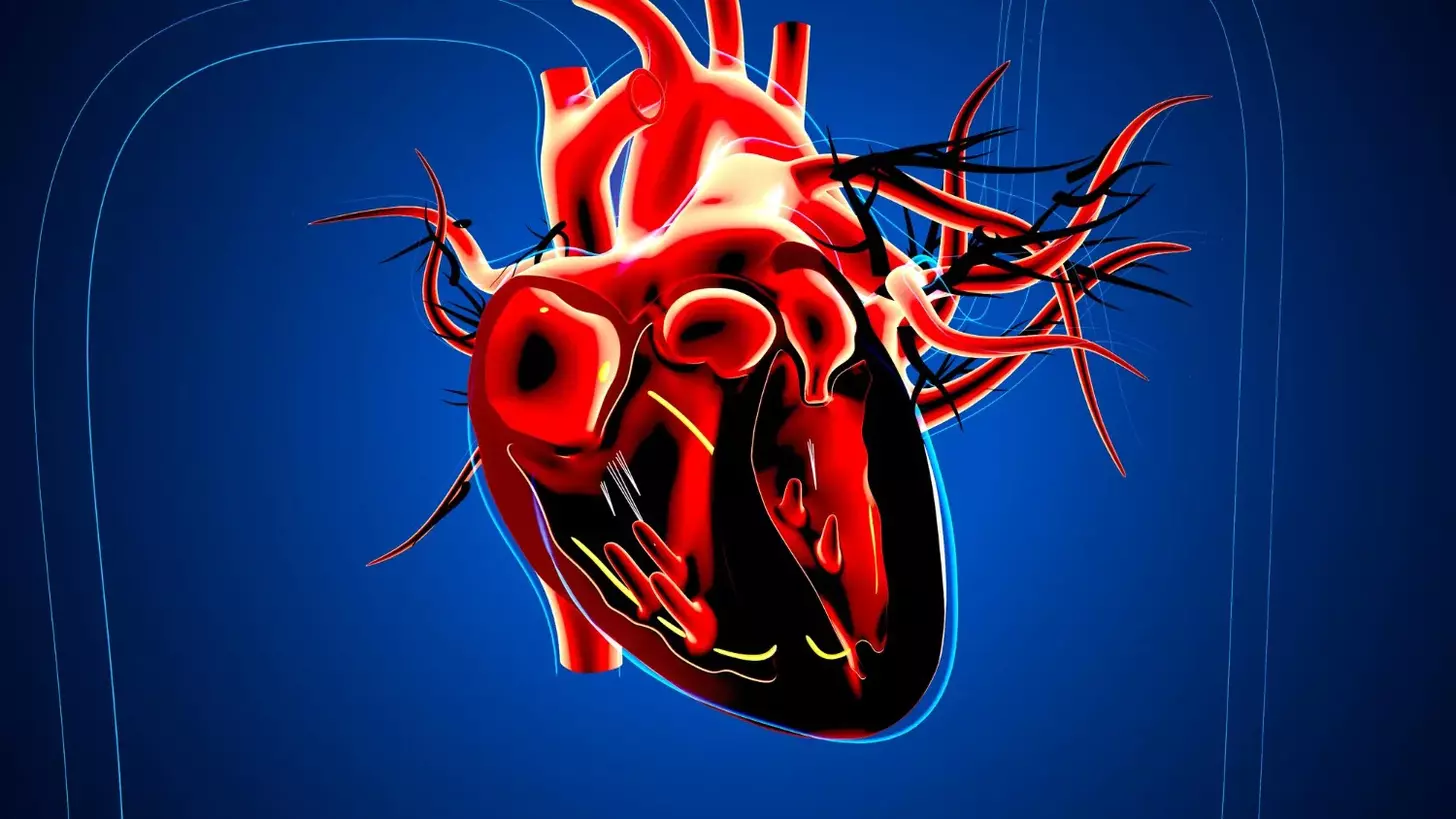
The atrioventricular (AV) valves are inside the heart. They let blood move from the atria to the ventricles but stop it from going back. The tricuspid and mitral valves are essential for blood to flow in one direction through the heart.
These valves help the heart pump blood well. They are important for the heart to work efficiently and keep the body healthy.
The AV valves are between the atria and ventricles. The tricuspid valve is on the right side, between the right atrium and ventricle. The mitral valve is on the left, between the left atrium and ventricle.
This placement helps the AV valves control blood flow. They make sure blood moves in the right direction.
The AV valves keep blood flowing in one direction. They open to let blood flow from the atria to the ventricles and then close to stop it from going back. This is key for the heart’s efficiency and overall health.
To better understand the AV valves, let’s look at their structure and how they work.
| Valve | Location | Function |
|---|---|---|
| Tricuspid Valve | Between right atrium and right ventricle | Regulates blood flow from right atrium to right ventricle |
| Mitral Valve | Between left atrium and left ventricle | Regulates blood flow from left atrium to left ventricle |
Knowing how AV valves work is important. It helps us understand their role in heart health and overall well-being.
The tricuspid valve is a key part of the heart. It has three leaflets that help blood flow smoothly. It’s important for blood to move right from the right atrium to the ventricle.
The tricuspid valve has three leaflets. These are thin, fibrous parts that open and close. This unique anatomy allows for efficient blood flow and stops backflow into the right atrium.
The leaflets are supported by chordae tendineae and papillary muscles. This setup makes sure the valve works right. It keeps the heart working well.
The tricuspid valve is key for blood to go from the right atrium to the right ventricle. This process is vital for keeping oxygen levels in the blood healthy. It makes sure blood only goes one way, keeping oxygen and deoxygenated blood separate.
There are several disorders that can affect the tricuspid valve. Tricuspid regurgitation is when the valve leaks, letting blood go back into the right atrium. Tricuspid stenosis is when the valve gets too narrow.
These conditions can cause serious problems if not treated, like right-sided heart failure. Knowing about these issues is key for finding and fixing them.
We know how important the tricuspid valve is for heart health. Understanding its role and how problems can arise helps us treat patients better. This improves their health outcomes.
The mitral valve is key to the heart’s function. It sits between the left atrium and ventricle. It makes sure blood flows only one way.
The mitral valve has two leaflets, a bicuspid design. This setup controls blood flow well. It helps the heart pump blood efficiently to the body. Chordae tendineae and papillary muscles support it, stopping blood from flowing back.
The mitral valve is vital for systemic circulation. It ensures oxygen-rich blood from the lungs reaches the body. Its proper working is key for healthy blood pressure and oxygen supply to tissues.
Mitral valve disorders, like mitral regurgitation and stenosis, affect life quality. They cause symptoms like shortness of breath and fatigue. Knowing about these disorders helps in finding better treatments.
| Condition | Description | Prevalence |
|---|---|---|
| Mitral Regurgitation | Backflow of blood from the left ventricle to the left atrium | Common in older adults |
| Mitral Stenosis | Narrowing of the mitral valve opening | Less common, often associated with rheumatic fever |
We understand the mitral valve’s role in heart health. Knowing its structure, function, and disorders helps us treat conditions better. This improves patient outcomes.
The heart has two AV valves: the tricuspid and mitral. They are key for blood flow. These valves make sure blood moves in one direction, stopping backflow and keeping circulation smooth.
The tricuspid and mitral valves are similar but also different. They are both between the atria and ventricles. They have leaflets that open and close to control blood flow. But, the tricuspid has three leaflets, and the mitral has two.
These valves are strong because of chordae tendineae and papillary muscles. They keep the leaflets from going back into the atria when the ventricles contract. The arrangement and size of these support systems vary between the valves.
Both AV valves make sure blood flows from the atria to the ventricles without backflow. But, the tricuspid valve is in the right heart, pumping blood to the lungs. The mitral valve is in the left heart, pumping blood to the body.
The tricuspid valve works under lower pressure. The mitral valve, in the left heart, works under higher pressure. This is because the left ventricle needs to pump blood throughout the body.
The right and left hearts have different pressures. This affects the AV valves. The right heart’s lower pressure means the tricuspid valve is adapted for it. The left heart’s higher pressure requires a stronger mitral valve during systole.
| Characteristics | Tricuspid Valve | Mitral Valve |
|---|---|---|
| Location | Right heart (between right atrium and ventricle) | Left heart (between left atrium and ventricle) |
| Number of Leaflets | Three | Two |
| Pressure Conditions | Lower pressure | Higher pressure |
Knowing these differences is key for treating valve problems. The unique anatomy and function of each valve mean treatments must be specific. This ensures the best care for each valve and its conditions.
The chordae tendineae are key for the heart’s valves to work right. These thin strings help the atrioventricular (AV) valves open and close. This ensures blood flows only one way through the heart.
The chordae tendineae are fibrous strings linking the AV valves to the papillary muscles. These muscles are specialized and found in the ventricles. This connection keeps the valve cusps tight, stopping them from closing back into the atria when the ventricles contract.
The chordae tendineae’s anatomy is complex. Many strings attach to each valve cusp. This setup allows for precise control over the valves’ opening and closing. The different types of chordae tendineae have specific roles and attachments.
When the ventricles contract, the papillary muscles pull the chordae tendineae. This stops the AV valve cusps from bulging back into the atria. It ensures blood flows forward through the heart and into the body.
The chordae tendineae’s role is vital for the heart’s efficiency. Without them, the AV valves can’t close right, causing backflow and serious heart problems.
A chordae tendineae rupture or dysfunction can severely affect the heart. If a chord ruptures, the valve cusp can bulge into the atrium, causing valvular regurgitation. This can lead to:
The effects of chordal rupture or dysfunction highlight their importance. They are key to keeping the valves working right and the heart healthy.
It’s key to know how AV valves work in the cardiac cycle for good heart health. The cycle has phases of diastole and systole. These phases are closely tied to how these valves function.
The AV valves, like the tricuspid and mitral, open and close with heart pressure changes. In diastole, when the ventricles relax, atrial pressure is higher. This lets the AV valves open, allowing blood to flow into the ventricles.
When the ventricles fill and pressure goes up, the AV valves start to close. They fully close when the ventricles contract in systole. This ensures blood flows only from the atria to the ventricles.
The AV valves work due to pressure differences between the atria and ventricles. In diastole, atrial pressure is higher, opening the valves for ventricular filling. In systole, ventricular pressure is higher, closing the valves to stop backflow.
This pressure difference is vital for efficient blood flow. It’s a key part of the cardiac cycle.
The movement of AV valves is linked to heart sounds during auscultation. The first heart sound (S1) happens when the AV valves close at systole start. This sound shows the shift from diastole to systole and is important for checking valve health.
Knowing how heart sounds relate to AV valve movement helps in diagnosing heart issues. It’s essential for checking heart health.
Checking AV valves is key to heart health. It helps spot and treat heart problems.
First, doctors use physical checks to look at AV valves. They listen with a stethoscope and feel the chest. This helps find any heart rhythm or valve issues.
Listening with a stethoscope is very important. It can pick up murmurs that show valve problems. For example, a certain murmur might mean the mitral valve is not working right.
Advanced imaging gives clear views of AV valves. Echocardiography shows the heart’s inner workings in real-time.
Other tools include:
Measuring heart pressures is key to seeing how AV valve problems affect the heart. Cardiac catheterization lets doctors measure these pressures.
| Hemodynamic Parameter | Normal Value | Abnormal Value Indicating Dysfunction |
|---|---|---|
| Left Ventricular End-Diastolic Pressure (LVEDP) | 5-12 mmHg | >12 mmHg (Possible mitral stenosis or regurgitation) |
| Mean Pulmonary Capillary Wedge Pressure (PCWP) | 6-12 mmHg | >12 mmHg (Possible mitral valve disease) |
Understanding these measurements is complex. But, by combining them with other data, doctors can make the best care plans for patients.
The atrioventricular (AV) valves are key to the heart’s function. Yet, they can face many disorders. These issues can harm the heart’s performance and overall health.
Stenosis is when the AV valves narrow, blocking blood flow. This can put extra pressure on the heart. If not treated, it might cause the heart to work too hard and fail.
Tricuspid stenosis often comes from rheumatic fever. Mitral stenosis can be caused by rheumatic heart disease or calcification. Echocardiography helps find how severe stenosis is, helping doctors decide on treatment.
Regurgitation happens when the AV valves don’t close right, letting blood leak back. This can make the heart work less efficiently. We’ll look at what causes it, its symptoms, and how to manage it.
Mitral regurgitation is common and can be due to mitral valve prolapse or other issues. Tricuspid regurgitation often comes from right ventricular problems. Treatment can range from medication to surgery.
AV valve problems from birth or structural issues can affect the heart a lot. Conditions like Ebstein’s anomaly or AV septal defects can mess with valve function. We’ll talk about how to diagnose and manage these issues.
| Condition | Description | Clinical Impact |
|---|---|---|
| Ebstein’s Anomaly | A congenital defect where the tricuspid valve is abnormally formed and the right ventricle is small. | Can lead to heart failure and arrhythmias. |
| AV Septal Defect | A congenital defect involving a hole in the septum between the heart’s chambers and often associated with AV valve abnormalities. | Can cause heart failure and pulmonary hypertension. |
Infective endocarditis is a serious infection of the AV valves. It can destroy the valves and be life-threatening. Inflammatory conditions like rheumatic fever can also affect the valves. We’ll cover how to diagnose, treat, and prevent these issues.
Infective endocarditis needs quick antibiotic treatment and sometimes surgery. To prevent it, people at high risk should get antibiotics before certain medical procedures.
At Liv Hospital, we lead in treating AV valve disorders with cutting-edge care. Our methods range from medical treatments to advanced surgeries. We focus on what’s best for each patient.
For many, treatment starts with medicine. This helps manage symptoms and slow disease. We tailor treatments to fit each patient’s needs.
Our cardiologists create detailed treatment plans. This might include medicines like anticoagulants and diuretics. Regular check-ups help adjust the plan as needed.
When medicine isn’t enough, we use less invasive repairs. These, like transcatheter valve repair, cut down on recovery time and risks.
At Liv Hospital, we use the latest in cardiac care. Our team works together to find the best treatment for each patient. This ensures the best results.
When repair isn’t possible, we replace the valve. We offer mechanical and biological options, each with its own benefits.
Mechanical valves last long but need lifelong blood thinners. Biological valves don’t need blood thinners but last shorter. Our surgeons help choose the best valve based on the patient’s life and health.
Liv Hospital is dedicated to top-notch cardiac care. We use the latest technology and focus on the patient. Our team aims for the best results.
We strive for international excellence in cardiac care. Our team works together to give each patient full care, from start to finish.
Keeping AV valves healthy is key for a good heart. The tricuspid and mitral valves control blood flow. They make sure blood moves in the right direction.
Knowing how these valves work helps us take care of our heart. This is why we need to look after them well.
Healthy AV valves prevent problems like stenosis and regurgitation. Regular check-ups and new tests can spot issues early. This means we can treat them quickly.
At Liv Hospital, we focus on top-notch heart care. We diagnose and treat AV valve problems. Our team works hard to give patients the best care for their heart and overall health.
The heart has two AV valves: the tricuspid and mitral valves. They let blood flow from the atria to the ventricles.
AV valves stop blood from flowing backward when the ventricles contract. This helps blood move efficiently around the body. They are supported by chordae tendineae.
The tricuspid valve has three leaflets and is on the right side. The mitral valve has two leaflets and is on the left.
Disorders include stenosis and regurgitation. Other issues are structural problems, congenital defects, and infections like endocarditis.
Doctors use physical exams and imaging like echocardiography. They also measure blood flow to check valve health.
Treatments include medicines and minimally invasive repairs. Sometimes, valves need to be replaced with prostheses.
Chordae tendineae connect the valve leaflets to papillary muscles. They stop blood from flowing backward during contraction.
AV valves open and close with pressure changes. This ensures blood flows in one direction. It also makes the sounds we hear when the heart beats.
Healthy AV valves are key for the heart to work well. They help blood flow efficiently and prevent problems from valve issues.
Subscribe to our e-newsletter to stay informed about the latest innovations in the world of health and exclusive offers!