Last Updated on November 25, 2025 by Ugurkan Demir

The human heart is a complex and vital organ. It pumps blood throughout the body. At Liv Hospital, we focus on advanced cardiac care. Knowing the human heart well is key to our mission.Learn key facts about heart chambers diagram, size, and their role in blood flow.
The heart has four chambers: two atria and two ventricles. They work together to circulate blood well. Its size is like a closed adult fist, and it’s a muscular, cone-shaped organ.
It’s important to understand the heart’s structure. This helps us see its function and why it’s so vital for our health. In this article, we’ll dive into thirteen key facts about the heart’s anatomy. We’ll look at its size, shape, and structure.

The human heart is a true marvel of anatomy. It has a complex structure and plays a vital role in our bodies. As we dive into its anatomy, we see just how complex and important it is.
The heart is known scientifically as “cor” or “cor humanum.” These Latin names are used worldwide in medicine and science. They highlight the heart’s importance across cultures and languages.
“The heart is the first organ to develop and the last to stop functioning in the human body.”
— A testament to its vital importance.
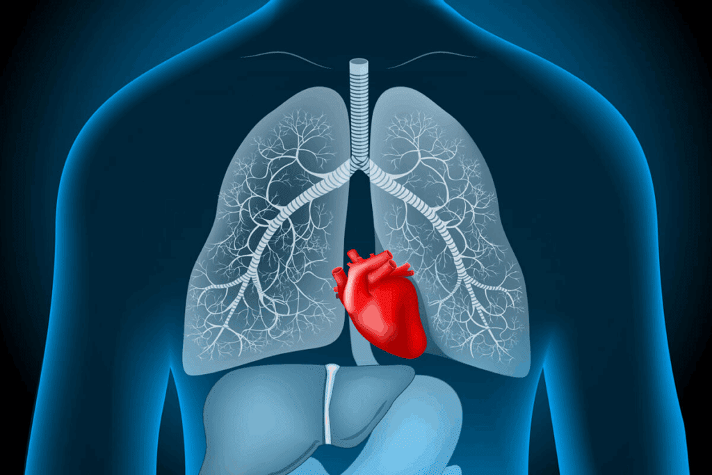
The heart sits in the thoracic cavity, between the lungs, in the mediastinum. This spot lets it pump blood efficiently around the body. It’s also near other key structures like the trachea, esophagus, and major blood vessels.
| Anatomical Feature | Description |
| Location | Thoracic cavity, between the lungs |
| Region | Mediastinum |
| Scientific Name | Cor or Cor Humanum |
The heart’s spot in the thoracic cavity is key for its work. It also affects its relationship with nearby structures. Knowing this anatomy is vital for diagnosing and treating heart issues.

Learning about the heart starts with its chambers. The heart chambers diagram is key in cardiology. It shows the heart’s inside parts clearly.
The heart has four chambers: the right and left atria, and the right and left ventricles. This setup is vital for blood circulation in the body.
The right atrium gets blood without oxygen from the body. The left atrium gets blood full of oxygen from the lungs. The right ventricle sends blood to the lungs for oxygen. The left ventricle sends oxygen-rich blood to the body.
Diagrams use colors to show blood types. Oxygenated blood is red, and deoxygenated blood is blue. This makes it easier to see blood flow in the heart.
This color system is essential for students and doctors. It helps them understand the heart’s work and explain it to patients.
The human heart is actually a deep, dark red. This is different from the bright red and blue colors often seen in textbooks. These colors are used to show oxygenated and deoxygenated blood.
So, why does the heart look different in real life than in diagrams? It’s because of its rich blood supply and strong muscles.
The heart is a muscular organ that pumps blood. Its dark red color comes from lots of blood vessels and oxygen-depleted blood. Diagrams, on the other hand, use bright colors to show the heart’s parts and blood flow.
The heart’s real color is not as bright as diagrams show. This is because it’s a living organ with lots of blood. This gives it a deeper, richer color.
There are a few reasons why the heart’s color in diagrams is different:
| Characteristics | Actual Heart | Diagram Representations |
| Color | Dark Red | Bright Red and Blue |
| Purpose of Color | Natural Color due to Blood Supply | To Differentiate Between Chambers and Vessels |
| Representation | Realistic | Simplified for Educational Purposes |
In conclusion, the human heart is actually a deep, dark red. This is because of its blood and muscles. Diagrams show the heart in bright colors for learning and to simplify complex details.
The human heart is often compared to a closed fist. This simple analogy helps us understand its size. It makes it easier to grasp its role in our body’s circulatory system. We’ll dive deeper into this comparison, looking at the average heart size and how it relates to body size.
The average adult heart is about the size of a closed fist. It measures around 12 cm in length, 8 cm in width, and 6 cm in thickness. These numbers can vary, but they give us a general idea of the heart’s size.
Studies show that heart size and body size are linked. Larger people usually have bigger hearts. This is because their hearts need to work harder to pump blood to their bigger bodies. But, this isn’t always the case and can be affected by health and fitness.
For example, athletes or those who exercise a lot might have bigger hearts. This is because their hearts have to work harder to meet the body’s needs. On the other hand, people with certain health issues might have hearts that are larger or smaller than usual.
The human heart’s dimensions are key to understanding its health and function. We’ll look at the average heart measurements. These are important for grasping its size and structure.
An adult human heart is about 12 cm long. This size is important for knowing the heart’s size and where it fits in the chest.
The heart is usually 8 cm wide. This, along with its length, helps us see its shape and where it fits in the body.
The heart is about 6 cm thick. This shows how strong the heart is and how well it can pump blood.
These exact measurements – length, width, and thickness – are key for doctors to check heart health. They help in diagnosing and treating heart issues. Knowing these details helps us appreciate the heart’s complex anatomy.
The heart’s shape is like a cone or pyramid. This shape is key for pumping blood all over the body. It helps blood flow well and is a big part of the heart’s design.
The heart looks like a cone or pyramid because it tapers from the base to the tip. This shape is not just for looks; it’s essential for pumping blood. The cone shape helps the heart contract and relax to move blood well.
As a cardiologist, notes, “The heart’s shape is linked to its function. The cone shape makes pumping blood more efficient. This ensures blood flows all over the body with little resistance.”
The heart has different areas, each with its own boundaries. The base of the heart is at the top, made mainly by the left atrium. The apex is the bottom tip, made by the left ventricle. Knowing these areas helps doctors diagnose and treat heart problems.
The heart’s edges are set by its surroundings, like the sternum, diaphragm, and lungs.
The heart’s apex points downward and to the left. This is not just a random direction. It’s key for understanding how the heart works and when it doesn’t.
The heart’s apex is directed downward and leftward. This is due to the heart’s structure and where it sits in the chest. This direction affects how the heart functions and how it interacts with other parts of the body.
Anatomical Basis: The heart’s apex is at the tip of the left ventricle. This ventricle pumps blood all over the body. The muscle and how it contracts shape the apex’s direction.
The heart’s apex position is very important for doctors. It helps them diagnose heart problems and check heart health.
“The apex beat, also known as the point of maximal impulse, is a key sign for doctors. It’s usually found in the fifth intercostal space, mid-clavicular line. If it’s off, it might mean there’s something wrong with the heart,” as noted in clinical cardiac examination guidelines.
Diagnostic Significance: The apex’s position can show if the heart is too big or too small. For example, if the apex beat is off, it could mean the ventricle is too big.
| Condition | Apex Beat Location | Clinical Implication |
| Normal Heart | Fifth intercostal space, mid-clavicular line | Normal cardiac function |
| Left Ventricular Hypertrophy | Displaced laterally | Suggests ventricular enlargement |
| Right Ventricular Hypertrophy | May be displaced or not palpable | Indicates right ventricular overload |
Knowing about the heart’s apex is vital for both learning about the heart and for doctors. It helps them diagnose and treat heart problems. This shows how important this part of the heart is.
The heart is a single muscle that connects its chambers and valves. This unity is key for pumping blood across the body.
The heart’s muscle cells work together to pump blood. This unity is vital for the heart to function as one organ.
The heart’s muscle is not broken but works as one. This unity keeps the heart pumping well and keeps the heart healthy.
The heart’s chambers and valves are part of its muscle. The four chambers and valves work together to move blood right.
The valves stop blood from flowing back. This ensures blood moves smoothly through the heart’s chambers.
| Component | Function |
| Right Atrium | Receives deoxygenated blood |
| Left Atrium | Receives oxygenated blood |
| Right Ventricle | Pumps deoxygenated blood to lungs |
| Left Ventricle | Pumps oxygenated blood to body |
The heart’s structure makes it a single, efficient pump. Knowing this helps us understand how it keeps us alive.
A healthy heart usually has a certain weight range. This range can change based on different factors. The average weight of a healthy adult heart is a key sign of good heart health.
The average weight of a healthy heart is between 250-350 grams. This can vary a bit. It depends on overall health, body size, and other factors.
Many things can affect a healthy heart’s mass. These include:
Knowing these factors is key to understanding heart health. It helps spot any possible issues.
The human heart is mostly the same but can change a lot. These changes show how the heart can adapt to different needs. It’s amazing how the heart can adjust to our lifestyle and health.
One area where hearts can differ is in their shape and size. Normal anatomical variations mean the heart can be slightly different in size and shape. These differences are okay and don’t usually cause problems.
Hearts can vary in size and shape, and even in how thick the walls are. Some people might have a bigger or smaller heart than others. This can be because of their genes.
A study found that the heart can change a lot. This is because of things like age, sex, and how fit someone is.
“The heart’s structure and function can vary significantly among healthy individuals, reflecting adaptations to different lifestyles and physiological conditions.”
Exercise and how we live can change our heart. For example, working out can make the heart muscle thicker. This is a normal response to more physical activity.
But, not moving much can make the heart less efficient. On the other hand, people who exercise a lot might have a heart that pumps blood better. This shows how the heart can change to meet our needs.
In summary, hearts are different from person to person. This is because of genetics, how we live, and our environment. Knowing this helps us understand how amazing the heart is. It also helps us spot health problems early.
The heart has four chambers, each with its own role. They work together to keep blood flowing well. Knowing what each chamber does helps us understand how the heart works.
The right and left atria collect blood. The right atrium gets blood from the body, and the left atrium gets blood from the lungs. They hold the blood until it’s needed.
The atria also push blood into the ventricles. This makes sure the ventricles are ready to pump. This teamwork is key for the heart to work well.
The right and left ventricles pump blood. The right ventricle sends blood to the lungs, and the left ventricle sends blood to the body. They have strong muscles to push blood through the body.
The heart’s design is amazing. It lets the heart meet the body’s needs efficiently.
We’ve looked into the human heart’s details, from its anatomy to its engineering. Its four-chamber setup, precise sizes, and cone shape help it work well.
The heart is a complex organ, key to our life. Its size, shape, and structure are made for its vital tasks. It shows amazing engineering.
Knowing the heart’s anatomy helps us see its importance and health needs. Recognizing its engineering makes us value heart health more. We should take steps to protect it.
Studying heart anatomy helps us understand this vital organ better. It shows the complex systems that keep us alive. As we learn more about the heart, we see its importance and the need to care for it.
The scientific name for the human heart is “Cor” or “Cor Humanum”.
The human heart is about the size of a closed fist. It is roughly 12 cm long, 8 cm wide, and 6 cm thick.
The human heart is actually dark red in color. This is different from the red and blue colors used in diagrams to show oxygenated and deoxygenated blood.
The human heart is shaped like a cone or pyramid. It has distinct parts and boundaries.
Yes, the human heart is a single muscular structure. Its chambers and valves work together as one organ.
A healthy heart weighs between 250-350 grams. Exercise and lifestyle can affect its mass.
Heart size usually matches body size. People with larger bodies tend to have bigger hearts.
The human heart is about 12 cm long, 8 cm wide, and 6 cm thick.
The heart’s apex points downward and leftward. This is important for medical assessments and diagnoses.
The right and left atria collect blood. The right and left ventricles pump blood throughout the body. They work together to circulate blood.
Subscribe to our e-newsletter to stay informed about the latest innovations in the world of health and exclusive offers!