Last Updated on November 25, 2025 by Ugurkan Demir

Explore labeled diagram of cardiac system with heart and circulation explained.
It’s key to understand the cardiac and circulatory system to grasp blood circulation. The heart pumps blood through arteries, veins, and capillaries. This makes it a critical organ for health.
At Liv Hospital, we know how important clear anatomical insights are. That’s why we’re looking at 11 essential diagrams. They show the heart’s structure and its link to the circulatory system.
These labeled diagrams give a full view of how the heart and vessels keep you healthy. By looking at these diagrams, people can better understand the circulatory system’s complexity.

Understanding the basics of cardiac and circulatory anatomy is key to knowing how our bodies work. This system, which includes the heart, blood vessels, and blood, is complex. It’s essential for delivering oxygen and nutrients to our tissues and organs.
We will look at why visual learning is important for grasping circulation. We’ll also guide you through this detailed guide.
Visual learning is a great way to understand complex systems like the heart and blood flow. Diagrams and labeled pictures help us see how blood moves through the heart, lungs, and body. Visual aids make complex info simpler, helping us understand circulation’s detailed paths.
This guide will walk you through the basics of the heart and blood flow. It’s set up to be easy to follow, covering all key parts of the circulatory system. You’ll learn about its components, how it works, and why visual learning is key.
The circulatory system has four main parts: blood, the heart, blood vessels, and lymph. It has two main loops: one for oxygen-rich blood and another for oxygen-poor blood. Knowing about these parts and their roles is essential for a full grasp of the circulatory system.
| Component | Function |
| Blood | Transports oxygen, nutrients, and waste products |
| Heart | Pumps blood throughout the body |
| Blood Vessels | Arteries, veins, and capillaries that transport blood |
| Lymph | Part of the immune system, aids in waste removal |

Seeing the heart’s structure through a detailed diagram helps us understand its role. The heart is a vital organ that acts as the body’s pump. It makes sure blood circulates throughout the body.
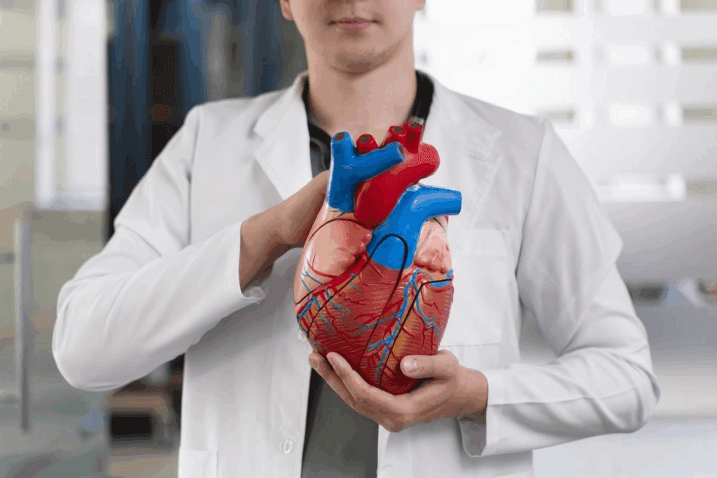
We will look at the heart’s detailed anatomy. This includes its four chambers and the role of its valves. A detailed diagram of the cardiac system shows the heart’s structure. It has four chambers: the right and left atria, and the right and left ventricles.
The heart’s four chambers work together to move blood efficiently. The atria take in blood coming back to the heart. The ventricles then push blood out into the body.
The heart’s valves make sure blood flows the right way. There are four main valves: the tricuspid, pulmonary, mitral, and aortic valves.
These valves are key for the heart’s efficiency and stopping backflow. A cardiovascular system labeled diagram helps us see these structures and their roles.
Knowing the heart’s structure is key to understanding the whole cardiovascular system. By seeing the heart’s chambers and valves in a detailed diagram, we can better grasp its complexity and importance.
Diagrams help us see how the human circulatory system works. It’s a closed network of blood vessels. These vessels include arteries, veins, and capillaries. They are key for moving blood around the body.
The circulatory system is a complex network of blood vessels. It helps move blood all over the body. Arteries carry oxygen-rich blood away from the heart. Veins bring back blood that’s low in oxygen. Capillaries are where oxygen and nutrients get to the cells and waste is removed.
The closed vascular network keeps blood flowing well. It’s vital for delivering oxygen and nutrients. It also helps get rid of waste.
A detailed diagram shows the main arteries and veins. The aorta is the biggest artery. It starts from the left ventricle of the heart. It then splits into smaller arteries that spread oxygen-rich blood everywhere.
The pulmonary arteries and pulmonary veins are also key. Pulmonary arteries take deoxygenated blood to the lungs. Pulmonary veins bring oxygen-rich blood back to the heart.
Knowing how the circulatory system works is important. It helps doctors find and treat heart problems.
Exploring the circulatory system shows us two key paths: the pulmonary and systemic circuits. These paths are key to grasping how blood moves in our bodies.
The circulatory system has two main paths: the pulmonary circuit and the systemic circuit. The pulmonary circuit helps exchange oxygen and carbon dioxide between the heart and lungs. The systemic circuit carries oxygen-rich blood to our body’s tissues and brings back oxygen-poor blood to the heart.
The pulmonary circuit is essential for gas exchange between the heart and lungs. It sends deoxygenated blood to the lungs. There, it picks up oxygen and lets go of carbon dioxide through breathing.
The process involves:
The systemic circuit carries oxygen-rich blood to our body’s tissues. It also brings back oxygen-poor blood to the heart. This circuit is vital for our body’s health and function.
| Circuit | Function | Blood Flow Path |
| Pulmonary | Oxygen exchange between heart and lungs | Right ventricle to lungs to left atrium |
| Systemic | Oxygen delivery to body tissues | Left ventricle to body to right atrium |
The diagram of the human circulatory system shows the pulmonary and systemic circuits. Knowing about these circuits helps us understand how the circulatory system works.
For those new to anatomy, a simple diagram of the circulatory system is very helpful. Visuals are key to understanding how blood moves through our bodies.
A simple diagram of the circulatory system shows the heart, arteries, veins, and capillaries. These parts work together to move blood around the body. They deliver oxygen and nutrients to our tissues and organs. Knowing these basics is key to understanding the circulatory definition biology.
The heart is like a pump, pushing blood through our blood vessels. Arteries carry blood full of oxygen away from the heart. Veins bring blood back to the heart without oxygen. Capillaries, the smallest vessels, help swap oxygen, nutrients, and waste.
Color-coded representations in diagrams make learning easier. They show different blood vessels and where blood flows. For example, arteries are red for oxygen-rich blood, and veins are blue for oxygen-poor blood.
This makes it simpler for beginners to grasp the labeled circulatory system. It helps us see how all the parts work together.
Using simple, labeled diagrams and colors helps us understand the circulatory system better. It shows us how it affects our health.
Anatomical terms are key to understanding the circulatory system. They help us identify its parts clearly. This is important in both learning and medical settings.
The circulatory system has many important parts. The heart has four chambers: the right and left atria, and the right and left ventricles. Knowing these terms is essential.
Arteries and veins also have specific names. The aorta, the biggest artery, starts from the left ventricle. The superior and inferior vena cava carry deoxygenated blood back to the heart.
Diagrams with labels show blood flow direction. They use arrows and markers to illustrate the path. This helps us understand how blood moves.
Knowing blood flow direction is important. It shows how oxygen and deoxygenated blood move around the body. The pulmonary circuit and systemic circuit are key paths shown in these diagrams.
Clinical annotations on a human cardiovascular system diagram offer insights into where to look for problems. They help doctors and students understand the heart better. This is key for learning about its structure and how it works.
Looking at a cardiovascular system labeled diagram helps spot important points for diagnosis. These include major arteries and veins. They are key for spotting heart problems.
On a blood circulatory system diagram, certain points are vital for making accurate diagnoses. These include:
Knowing these points is essential for reading ECGs and spotting irregular heart rhythms.
A human cardiovascular system diagram with clinical notes also points out common problem areas. These include spots prone to:
By knowing these areas, doctors can plan better treatments for heart diseases.
In summary, a cardiovascular system labeled diagram is a vital tool in healthcare. It helps us understand the heart system better. It also helps in diagnosing and treating heart conditions.
Seeing how the blood circulatory system works helps us understand oxygen transport. We’ll look at how oxygen and deoxygenated blood move through the body. We’ll also see why gas exchange is key at tissue and cellular levels.
Oxygen-rich blood goes from the lungs to the body’s tissues through the arterial system. Deoxygenated blood, full of carbon dioxide, goes back to the lungs via the venous system. This balance is vital for our health.
The arterial system has arteries that split into smaller arterioles. These lead to capillaries where gas exchange happens. The venous system collects deoxygenated blood from capillaries and brings it back to the heart.
Gas exchange happens in the capillaries. Oxygen moves into tissues, and carbon dioxide moves into the blood. This is key for cells to make energy.
The thin walls of capillaries help gases move efficiently. Oxygen fuels cell activities, while carbon dioxide is carried back to the lungs to be exhaled.
Understanding the blood circulatory system is key to grasping oxygen transport. By seeing how oxygenated and deoxygenated blood move, we see the circulatory system’s life-giving role.
The vascular system is key for delivering oxygen and nutrients to our bodies. It’s made up of arteries, veins, and capillaries. This network ensures blood flows everywhere it needs to.
We’ll look at how arteries branch out and veins bring blood back. This will help us understand how the vascular system works.
The arterial system carries oxygen-rich blood from the heart to the rest of us. It splits into smaller arteries and arterioles. These lead to capillaries, where oxygen and nutrients are exchanged.
| Arterial Branch | Description | Function |
| Aorta | Main artery arising from the heart | Distributes oxygenated blood |
| Arteries | Branches of the aorta | Carry blood to organs and tissues |
| Arterioles | Small branches of arteries | Regulate blood pressure and flow |
The venous system brings deoxygenated blood back to the heart. It starts with capillaries merging into venules. These then join into larger veins.
Knowing how the venous system works is important for treating blood circulation problems.
A detailed human vascular system diagram shows the complex paths of arteries and veins. It helps us understand the circulatory system better.
Looking at a diagram circulatory system helps us see the different circulatory systems. We learn how they keep us healthy.
The human circulatory system is a vast network that stretches over 60,000 miles. It’s essential for delivering oxygen and nutrients to tissues all over the body. We’ll dive into the details of this system, from the big structures like arteries and veins to the tiny capillary networks.
At the micro-level, the circulatory system breaks down into capillary networks. These networks are key for exchanging oxygen, nutrients, and waste with tissues. Microcirculation is the blood flow within these networks. It’s vital for keeping tissues healthy and working right.
Capillary networks are so dense that almost every cell is near a capillary. This ensures efficient exchange. The thin walls of capillaries make this exchange possible.
Calculating the total length of the circulatory system is a challenge. It’s because of its complex branching and the huge number of capillaries. Yet, it’s estimated that an adult human’s blood vessels, if laid end to end, would stretch for about 60,000 miles.
To understand this better, the Earth’s equator is about 24,901 miles around. So, the circulatory system’s length is like circling the Earth more than twice.
| Component | Estimated Length (miles) | Description |
| Major Arteries | Several hundred | Primary vessels carrying oxygenated blood away from the heart |
| Capillary Networks | Tens of thousands | Dense networks facilitating exchange with tissues |
| Major Veins | Several hundred | Vessels returning deoxygenated blood to the heart |
This amazing length shows how complex and vital the circulatory system is. Knowing about its structure, from big to small, helps us understand its role in our health.
Understanding the cardiac and circulatory systems is key to grasping blood circulation. A diagram of the cardiac system shows us the heart’s structure. It also tells us how it keeps the body balanced.
The circulatory system is a complex network. It carries oxygen and nutrients to tissues and takes away waste. A diagram of the human cardiovascular system shows the blood’s journey, pointing out key arteries and veins.
It’s vital to know about the heart, the blood’s path, and the vascular system’s network. This knowledge helps in both medical care and teaching. It improves patient care and results.
Studying the cardiac and circulatory systems helps us see how the body stays healthy. It supports our overall health and well-being.
The heart acts as the body’s pump. It pushes blood all over the body. It takes in blood without oxygen, sends it to the lungs to get oxygen, and then sends oxygen-rich blood to the body.
The heart has four chambers: the right and left atria, and the right and left ventricles. The atria catch blood coming back to the heart. The ventricles push blood out to the body.
The pulmonary circuit helps the heart and lungs exchange oxygen and carbon dioxide. The systemic circuit carries oxygen-rich blood to the body’s tissues and brings back deoxygenated blood to the heart.
A labeled diagram uses special terms to explain the circulatory system. It shows how blood moves through the body. This makes it easier to understand.
Knowing how long and complex the circulatory system is shows its importance. It helps us see how it keeps the body balanced.
Capillary networks are key for exchanging oxygen and nutrients with tissues. They help deliver what the body’s cells need.
Visual aids are great for learning about the circulatory system. They make it easier to see how blood moves through the heart, lungs, and body.
A detailed diagram shows the main arteries and veins, like the aorta and pulmonary veins. Knowing the circulatory system’s layout helps us understand blood flow.
The circulatory system sends oxygen-rich blood to tissues through the systemic circuit. It then brings back deoxygenated blood to the heart for oxygen.
The heart’s valves, like the tricuspid and mitral valves, are vital. They ensure blood flows the right way, preventing backflow and keeping circulation smooth.
Subscribe to our e-newsletter to stay informed about the latest innovations in the world of health and exclusive offers!