Did you know that cancerous lymph nodes are a key sign of cancer spreading? CT Scan Detection of Cancerous Lymph Nodes can spot enlarged lymph nodes, which might mean cancer. This is important for figuring out how far and how fast cancer has grown.
We know how important it is to check lymph nodes right for cancer diagnosis. CT scans help find lymph node metastasis. This helps doctors make better treatment plans.
Key Takeaways
- Cancerous lymph nodes can be detected using CT scans.
- Enlarged lymph nodes may indicate cancer.
- CT imaging is key for checking lymph node metastasis.
- Getting lymph nodes right is essential for cancer staging.
- Good treatment plans need a clear diagnosis.
Understanding Lymph Nodes and Their Role in Cancer
To understand how cancer affects the body, knowing about lymph nodes is key. These small, bean-shaped structures are vital for the immune system.
The Lymphatic System’s Function in the Body
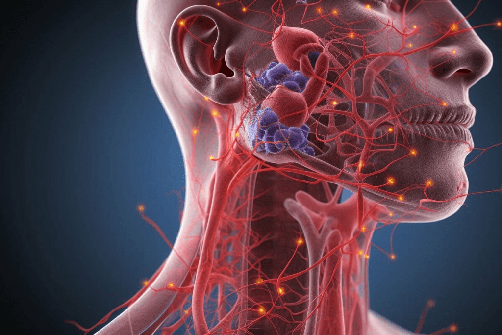
The lymphatic system is a network that helps defend against infections and diseases. It filters out harmful substances and moves immune cells around the body. The lymphatic system’s function is vital for overall health, and problems with it can lead to cancer.
How Cancer Affects Lymph Nodes
Cancer can spread to lymph nodes, a process called metastasis. When this happens, cancer cells build up, making the nodes bigger. This enlargement can be an indicator of cancer, and finding these changes is key for diagnosis and staging. We will look at how CT scans help spot cancerous lymph nodes.
Cancer in lymph nodes not only harms the lymphatic system but also affects cancer staging and treatment plans. Knowing how cancer impacts lymph nodes is critical for creating effective treatments.
What is a CT Scan and How Does it Work?

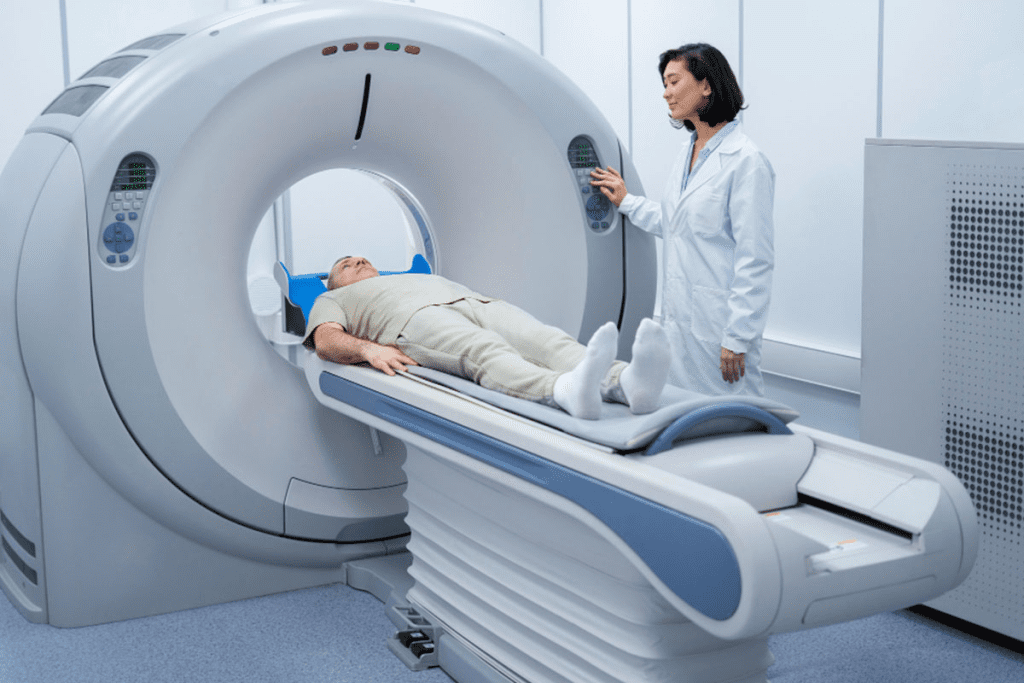
CT scans are a key tool for doctors. They use X-rays and computers to see inside the body. This helps us find and track many health issues.
Basic Principles of CT Imaging Technology
Basic Principles of CT Imaging Technology
CT scans work by using X-rays differently in each body part. An X-ray machine sends beams through the body. Sensors catch these beams and send them to a computer.
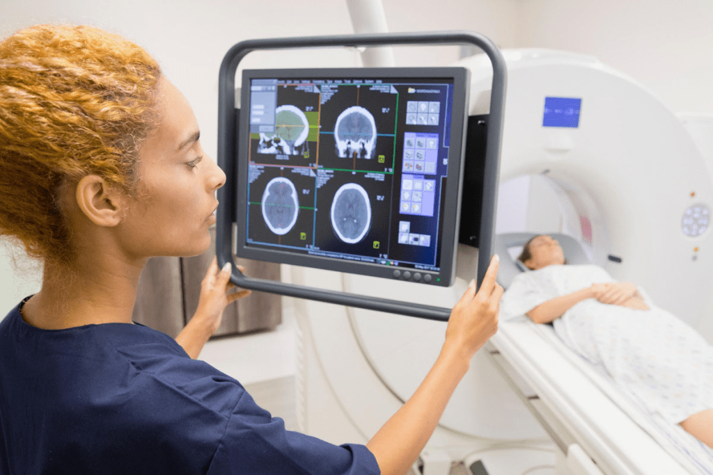
The computer then makes detailed pictures from these images. The patient lies on a table that moves into a CT scanner. As the scanner moves, it takes pictures from all sides.
The computer puts all these pictures together. This creates a full view of what’s inside the body.
Differences Between CT and Other Imaging Methods
CT scans are different from MRI and ultrasound. MRI uses magnetic fields, while CT scans use X-rays. This makes CT scans better for seeing bones and certain tissues.
CT scans show more detail than regular X-rays. They can spot problems in soft tissues, like in lymph nodes. This is key for finding and understanding cancer.
CT scans are fast, give clear images, and show complex structures well. These qualities make them vital in medical care, even in emergencies.
Knowing how CT scans work helps us see their value in health care. They are key in diagnosing and managing diseases, including cancer.
CT Scan Detection of Cancerous Lymph Nodes

CT scans are key in finding cancer in lymph nodes. They help doctors plan treatment by
showing detailed images. These images are important for spotting and treating cancer.
How CT Scans Visualize Lymph Node Abnormalities
CT scans show lymph node problems by taking detailed pictures inside the body. Contrast-enhanced CT scans are better because they show differences in tissues. This makes it easier to see if lymph nodes are too big or look wrong.
Research shows contrast CT scans are better at finding cancer in lymph nodes. They help see lymph nodes clearly by showing them against other tissues. This makes finding problems easier.
- Big lymph nodes might mean cancer.
- Abnormal shapes or density in lymph nodes also suggest cancer.
- CT scans are a safe way to check these signs.
Contrast vs. Non-Contrast CT for Lymph Node Imaging
Choosing between contrast and non-contrast CT scans depends on the situation. Contrast-enhanced CT is often better for clear images of lymph node issues.
But, non-contrast CT scans are good when contrast is not safe. We need to think about what’s best for each patient.
| Imaging Type | Advantages | Limitations |
| Contrast-Enhanced CT | Shows lymph node problems well | May cause allergic reactions |
| Non-Contrast CT | Safe for those who can’t have contrast | May not show soft tissue issues well |
Characteristics of Cancerous Lymph Nodes on CT Images
Cancerous lymph nodes show clear signs on CT scans that help doctors diagnose them. These signs are key for radiologists to spot and understand during scans.
Size and Shape Changes
Lymph nodes change size and shape when cancer is present. Normally, they are small and bean-shaped. But, cancer can make them grow and become irregular.
Getting bigger is a big warning sign of cancer. We look for nodes that are too big for their usual size. This could mean cancer is there.
The shape of lymph nodes also changes with cancer. They can turn from oval to round or irregular. This change is a key thing radiologists check on CT scans.
Density and Enhancement Patterns
The density and how they react to contrast on CT scans tell us a lot. Cancerous nodes might look different than normal ones. They could be more heterogeneous because of dead tissue or calcium inside.
When contrast is used in the scan, how nodes react can tell us if they’re cancerous. Cancerous nodes often don’t take the contrast evenly. This uneven reaction is a clue to cancer.
Knowing these signs is vital for doctors to correctly read CT scans for cancer. By looking at size, shape, density, and how they react to contrast, doctors can spot cancerous lymph nodes. This helps them plan the best treatment.
Accuracy and Limitations of CT Scans for Lymph Node Cancer Detection
It’s important to know how well CT scans work in finding lymph node cancer. They help us see if cancer is in the lymph nodes and how far it has spread.
Sensitivity and Specificity Rates
The sensitivity and specificity of CT scans are key. Sensitivity means the test can find cancer in lymph nodes correctly. Specificity means it can also find who doesn’t have cancer in lymph nodes correctly.
Research shows CT scans can spot lymph node cancer 70% to 90% of the time. This depends on how big the lymph nodes are and if there’s necrosis or calcification.
Factors Affecting Detection Accuracy
Several things can change how well CT scans find lymph node cancer. These include:
- Contrast agents make lymph nodes easier to see.
- The quality of the CT scan matters, depending on the technology and the radiologist’s skill.
- It’s harder to spot small or tricky-to-reach lymph nodes.
| Factor | Impact on Detection Accuracy |
| Contrast Agents | Make lymph nodes more visible |
| Scan Quality | It affects how clear and detailed the images are |
| Lymph Node Size and Location | It’s harder to find small or hard-to-reach nodes |
Knowing these factors and the limits of CT scans helps doctors make better choices for patients. This includes diagnosing and planning treatments.
Types of Cancer That Commonly Spread to Lymph Nodes
Lymph nodes are often where cancer spreads to. This spread helps doctors figure out the cancer’s stage and how it might progress. We’ll look at cancers that often spread to lymph nodes, including those that start in the lymph system and others that spread from other cancers.
Primary Lymphatic Cancers
Primary lymphatic cancers start in the lymph system. Lymphomas are a prime example. They grow in lymph nodes, spleen, or other lymphoid tissues. There are two main types: Hodgkin lymphoma and non-Hodgkin lymphoma.
These cancers can make lymph nodes swell and hurt. Another rare cancer, lymphoblastic lymphoma, is very aggressive and needs quick treatment. Knowing the type of lymphoma is key to choosing the right treatment.
Metastatic Cancers Affecting Lymph Nodes
Cancers like breast cancer, lung cancer, and melanoma often spread to lymph nodes. This means the cancer has reached a more advanced stage. Finding cancer in lymph nodes usually means changing treatment plans, possibly to more intense therapies.
In breast cancer, finding cancer in axillary lymph nodes is very important for treatment planning. For melanoma, checking the sentinel lymph node is a key step in diagnosing cancer spread.
Knowing which cancers spread to lymph nodes is vital for diagnosis and treatment. We’re always learning more and improving treatments for these complex cases, helping patients get better.
Regional Differences in Lymph Node Imaging
Lymph node imaging varies by region for accurate diagnosis and treatment. Lymph nodes look and act differently in different parts of the body. Knowing these differences is key to good patient care.
Neck and Head Lymph Nodes
Lymph nodes in the neck and head are checked for cancers like thyroid and head and neck tumors. These nodes are smaller and more numerous than others. On CT scans, they’re suspicious if they’re big, round, or show dead tissue in the middle.
We look at size, shape, and how they change after contrast on CT scans. For example, nodes over 1.5 cm in the jugulodigastric area are seen as abnormal.
Thoracic and Abdominal Lymph Nodes
In the chest, lymph nodes are checked for lung cancer and lymphoma. The size that’s considered abnormal varies by location. For instance, nodes over 1 cm in short axis are usually suspicious.
In the belly, nodes are looked at for cancers like those in the gut and kidneys. Nodes bigger than 1 cm in short axis are seen as enlarged. Dead tissue or calcium inside these nodes can also mean cancer.
Pelvic and Inguinal Lymph Nodes
Pelvic nodes are checked for cancers like prostate and bladder tumors. Big pelvic nodes are common in these cancers. Finding them on CT scans helps decide what to do next.
Inguinal nodes are checked for cancers in the lower leg, genitals, and anus. Nodes bigger than 1.5 cm in the short axis are considered enlarged.
We summarize the regional differences and characteristics of lymph nodes in the table below:
| Region | Size Criteria for Suspicion | Common Cancers Involved |
| Neck and Head | >1.5 cm | Thyroid, Head and Neck cancers |
| Thorax | >1 cm | Lung cancer, Lymphoma |
| Abdomen | >1 cm | Gastrointestinal, Genitourinary cancers |
| Pelvis | >1 cm | Prostate, Bladder, Gynecological cancers |
| Inguinal | >1.5 cm | Lower limb, Genital, Anal cancers |
Understanding these regional differences is key for accurate CT scan interpretation and guiding cancer treatment.
The Patient Experience: Undergoing a CT for Lymph Node Assessment
Getting a CT scan can seem scary, but we’re here to help. A CT scan for lymph nodes is a big step in finding and treating health issues.
Preparation and Procedure Details
Getting ready for a CT scan is important. Patients are usually told to:
- Wear comfy, loose clothes
- Take off metal things like jewelry or glasses
- Tell their doctor about any allergies, like to contrast dye
- Follow special rules about eating and drinking before the scan
During the scan, you’ll lie on a table that moves into a big machine. The CT scanner takes pictures of your body’s inside parts. These pictures are then checked by doctors. The whole thing is usually painless and takes 15-30 minutes. But, you might be there longer.
What to Expect During and After the Scan
During the scan, you might need to hold your breath sometimes. This helps get clear pictures. The machine will make sounds as it works. After it’s done, you can usually go back to your normal day unless your doctor says not to.
It’s okay to feel a bit worried about the results. But, the CT scan is a key tool for doctors. It helps them make good plans for your care. They’ll look at the results and tell you what’s next.
Knowing what to expect from a CT scan can help you feel less scared. Our medical team is here to support you every step of the way.
When CT Scans Might Miss Cancerous Lymph Nodes
CT scans can struggle to find cancerous lymph nodes. This is due to technical and interpretive issues. The size of the nodes and the scan technology play big roles in how accurate they are.
Size Limitations in Detection
One big problem with CT scans is their ability to see small lymph nodes. Even if these nodes are cancerous, they might not show up on a scan. This is because they are too small for the scan to detect.
Here’s a table that shows how hard it is to spot cancerous lymph nodes based on their size:
| Lymph Node Size | Detection Likelihood | Challenge |
| < 5 mm | Low | Resolution limitations |
| 5-10 mm | Moderate | Variable appearance |
| > 10 mm | High | Easier to detect |
Technical and Interpretive Challenges
There are also technical and interpretive hurdles with CT scans. Things like patient movement or metal objects can mess up the scan. Radiologists also face challenges in telling apart benign and malignant nodes.
To tackle these issues, new CT tech and contrast agents are being used. Artificial intelligence and machine learning are also helping improve lymph node detection on CT scans.
It’s key for radiologists and doctors to know these limitations. If a CT scan is unclear, more tests like PET-CT or biopsies might be needed to confirm cancer.
Alternative and Complementary Imaging Methods
Many imaging techniques are used to check lymph nodes, adding to what CT scans show. CT scans give a good start, but other methods can add important details for diagnosis and planning treatment.
PET-CT Scans for Lymph Node Assessment
PET-CT scans mix PET’s functional info with CT’s body details. This combo is great for spotting active lymph nodes, which might mean cancer.
Benefits of PET-CT:
- It shows both how things work and their shape
- It’s very good at finding cancer in lymph nodes
- Helps in knowing how far cancer has spread and how well it’s responding to treatment
| Imaging Modality | Strengths | Limitations |
| PET-CT | Combines functional and anatomical information, high sensitivity for cancer detection | Involves radiation exposure, can be costly |
MRI and Ultrasound Applications
MRI and ultrasound are also key for checking lymph nodes. MRI is top-notch for soft tissue, perfect for tricky spots.
“MRI is a must-have for lymph node checks, like in the neck and head, because it’s super sensitive and shows details without radiation.”
Ultrasound is fast and doesn’t hurt, great for looking at surface lymph nodes. It’s also good for guiding biopsies.
Key Features of MRI and Ultrasound:
- MRI: Great for soft tissue, no radiation
- Ultrasound: Quick, no harm, good for biopsies
The Role of Lymph Node Biopsy After CT Detection
When CT scans show possible cancer in lymph nodes, a biopsy is key. A biopsy removes cells or tissue from a lymph node for testing.
We use biopsies to check if lymph nodes seen as abnormal on a CT scan are cancerous. This is important because CT scans can’t always tell for sure if something is cancer or not.
Fine Needle Aspiration vs. Excisional Biopsy
There are different biopsies, depending on the lymph node’s location and the situation. Fine needle aspiration (FNA) uses a thin needle to get cells. It’s less invasive and done under local anesthesia.
Excisional biopsy removes the whole lymph node or a big part of it. It gives more tissue for tests and is good when lymphoma is thought of.
When Biopsy is Necessary Following Imaging
A biopsy is usually needed when CT scans show big or abnormal lymph nodes. The decision also looks at the patient’s health and other tests.
More tests like PET-CT scans might be used with CT scans to decide on a biopsy. The choice between FNA and excisional biopsy depends on the lymph node’s size, location, and suspected cancer type, and the patient’s health.
Advances in CT Technology for Improved Lymph Node Cancer Detection
Recent CT technology advancements are changing oncology, focusing on cancerous lymph nodes. Medical professionals now diagnose and treat cancer differently, thanks to new tech.
Artificial Intelligence and Machine Learning Applications
Artificial intelligence (AI) and machine learning are making CT scans better for finding lymph node cancer. These tools look at lots of data to spot patterns we can’t see.
AI can tell the difference between normal and cancerous lymph nodes. This means fewer mistakes in diagnosis. Machine learning also helps measure lymph nodes, which is key for cancer staging and treatment planning.
Next-Generation CT Scanners and Techniques
New CT scanners have spectral imaging and iterative reconstruction. These features make images clearer and give more info on lymph node issues. They help see cancer changes better.
Dual-energy CT can also tell different tissues apart. This helps in understanding lymph nodes better. The new scanners are more accurate, helping catch cancer early.
With these tech advances, the future of finding lymph node cancer looks bright. It’s all about combining the latest CT tech with medical know-how. This way, we can give patients better care and outcomes.
Conclusion
Spotting and diagnosing cancer in lymph nodes is key for good treatment and patient care. We’ve looked at how CT scans help see these issues. They are very important for finding and diagnosing cancer.
CT scans help doctors find cancer in lymph nodes. This lets them see how far the cancer has spread. They can then plan the best treatment.
CT scans have their limits, but new tech is making them better. Things like artificial intelligence help them find cancer more accurately. It’s also important to use CT scans with other tests like PET-CT scans and biopsies. This way, doctors can make sure they have the right diagnosis and treatment plan.
By using CT scans and other tests together, we can help patients get better care. This approach helps us provide top-notch healthcare to patients from around the world.
FAQ
What is the role of lymph nodes in the body, and how does cancer affect them?
Lymph nodes are key to the lymphatic system, which fights off infections and diseases. When cancer spreads to lymph nodes, they can grow or change. This often means cancer is present.
How do CT scans help in detecting cancerous lymph nodes?
CT scans use X-rays and computer tech to show the body’s inside, like lymph nodes. They spot oddities in lymph nodes, like growths or density changes. These signs might mean cancer.
What are the benefits of using CT scans for lymph node assessment?
CT scans are quick, easy, and show detailed lymph node images. They help find cancerous lymph nodes. This guides further tests and treatments.
How accurate are CT scans in detecting cancerous lymph nodes?
CT scans’ accuracy depends on lymph node size, location, and cancer type. They’re usually good but might miss tiny or early cancers.
What are the limitations of CT scans in detecting lymph node cancer?
CT scans might miss small or early cancers. They can also face technical or reading challenges.
Are there alternative imaging methods for assessing lymph nodes?
Yes, options like PET-CT, MRI, and ultrasound offer different views. They help in various situations.
What is the role of lymph node biopsy after CT detection?
After CT finds cancer, a biopsy is key to confirm it. Techniques like fine-needle aspiration or excisional biopsy give a clear diagnosis.
How do advances in CT technology improve lymph node cancer detection?
New CT tech, like AI and machine learning, boosts image analysis. Next-gen scanners offer clearer, more precise images.
What can patients expect during a CT scan for lymph node assessment?
Patients face a quick, non-invasive test. They must stay calm during the scan. Contrast agents might be used for better images.
How do regional differences in lymph node imaging affect diagnosis and treatment?
Different areas have unique lymph nodes linked to various cancers. This impacts diagnosis and treatment plans.
References
- Lee, J. K., et al. (2012). Accuracy of CT in detecting intra-abdominal and pelvic lymph node metastases. American Journal of Roentgenology, 139(6), 1151-1157. https://ajronline.org/doi/10.2214/ajr.131.4.675
- Pandeshwar, P., et al. (2022). Diagnostic accuracy of CT scan for detection of cervical lymph node metastasis in head and neck cancers. Pakistan Armed Forces Medical Journal, 72(3), 565-570. https://pafmj.org/PAFMJ/article/download/5650/3018/36107
- Widschwendter, P., et al. (2020). CT scan in the prediction of lymph node involvement in ovarian cancer. Journal of Clinical Medicine, 9(3), 931. https://pmc.ncbi.nlm.nih.gov/articles/PMC7234823/


