Last Updated on October 22, 2025 by mcelik

A SPECT scan is a diagnostic tool that helps doctors assess the function of organs within the body. It’s different from other tests that just show what organs look like. A SPECT scan uses a special camera and a radioactive substance to make 3D pictures of organ function.
Doctors might order a SPECT CT scan to find and track different health issues. They use hybrid imaging technology. This combines the best of CT vs SPECT CT methods.
SPECT imaging uses nuclear medicine to show how the body works. It involves giving a radioactive tracer and scanning with a SPECT machine. This helps see what’s happening inside the body.
Nuclear medicine, like SPECT, uses tiny amounts of radioactive tracers. These tracers go to areas of the body that are very active, like tumors. This helps doctors find and treat diseases.
SPECT scanners use gamma cameras to see where the tracer is. They move around the body to get images from all sides. Then, they make a 3D picture of what they find.
Technetium-99m is a key radiotracer because it works well with the body. It can attach to many things, making it useful for many tests.
| Radiotracer | Application |
| Technetium-99m | Myocardial perfusion imaging, bone scans |
| Iodine-123 | Thyroid function assessment |

CT and SPECT technologies have changed how we do diagnostic imaging. They give us a better look at the body’s structures and how they work. This mix of technologies makes diagnosis more accurate and helps patients more.
CT scans show us detailed anatomical images of the body’s inside. On the other hand, SPECT scans show us how different body parts work. Together, SPECT CT gives us a full view of both the body’s shape and how it functions.
SPECT CT uses CT’s high resolution and SPECT’s ability to see function. This mix makes diagnostic accuracy better by showing detailed body images and how they work. The attenuation correction from CT makes SPECT images clearer, cutting down on errors.
By combining CT and SPECT images, doctors can see how body parts work together. This is key for diagnosing and treating complex health issues. Knowing both the body’s structure and function is essential.
In summary, CT and SPECT CT together offer a better way to diagnose and treat patients. They combine the best of both worlds, making diagnosis more accurate and patient care better.

SPECT CT technology has greatly improved medical imaging. It combines functional and anatomical information in one tool.
Hybrid imaging, like SPECT CT, has grown a lot. This is thanks to better SPECT and CT tech. It has made diagnosing diseases more accurate and helped patients more.
Making SPECT CT involved merging two imaging types into one. This needed new tech, like better detectors and advanced algorithms.
SPECT CT fuses images, giving a full view of a patient’s health. This fusion boosts confidence in diagnoses and helps plan treatments.
A leading researcher said, “Fusing SPECT and CT images helps pinpoint problems. This makes diagnoses much more accurate.”
“The mix of functional and anatomical imaging has changed nuclear medicine a lot.”
SPECT CT also corrects for gamma ray loss as they go through the body. This makes SPECT images clearer, even in complex areas.
| Feature | SPECT | SPECT CT |
| Functional Imaging | Yes | Yes |
| Anatomical Imaging | No | Yes |
| Attenuation Correction | Limited | Yes |
Combining SPECT and CT has made diagnosing better. Hybrid imaging keeps getting better, promising more accurate diagnoses and better care for patients.
SPECT imaging is versatile and widely used. It helps diagnose many conditions. This is because it gives detailed information about the body’s organs and tissues.
Doctors often pick SPECT scans for specific tasks. They use them to check heart function, find infections, or look at thyroid issues. Cardiac perfusion SPECT is key for checking heart blood flow and health.
In oncology staging, SPECT scans help spot and understand tumors. They also check for cancer spread. SPECT’s ability to use specific tracers makes it great for infection imaging and finding conditions like bone infections.
SPECT scans have main and secondary uses. Mainly, they’re for cardiac perfusion SPECT and thyroid imaging. They also help other tests in tricky cases by adding extra details.
| Clinical Application | Description | Benefits |
| Cardiac Perfusion SPECT | Evaluates myocardial blood flow and viability | Assesses coronary artery disease, guides intervention |
| Oncology Staging | Identifies and characterizes tumors, assesses metastasis | Supports treatment planning and monitoring |
| Infection Imaging | Detects infections, such as osteomyelitis | Guides antibiotic therapy and surgical intervention |
| Thyroid Imaging | Evaluates thyroid function and pathology | Aids in diagnosing thyroid disorders, guides treatment |
Insurance for SPECT scans varies. Many cover it for approved uses. Reimbursement depends on the situation and SPECT’s use.
The mix of SPECT and CT has changed how we diagnose and treat heart diseases. It combines SPECT’s function info with CT’s body details. This gives doctors a full view of heart problems.
Myocardial perfusion imaging (MPI) with SPECT CT is key for checking heart artery disease. It uses stress and rest images with special tracers. Stress can come from exercise or medicine. Good MPI protocols help spot perfusion problems, showing ischemia or infarction.
SPECT CT in MPI boosts accuracy by fixing image issues and pinpointing defects. This is very helpful for those with heart artery disease.
SPECT CT is essential for checking heart artery disease and determining risk. It looks at heart blood flow under stress and at rest. This helps find ischemia and scar tissue and guides treatment, like needing heart surgery.
It also measures heart blood flow and function. This makes SPECT CT better for figuring out risk. It helps find who needs strong treatment.
Checking if heart muscle can recover is a big use of SPECT CT. It finds heart muscle that can get better with treatment. SPECT CT checks for alive heart muscle and tells apart it from dead tissue.
This info is key for planning treatments like heart surgery or stents. It helps pick the best treatment for each patient, leading to better results.
SPECT imaging has greatly improved our understanding of brain function. It helps us see how different parts of the brain work. This is key for diagnosing and treating many brain-related issues.
SPECT imaging is great for checking brain health in dementia and Alzheimer’s patients. It uses special tracers to see how well brain areas are working. This helps doctors figure out what’s wrong and how the disease is progressing.
In epilepsy, SPECT imaging is essential for finding where seizures start. It uses scans before and during seizures to pinpoint the problem areas. This helps doctors plan surgeries better, which can lead to better results.
SPECT imaging also helps with traumatic brain injuries and some mental health issues. It shows how brain blood flow changes. This info helps doctors decide on the best treatments and how to help patients recover.
| Neurological Condition | SPECT Imaging Application | Clinical Benefit |
| Dementia and Alzheimer’s | Brain perfusion studies | Aids in differential diagnosis and monitoring disease progression |
| Epilepsy | Seizure focus localization | Assists in pre-surgical planning and improving surgical outcomes |
| Traumatic Brain Injury | Assessment of brain perfusion changes | Guides treatment decisions and rehabilitation strategies |
SPECT imaging in neurology shows how nuclear medicine helps us understand brain function. As technology advances, SPECT’s role in diagnosing and treating brain issues will grow. It will give us even more insights into how our brains work.
SPECT CT has greatly improved how we find tumors and check for cancer spread. It mixes SPECT’s functional info with CT’s body details. This gives a full view of tumor biology and size.
SPECT CT is great for spotting and understanding tumors, even when CT alone can’t. SPECT shows tumor activity, and CT pinpoints its location. Together, they help in precise tumor staging and treatment planning.
SPECT CT excels in checking for cancer spread through whole-body scans. This is key for cancers that spread a lot, like lymphoma or melanoma. It gives detailed images of disease extent, vital for staging and predicting outcomes.
SPECT CT is also key in tracking treatment success and catching cancer return early. It watches for changes in tumor activity and shape over time. This lets doctors tweak treatment plans for better results and fewer unnecessary steps.
| Application | Benefits | Clinical Impact |
| Tumor Detection and Characterization | Accurate diagnosis, staging, and treatment planning | Improved patient outcomes through targeted therapy |
| Metastatic Disease Assessment | Comprehensive evaluation of disease extent | Enhanced prognostication and treatment stratification |
| Treatment Response Monitoring | Early assessment of treatment efficacy | Timely adjustment of treatment plans, reducing unnecessary interventions |
SPECT CT imaging is key in diagnosing and treating bone and joint issues. It combines SPECT’s functional data with CT’s detailed images. This gives a full view of bone and joint problems.
SPECT CT is great for finding and understanding bone lesions. It mixes SPECT’s metabolic info with CT’s detailed images. This helps doctors pinpoint and study bone lesions, leading to better treatment plans.
SPECT CT is also good for chronic pain and joint disease. It spots the root causes and shows how far the disease has spread. This info is vital for making treatment plans, like surgery or other therapies.
SPECT CT is useful for sports injuries and stress fractures, too. It offers both functional and anatomical insights. This helps athletes get back to their sports safely and quickly.
The role of SPECT CT in bone and joint issues is a big step forward in imaging. It gives doctors a strong tool for managing tough cases and improving patient results.
SPECT CT has changed how we look at the endocrine system. It gives us detailed pictures of how the system works, along with where everything is. This is key for diagnosing and treating many endocrine problems.
SPECT CT is very important for thyroid issues. It helps us see how thyroid nodules work and helps with thyroid cancer treatment. Thanks to Technetium-99m, we can get clear images of the thyroid.
SPECT CT is also great for finding and pinpointing parathyroid adenomas. It uses Technetium-99m sestamibi to help surgeons plan better surgeries. This makes parathyroid surgery more successful.
For neuroendocrine tumors, SPECT CT is very effective. It uses Octreotide to spot these tumors. This helps doctors plan treatments.
Combining SPECT with CT makes diagnosing endocrine issues more accurate. It gives us both the function and location of the system. This is vital for taking care of patients well.
SPECT imaging is key in finding and tracking infections and inflammatory diseases. This nuclear medicine method gives important information for managing patients.
Osteomyelitis, a bone infection, is hard to spot with regular imaging. SPECT scans, with special tracers like Technetium-99m, show where bone is most active. These points to infection areas.
When the cause is unknown, SPECT imaging can find the problem. It uses the right tracers to pinpoint where things are off. This helps doctors decide what to do next.
SPECT is also great for checking and tracking inflammatory diseases. It lets doctors see how active the disease is. This helps them see if treatments are working and make better care plans.
| Condition | SPECT Application | Diagnostic Benefit |
| Osteomyelitis | Diagnosis and monitoring | Enhanced diagnostic accuracy |
| Fever of Unknown Origin | Investigation protocols | Localization of the infection source |
| Inflammatory Diseases | Assessment and activity measurement | Measurement of disease activity |
Patients getting a SPECT CT scan have to follow some guidelines for a smooth experience. Knowing what to expect can help lower anxiety. It also makes sure the scan goes well.
Getting ready for a SPECT CT scan involves a few steps. You might need to skip certain foods or meds beforehand. Always listen to what your healthcare provider or the imaging center tells you.
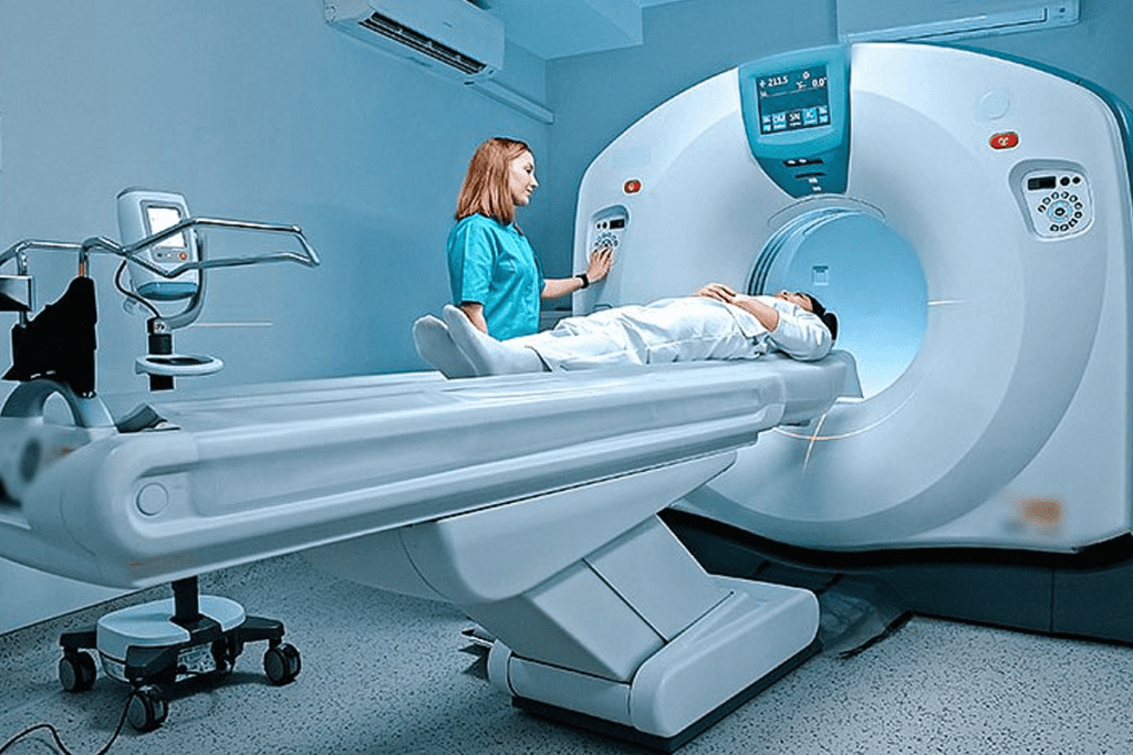
During the SPECT CT scan, you’ll lie on a table that slides into the scanner. The scan is usually painless. It can last from 30 minutes to several hours. This depends on the scan’s complexity and the specific protocol.
SPECT CT scans expose you to a small amount of radiation. Though the risk is low, it’s important to talk about any worries with your healthcare provider. The scan’s benefits usually outweigh the risks, even if it involves some radiation.
| Radiation Exposure | SPECT CT | Standard CT |
| Effective Dose (mSv) | 4-12 | 2-10 |
| Risk Level | Low to Moderate | Low |
The future of SPECT CT looks bright, thanks to new tech in hybrid imaging. This tech boosts how well doctors can diagnose, helping them make better choices. By combining SPECT and CT, doctors get a full picture of the body’s function and structure, leading to better care for patients.
SPECT CT scans are getting more important in many areas of medicine. They help in checking heart health, cancer, and more. This accuracy is key for planning the right treatment and improving care for patients.
SPECT CT is set to stay a key tool in medicine, giving deep insights into the body. As tech keeps getting better, we’ll see even clearer images and better care for patients. This makes SPECT CT even more essential in today’s healthcare.
A SPECT scan uses a tiny amount of radioactive material to see how different parts of the body work. It’s not like a CT scan, which shows detailed pictures of the body’s structure. Instead, SPECT scans focus on how the body functions.
Technetium-99m is a key part of SPECT scans. It helps show how the body works, like blood flow and organ function. It’s great for looking at the heart, bones, and tumors.
SPECT CT combines the best of both worlds. It uses SPECT to see how the body works and CT to show detailed pictures. This combo gives doctors a clearer view of what’s going on inside the body. It’s super helpful for diagnosing diseases in the heart, bones, and brain.
Image fusion in SPECT CT helps doctors pinpoint problems more accurately. It mixes SPECT and CT images to show how body parts work together. This helps doctors make better treatment plans.
SPECT scans are used for many things. They help find and track diseases like heart problems, cancer, bone issues, and brain disorders. They also check if treatments are working and how diseases are changing.
SPECT CT is key for heart disease checks. It looks at blood flow to the heart and finds blockages. This helps doctors plan treatments and figure out patient risks.
In cancer care, SPECT CT helps find and size tumors. It also checks if cancer has spread and how treatments are doing. This info helps doctors decide the best treatment plans.
SPECT CT is great for bone and joint problems. It finds and describes bone issues, checks chronic pain, and spots injuries and fractures. This helps doctors understand and treat these problems better.
SPECT CT is used for the endocrine system, too. It helps diagnose thyroid issues, find parathyroid adenomas, and spot neuroendocrine tumors. It gives doctors a clear picture of how these organs are working.
SPECT CT is also used for infections and inflammation. It shows where and how severe these issues are. This helps doctors treat them more effectively.
Mettler, F. A., et al. (2017). Effective doses in radiology and diagnostic nuclear medicine: a catalog. Radiology, 248(1), 254-263.
Subscribe to our e-newsletter to stay informed about the latest innovations in the world of health and exclusive offers!