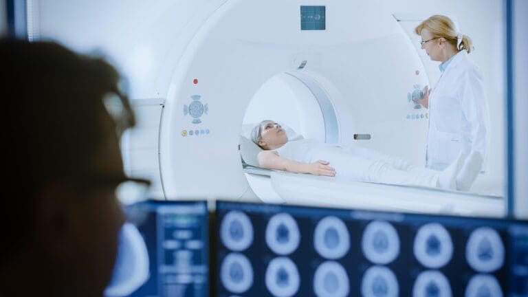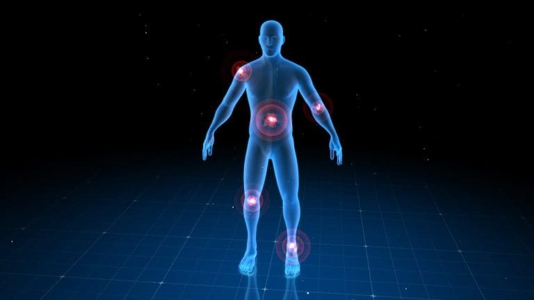
We are always improving nuclear medicine, thanks to the gamma camera scan, also known as scintillation or Anger camera imaging. This method lets us see how organs work inside the body. It does this by catching gamma radiation from special medicines.
The medical imaging market is getting bigger, with new tools like AI-based orthopedic imaging and hyperspectral imaging systems. At places like Liv Hospital, we use gamma imaging to meet global care standards. We focus on caring for our patients with compassion and understanding.
Key Takeaways
- Gamma camera scans are a key tool in nuclear medicine for checking how organs work.
- Radiopharmaceuticals send out gamma radiation, which gamma cameras catch.
- This tech is vital for finding and tracking many health issues.
- New imaging tech is helping the global medical imaging market grow.
- Places like Liv Hospital use gamma imaging to ensure top patient care.
The Fundamentals of Nuclear Medicine Imaging
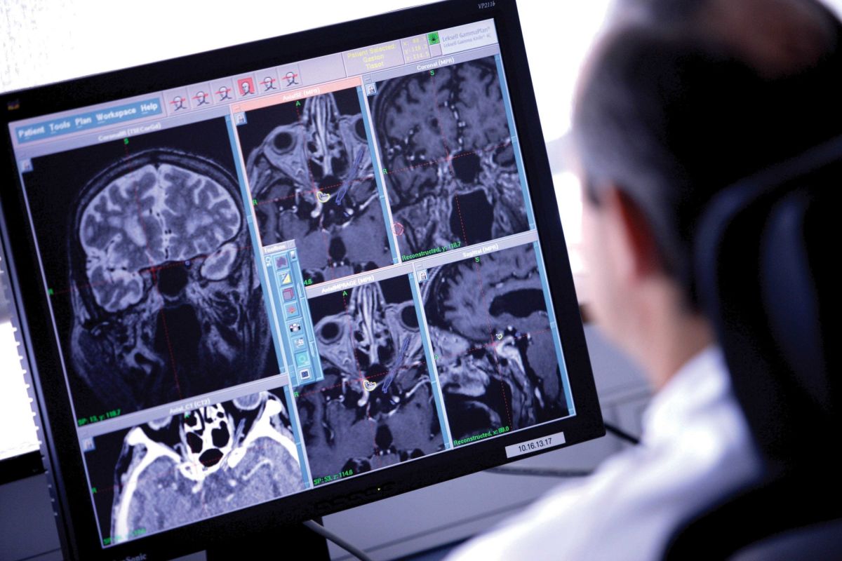
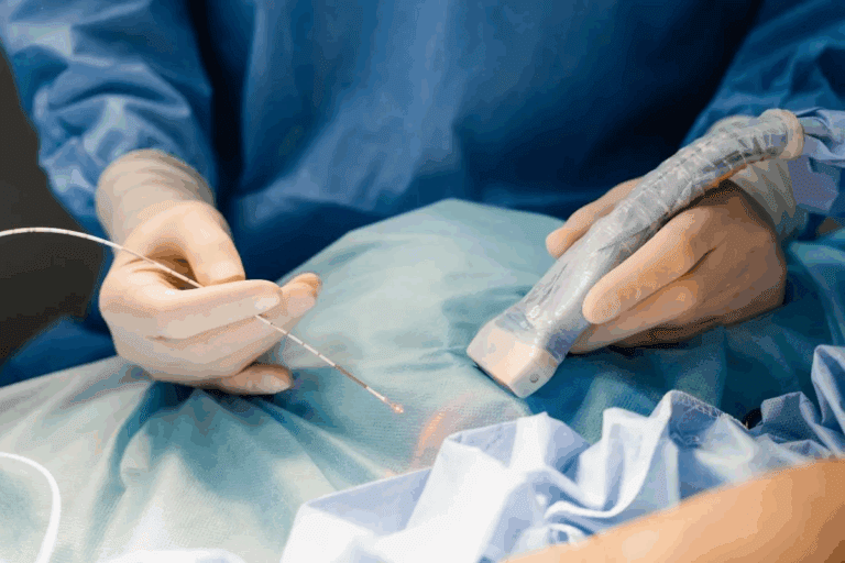
Nuclear medicine imaging is key in healthcare. It uses small amounts of radioactive materials. These help diagnose and treat diseases like cancers and heart issues.
The Role of Radiopharmaceuticals in Diagnostic Imaging
Radiopharmaceuticals are vital in nuclear medicine. They contain radioactive elements that target specific body areas. For example, technetium-99m is used in bone scans to find bone problems.
The right radiopharmaceutical is chosen based on the study type. A detailed review on nuclear medicine shows how these compounds have improved imaging nuclear medicine resources.
Evolution of Nuclear Medicine Techniques
Nuclear medicine techniques have grown a lot over time. This has made diagnosis better and care for patients more effective. Gamma cameras are a big part of this progress.
Gamma cameras use flat detector heads to catch gamma photons from tracers, like technetium-99m. Better detectors and image processing have made nuclear medicine images clearer. This helps doctors find problems sooner, leading to better patient care.
Understanding Gamma Camera Scans

In nuclear medicine, gamma camera scans are key for diagnosing and tracking health issues. They give insights into how organs and tissues work. This helps doctors make better choices for patient care.
Definition and Basic Principles
A gamma camera scan, or scintigraphy, uses tiny amounts of radioactive materials. It shows how different parts of the body work. The scan catches gamma rays from a special medicine given to the patient. It turns these rays into images that show where the medicine is in the body.
These images help doctors see if the heart, thyroid, bones, and more are working right. It’s a way to see how organs function, not just their shape. This is different from X-rays or CT scans.
Historical Development: From Anger to Modern Cameras
The first gamma camera was made in the 1950s by Hal Anger. It used a scintillation crystal and photomultiplier tubes to detect gamma rays. This was a big step forward in nuclear medicine.
Over time, gamma cameras have gotten better. They can now detect more, see clearer, and do more things. This is because of new materials, designs, and ways to process images.
Today’s gamma cameras are more sensitive and detailed. They help doctors diagnose and treat patients better. This is thanks to the ongoing improvement in technology.
Alternative Names: Scintillation and Anger Camera Imaging
Gamma camera imaging is also called scintillation camera or Anger camera imaging. These names honor Hal Anger’s work. They show how gamma rays are detected in the camera.
Knowing these names helps us understand nuclear medicine better. It shows the history and technology behind gamma camera scans.
| Terminology | Description |
|---|---|
| Gamma Camera Scan | A diagnostic imaging technique using gamma rays |
| Scintillation Camera | Emphasizes the detection of scintillations produced by gamma rays |
| Anger Camera | Named after Hal Anger, the inventor of the first practical gamma camera |
The Technology Behind Gamma Cameras
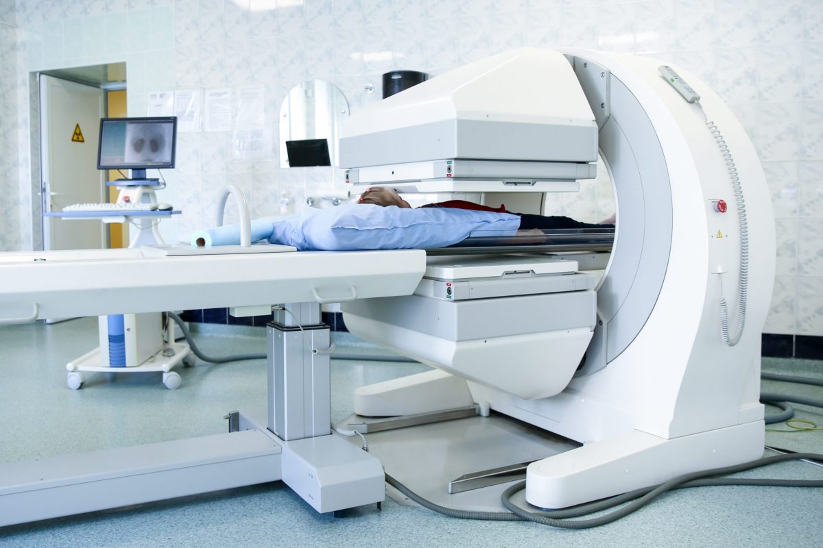
Gamma cameras are key in nuclear medicine. They use gamma radiation to show what’s inside the body. This is done with special devices called gamma ray cameras or scintillation cameras.
We’ll look at what makes up a gamma camera. This will help us understand how it creates detailed images.
Components of a Gamma Camera System
A gamma camera has important parts. These parts work together to detect and process gamma radiation. The main parts are detector heads, collimators, scintillation crystals, and photomultiplier tubes.
Detector Heads and Collimators
Detector heads are flat and close to the patient. They catch gamma photons from tracers. Collimators help direct these rays to the detector. This makes the image clearer by cutting down on scattered radiation.
Together, detector heads and collimators make the gamma camera better. New technology has made these cameras more sensitive and clear.
Scintillation Crystal and Photomultiplier Tubes
The scintillation crystal turns gamma radiation into light. This light is then boosted by photomultiplier tubes. These tubes are very sensitive to light. The crystal and tubes work together to catch the radiation and make an image.
| Component | Function |
|---|---|
| Detector Heads | Capture gamma photons emitted by tracers |
| Collimators | Direct gamma rays onto the detector, reducing scatter radiation |
| Scintillation Crystal | Convert gamma radiation into visible light |
| Photomultiplier Tubes | Amplify visible light into electrical signals |
These parts work together to make gamma cameras very good at creating images. They are very important in nuclear medicine.
How Gamma Imaging Works in Nuclear Medicine
Gamma camera technology is key in nuclear medicine. It lets us see how the body works by detecting gamma rays. We use it to find and track many health issues in different organs.
Gamma Ray Detection Process
The process starts with giving the patient a special substance. This substance sends out gamma rays. The gamma camera then catches these rays.
The camera has a few parts. The collimator focuses the rays. The scintillation crystal turns them into light. Photomultiplier tubes make this light stronger. This light is then turned into an image.
The Role of Technetium-99m and Other Radiopharmaceuticals
Technetium-99m (Tc-99m) is a top choice for nuclear medicine. It has the right half-life and gamma energy. It can attach to different substances, like for bone or thyroid scans.
Other substances, like iodine-123 for thyroid scans, are also used. But fluorine-18 is mainly for PET scans, not gamma cameras.
| Radiopharmaceutical | Application |
|---|---|
| Technetium-99m | Bone scans, cardiac studies, and various organ imaging |
| Iodine-123 | Thyroid function and structure assessment |
| Fluorine-18 | PET scans for oncology, neurology, and cardiology |
From Emission to Digital Image
Turning gamma rays into digital images takes a few steps. First, the gamma rays from the substance are caught by the camera. Then, they’re turned into electrical signals.
These signals are then fixed for things like energy and position. After that, they’re made into a digital image. This image shows up on a computer screen for doctors to look at.
Types of Gamma Camera Scan Procedures
Gamma camera scans are used in nuclear medicine to find and track many health issues. These scans fall into different types based on what they show and how they’re used.
Static Imaging Techniques
Static imaging takes one or more pictures at a set time. It’s great for seeing where certain medicines go in the body. Static imaging helps find tumors, check how organs work, and see how far diseases have spread.
Dynamic Imaging and Functional Studies
Dynamic imaging shows how organs work over time with moving pictures. It’s key for checking the kidneys, liver, and heart. Dynamic imaging lets doctors see how medicines move in the body, helping them understand organ health.
Whole-Body Scanning Protocols
What Tests Rule Out Ovarian Cancer?Whole-body scans look at the whole body or big parts of it in one go. They’re good for finding cancer spread, checking for diseases, and seeing how far cancer has gone. These scans give a big picture of where diseases are, helping plan treatments.
SPECT Imaging: 3D Gamma Camera Technology
SPECT imaging is a big step forward in nuclear medicine. It gives us three-dimensional views of how the body works. This helps doctors see more clearly what’s going on inside us.
How Single Photon Emission Computed Tomography Works
SPECT uses gamma cameras to detect rays from a special medicine given to the patient. The camera moves around the patient, taking pictures from different sides. Then, these pictures are put together to show where the medicine is in the body.
The steps are:
- The patient gets a special medicine that shows up in certain areas.
- The gamma camera moves around, catching the rays it sends out.
- These rays help make two-dimensional pictures.
- A computer turns these pictures into a three-dimensional view.
SPECT vs. Planar Gamma Imaging
SPECT is better than planar gamma imaging because it shows more. Planar imaging is flat, but SPECT is three-dimensional. This is great for tricky cases where it’s hard to see what’s going on.
Comparison of SPECT and Planar Gamma Imaging
| Feature | SPECT Imaging | Planar Gamma Imaging |
|---|---|---|
| Dimensionality | 3D | 2D |
| Diagnostic Accuracy | Higher | Lower |
| Complexity of Cases | Suitable for complex cases | Limited in complex cases |
Hybrid Imaging: SPECT/CT Applications
Hybrid imaging, like SPECT/CT, combines SPECT’s function with CT’s anatomy. This mix gives doctors a clearer picture of what’s happening inside the body.
SPECT/CT is used in many ways, such as:
- Oncology: to find and check tumors.
- Cardiology: for heart studies.
- Infectious diseases: to spot infections.
By combining SPECT with CT, doctors get a better view of the patient’s health. This leads to more accurate diagnoses and better treatment plans.
Clinical Applications of Gamma Camera Nuclear Medicine
Gamma camera nuclear medicine has changed how we diagnose diseases. It helps us see inside the body in new ways. This technology improves patient care by helping doctors make better decisions.
Cardiac Imaging and Myocardial Perfusion Studies
Cardiac imaging is a key use of gamma camera nuclear medicine. It lets us see how well the heart is working. We can spot problems like coronary artery disease and check if treatments are working.
Myocardial perfusion studies show us where blood flow to the heart is low. This helps doctors decide the best course of action.
Bone Scintigraphy for Orthopedic Conditions
Bone scintigraphy is another important use. It shows how bones are working and helps find issues like bone cancer or fractures. This method is great for catching problems early, before they show up on other scans.
Thyroid and Parathyroid Imaging
Gamma camera nuclear medicine also helps with thyroid and parathyroid imaging. We use special medicines to see how these glands are working. This is key for finding and treating problems like thyroid nodules or hyperfunctioning parathyroid glands.
Renal Function Assessment
Renal function assessment is another big use. It lets us check how well the kidneys are working. We can spot blockages and see if treatments are working. This helps doctors take better care of patients with kidney disease.
| Clinical Application | Description | Benefits |
|---|---|---|
| Cardiac Imaging | Assesses myocardial perfusion and diagnoses coronary artery disease | Guides treatment decisions, improves patient outcomes |
| Bone Scintigraphy | Diagnoses bone metastases, fractures, and infections | Early detection, monitors treatment response |
| Thyroid and Parathyroid Imaging | Assesses thyroid function and identifies abnormalities | Diagnoses and manages thyroid and parathyroid disorders |
| Renal Function Assessment | Evaluates kidney function and diagnoses obstruction | Manages renal disease, assesses treatment effectiveness |
In conclusion, gamma camera nuclear medicine has many uses in medicine. It helps doctors diagnose and treat diseases better. We keep improving this technology to help patients even more.
Interpreting Gamma Camera Scan Results
Nuclear medicine physicians are key in understanding gamma camera scan data. They need to know a lot about nuclear medicine and how gamma imaging works. This is a complex task.
Understanding Scintigraphic Images
Scintigraphic images show how radiopharmaceuticals spread in the body. They help doctors see problems in the heart, thyroid, bones, and more. These images are two-dimensional.
They are made by detecting gamma rays and turning them into pictures. The brightness of each area shows how much radiopharmaceutical is there. This helps doctors see how organs are working.
Normal vs. Abnormal Findings
It’s important to know if something looks normal or not in gamma camera scans. Normal scans show the radiopharmaceutical evenly spread. But, if it’s not even, it might mean something’s wrong.
- More uptake can mean high activity or inflammation.
- Less uptake might show reduced function or damage.
For example, in heart scans, less uptake during stress tests can point to heart disease. In bone scans, more uptake can show tumors or fractures.
The Role of Nuclear Medicine Physicians
Nuclear medicine physicians are vital in reading gamma camera scan results. They use their knowledge to understand how the radiopharmaceutical spreads, the patient’s history, and the images.
“The interpretation of nuclear medicine images requires a deep understanding of the underlying physiology, pathology, and the specific characteristics of the radiopharmaceutical used.” – Expert in Nuclear Medicine
They match the images with the patient’s history to make accurate diagnoses. Their work is essential for using gamma camera scan data to help patients.
Patient Experience and Safety Considerations
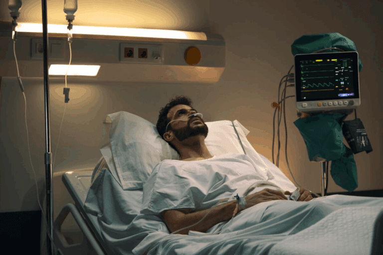
Keeping patients safe and happy is our main goal with gamma camera scans. We know that getting a medical scan can make people nervous.
Preparation and Procedure Steps
We give patients clear steps to follow before the scan. This includes advice on hydration, medication, and getting ready. On scan day, patients might need to remove jewelry or metal objects to avoid interference.
- Arrive on time to fill out any needed paperwork.
- Wear a comfortable gown if asked.
- Listen carefully to the nuclear medicine team’s instructions.
Radiation Exposure and Safety Protocols
We’re very serious about keeping radiation exposure low. The scan’s radiation is controlled to get good images without harming you. Our team follows strict safety protocols to protect everyone.
For more info on radiation safety and gamma camera scans,
Duration and Comfort Factors
How long a gamma camera scan takes varies. It can be from 30 minutes to several hours. We try to make it as comfy as possible, with comfortable seating and regular breaks.
Contraindications and Special Populations
Gamma camera scans are usually safe, but there are exceptions. For example, pregnancy and breastfeeding need special attention. Our medical team checks each patient’s situation to decide the best approach.
- Tell your doctor if you’re pregnant or breastfeeding.
- Share any allergies or sensitivities to medications or contrast agents.
- Follow all pre-procedure instructions carefully.
We focus on safety and comfort to make the scan experience better for everyone. Our team is dedicated to providing top-notch care and support during the scan.
Conclusion: Advances and Future Directions in Gamma Camera Technology
We’ve seen big steps forward in gamma camera tech, making it key in nuclear medicine. It’s known for being accurate and safe, with updates that make it even better. These changes help doctors make more accurate diagnoses.
New research and tech are on the horizon. They promise to make gamma cameras even more useful. This will help doctors and patients alike, showing how important gamma cameras are in today’s healthcare.
Looking ahead, gamma camera tech could do even more. With new ideas, we’ll see tools that are more precise and helpful. This will help patients all over the world.
FAQ
What is a gamma camera scan?
How does gamma imaging work in nuclear medicine?
What is the role of technetium-99m in diagnostic imaging?
What are the different types of gamma camera scan procedures?
What is SPECT imaging and how does it work?
How do I prepare for a gamma camera scan?
What are the risks associated with gamma camera scans?
How long does a gamma camera scan take?
What is the difference between a gamma camera scan and other imaging modalities?
How are gamma camera scan results interpreted?
What is the role of nuclear medicine physicians in image interpretation?
Are there any contraindications for gamma camera scans?
References
- StatPearls. (n.d.). Nuclear Medicine Instrumentation “ Gamma Camera. In StatPearls. https://www.ncbi.nlm.nih.gov/books/NBK597384/


