Last Updated on November 27, 2025 by Bilal Hasdemir

A nuclear medicine bone scan is a detailed test that uses a tiny amount of radiotracer. It’s injected into the body to show where bone metabolism is off. This test is great for finding bone infections, fractures, tumors, and cancer spread.
By using a radioactive tracer, a bone scan can spot many bone diseases and conditions. It helps doctors diagnose and check on these issues.
The scan takes place in a hospital’s nuclear medicine unit. It’s known for catching problems early, before symptoms get bad. Liv Hospital offers advanced, patient-focused care. You can count on accurate tests and expert advice at every step.
Key Takeaways
- A nuclear medicine bone scan helps identify areas of abnormal bone metabolism.
- It’s useful for detecting bone infections, fractures, tumors, and cancer metastases.
- The procedure involves injecting a small amount of radiotracer.
- Side effects are generally minimal, including slight pain from the injection and rare allergic reactions.
- The scan is typically painless and doesn’t require anesthesia.
The Purpose and Function of Bone Scans

Bone scans are a key tool in modern medicine. They help doctors find and understand bone problems. This gives them important information about bone health.
Diagnostic Role in Modern Medicine
Bone scans are important for finding bone issues. They help spot fractures, infections, tumors, and metastases from cancers like breast, prostate, or lung. They also help find arthritis or trauma. The National Center for Biotechnology Information says bone scans are a key tool in nuclear medicine.
Bone scans work by showing where bone is active. This can point to problems that other tests might miss.
When Doctors Recommend Bone Scans
A doctor might suggest a bone scan if you have bone pain or signs of bone disease. They might recommend it for:
- Unexplained bone pain
- Suspected bone infection or inflammation
- Diagnosis of bone cancer or metastasis
- Monitoring treatment for bone issues
The table below shows why doctors might suggest a bone scan:
| Condition | Description |
| Bone Fractures | Detecting fractures that are not visible on X-rays |
| Bone Infections | Identifying areas of bone infection or inflammation |
| Cancer Metastasis | Detecting cancer spread to the bones |
Knowing how bone scans work helps patients understand their value. This makes them appreciate the role of nuclear medicine in diagnosis.
What Is a Nuclear Medicine Bone Scan?

A nuclear medicine bone scan is a test that uses small amounts of radioactive materials to see the bones. It helps find and track bone problems like cancer, infections, and fractures.
Definition and Basic Principles
Nuclear medicine bone scans use radiotracers, which are substances that give off small amounts of radiation. When injected, these radiotracers go to the bones. A special camera then takes detailed pictures of the bones.
The most used radiotracer is Tc-99m, a technetium-based compound that sticks to bone tissue.
The scan works by showing where the bones are most active. This is because disease or injury makes bones take up more radiotracer. This helps spot problems.
Historical Development and Current Applications
Nuclear medicine bone scans have been around for decades. Over time, the technology has gotten better. This means we get clearer images and more accurate diagnoses.
Now, these scans help in many ways. They find bone metastases in cancer patients, spot stress fractures, and track bone diseases like Paget’s. They also check if treatments for bone issues are working.
The use of nuclear medicine bone scans is growing. New radiotracers and imaging methods are being developed. These advancements help us detect and manage bone problems better and earlier.
How Nuclear Medicine Bone Scans Work
Nuclear medicine bone scans use special tracers to check bone health. They help doctors see how bones are working and find problems.
The Science Behind Radiotracers
Radiotracers are tiny, radioactive particles. They go to parts of the body that are very active. The most used one is Technetium-99m (Tc-99m) mixed with a compound called methylene diphosphonate (MDP).
This mix is drawn to bones that are growing or breaking down. It helps find issues like fractures, infections, or tumors.
Technetium-99m and Other Common Agents
Technetium-99m is the top choice for bone scans. It has a short life and the right kind of radiation for cameras to pick up.
| Radiotracer | Characteristics | Common Uses |
| Technetium-99m MDP | Short half-life, suitable gamma energy | Bone scans, detecting fractures, infections, tumors |
| Fluorine-18 Fluoride | High resolution, longer half-life | PET bone scans, detailed bone imaging |
The tracer in a bone scan is safe and goes away in a few days. It shows where bones are changing a lot. This helps doctors find and track bone problems.
The Nuclear Medicine Bone Scan Procedure
A nuclear medicine bone scan is a detailed process. It helps find bone problems by using a small amount of radioactive material. This method is key for getting accurate results.
Patient Preparation Guidelines
Before the scan, patients must follow certain steps. They should drink lots of water before and after the injection to remove the radioactive material. Also, they might need to avoid certain foods or medicines that could affect the scan.
It’s important to tell the doctor about any allergies. Wear comfortable clothes without metal to avoid any issues during the scan.
The Injection Process
The scan starts with a radiotracer injection into a vein. This is usually quick and might feel a bit uncomfortable. The radiotracer then spreads through the blood, focusing on the bones.
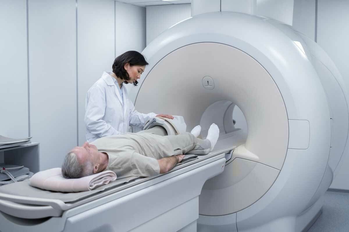
Scanning Process and Duration
After the radiotracer spreads, the scanning begins. The patient lies on a table that slides into a gamma camera. This scanning takes about 30 to 60 minutes, depending on the area scanned.
The whole process, from start to finish, takes several hours. But the actual scanning time is short. It’s important to stay very quiet during the scan for clear images.
| Procedure Step | Description | Duration |
| Patient Preparation | Guidelines to follow before the scan | Varies |
| Injection of Radiotracer | Administering Technetium-99m | Quick |
| Radiotracer Circulation | Allowing the tracer to accumulate in bones | 2-4 hours |
| Scanning Process | Imaging with a gamma camera | 30-60 minutes |
The scan uses low-level radiation, but the risks are small. Pregnant or breastfeeding women should talk to their doctor about any concerns.
Types of Nuclear Medicine Bone Scans
Nuclear medicine bone scans are divided into two main types: whole body scans and targeted area scans. These categories depend on the body area being checked and the patient’s specific needs.
Whole Body Nuclear Medicine Bone Scan
A whole body nuclear medicine bone scan shows images of the entire skeleton. It’s great for finding widespread bone issues, like cancer spread, infections, or trauma.
Benefits of Whole Body Scans:
- They can spot many problems in one go.
- They give a full view of the skeleton, helping diagnose systemic conditions.
This scan can find bone infections, fractures, tumors, and cancer spread. It’s also good for checking bone trauma that X-rays miss.
Targeted Area Scans
Targeted area scans focus on a specific part of the body. They’re used when there’s a known or suspected issue in that area.
Advantages of Targeted Area Scans:
- They give detailed pictures of the area of concern.
- They’re often better at finding problems in the focused area than whole body scans.
| Scan Type | Area Covered | Diagnostic Use |
| Whole Body Scan | Entire Skeleton | Detecting widespread abnormalities, metastases, infections. |
| Targeted Area Scan | Specific Region | Detailed examination of a known or suspected area of concern. |
What Does a Nuclear Medicine Bone Scan Show?
Understanding what a nuclear medicine bone scan shows is key for diagnosing bone issues. It can show areas of high or low bone activity. This can point to several conditions.
Normal vs. Abnormal Results
A normal scan shows the radiotracer evenly spread in the bones. But, abnormal results can mean different things. They might show fractures, infections, or even cancer.
Abnormal results can show “hot spots” or “cold spots.” Hot spots mean high activity, like in fractures or tumors. Cold spots suggest low blood flow or cancer.
“Hot Spots” and “Cold Spots” Interpretation
Understanding “hot spots” and “cold spots” is key. A hot spot might mean active bone changes, like in osteoarthritis. A cold spot could mean low blood flow or tumors.
- Hot Spots: Increased bone activity, often seen in fractures, infections, or tumors.
- Cold Spots: Reduced bone activity, potentially indicating reduced blood flow or certain cancers.
Limitations in Diagnostic Capabilities
Bone scans are useful but have limits. They might not tell apart conditions with similar results. A study by the Royal Australian College of General Practitioners shows they are sensitive but not always specific.
Also, bone scans use a small amount of radiation. But, the benefits of the information they provide usually outweigh the risks.
Detecting Bone Abnormalities and Conditions
Nuclear medicine bone scans are key in finding bone problems. They help spot a variety of bone issues. This info is vital for deciding how to treat patients.
Bone Fractures and Trauma
These scans are great at finding bone fractures, even when X-rays can’t. They’re perfect for stress or hairline fractures that are hard to see. The scan shows where bone activity is high, meaning there’s a fracture.
Early finding of fractures is key for good treatment and avoiding more harm. Doctors can plan a specific treatment by knowing exactly where and how bad the fracture is.
Bone Infections and Inflammatory Conditions
Bone infections and inflammatory diseases are hard to diagnose. Nuclear medicine bone scans are a sensitive way to find these issues. They show where bone activity is up, which often means infection or inflammation.
These scans help tell if it’s a bone infection or something else. Quick diagnosis is important to avoid lasting damage and help patients get better.
Arthritis and Joint Disorders
These scans are also useful for checking arthritis and joint problems. They help doctors see how much bone is involved. This lets them know how severe the condition is and if treatments are working.
The scans show where bone is breaking down or inflamed due to arthritis. This helps doctors make treatment plans that fit each patient’s needs. Tracking how the disease changes and how well treatments work is important for managing arthritis.
Nuclear Medicine Bone Scans in Cancer Diagnosis
Nuclear medicine bone scans are key in finding cancer, like bone metastases. They are very sensitive, spotting small changes in bone activity. This makes them very useful in fighting cancer.
These scans are great for finding cancer spread from places like the breast, prostate, or lung. For more details on bone scan results, check out the Prostate Cancer Research Institute website. It has lots of info on bone imaging and cancer.
With a nuclear medicine bone scan, doctors can see cancer spread all over the body. This helps them plan the best treatment. Knowing how far the cancer has spread is very important.
FAQ
What is a nuclear medicine bone scan?
A nuclear medicine bone scan is a test that uses tiny amounts of radioactive materials. It creates images of the bones to find areas where bone metabolism is not normal.
What does a nuclear medicine bone scan show?
A nuclear medicine bone scan shows where bone activity is high or low. This can help find bone infections, fractures, tumors, and cancer spread to the bones.
How does a nuclear medicine bone scan work?
The scan works by injecting a special dye, Technetium-99m, into the body. This dye goes to active bone areas, making detailed bone images possible.
What are the side effects of a nuclear medicine bone scan?
The side effects of a nuclear medicine bone scan are usually minor. The small amount of radioactive material used and the low risk of an allergic reaction to the dye are key reasons.
What is the difference between a whole body nuclear medicine bone scan and a targeted area scan?
A whole body scan looks at the entire skeleton. A targeted area scan focuses on a specific part, like the spine or hips.
How is a nuclear medicine bone scan used in cancer diagnosis?
Nuclear medicine bone scans help find bone metastases in cancer diagnosis. Bone metastases are when cancer spreads to the bones from other parts of the body.
What is the role of Technetium-99m in nuclear medicine bone scans?
Technetium-99m is a key dye used in bone scans. It goes to active bone areas, helping create detailed bone images.
How should I prepare for a nuclear medicine bone scan?
Before a scan, follow specific guidelines. This includes avoiding certain foods or medications and removing metal objects from your body.
What are “hot spots” and “cold spots” in a nuclear medicine bone scan?
“Hot spots” are areas with high bone activity. “Cold spots” are areas with low activity. Both can indicate bone-related conditions.
What are the limitations of a nuclear medicine bone scan?
While very sensitive, bone scans may not always be specific. Results might need further testing or confirmation with other diagnostic tools.
References
- Love, C., Palestro, C. J. (2023). Bone Scan – StatPearls. National Center for Biotechnology Information. https://www.ncbi.nlm.nih.gov/books/NBK531486/
- Radiopaedia. (2024). Bone scintigraphy. https://radiopaedia.org/articles/bone-scintigraphy-1
- Van den Wyngaert, T., et al. (2016). The EANM practice guidelines for bone scintigraphy. European Journal of Nuclear Medicine and Molecular Imaging. https://www.ncbi.nlm.nih.gov/pmc/articles/PMC4932135/
- Insideradiology. (2024). Nuclear Medicine Bone Scan. https://www.insideradiology.com.au/nuclear-medicine-bone-scan/


