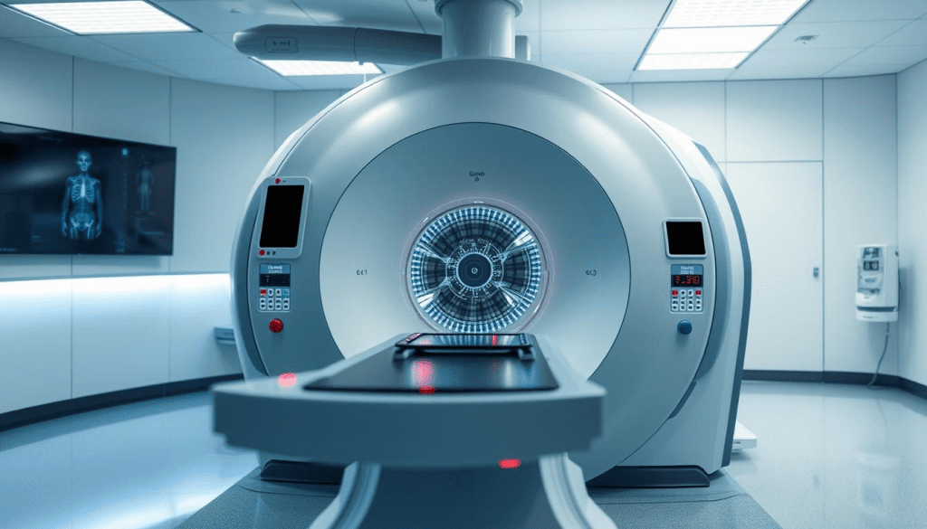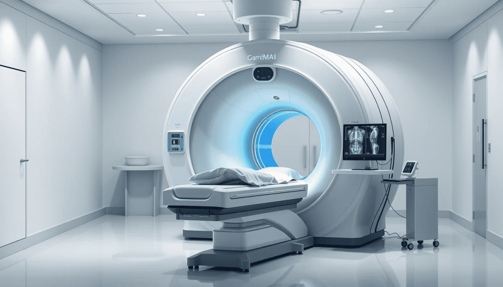
In modern medicine, diagnostic imaging tools are key for diagnosing and treating diseases. The gamma camera is a vital tool in nuclear medicine imaging. It helps us see how the body works by finding gamma rays from special medicines given to patients.
The gamma camera is very important for finding and watching diseases. With the latest technology and care for each patient, we can spot diseases early. This helps us find the best ways to treat them.
Key Takeaways
- Gamma cameras are key in nuclear medicine for finding and watching diseases.
- They find gamma rays from special medicines to see how the body works.
- Finding diseases early helps us find better treatments.
- The latest technology and caring for each patient are very important in medicine.
- Liv Hospital’s skill with gamma camera technology helps care for patients better.
The Fundamentals of Nuclear Medicine Imaging

To understand how gamma cameras work, we first need to grasp the basics of nuclear medicine imaging. This field uses radioactive tracers to diagnose and treat diseases. It includes many types of cancers, heart disease, and more.
Radioactive tracers, or radiopharmaceuticals, target specific parts of the body. They emit gamma rays when given to a patient. A gamma camera then detects these rays to create images for diagnosis.
The Role of Radioactive Tracers in Diagnostic Imaging
Radioactive tracers are compounds with a radioactive element. They are picked for their ability to gather in certain body areas, like tumors. For example, some tracers are absorbed by bone, making them great for bone scans.
The right tracer is key for a successful nuclear medicine imaging test. It’s usually given through an IV, but can be taken orally or inhaled for some tests. This is based on the specific needs of the test.
Principles of Gamma Ray Emission and Detection
Nuclear medicine imaging starts with gamma rays from the tracer. These rays are detected by the gamma camera, which moves around the patient. This helps create detailed images of the body’s inside.
The detection of gamma rays shows how different tissues and organs absorb or emit radiation. By analyzing these differences, doctors can understand the body’s internal workings better.
The Gamma Camera: Essential Components and Working Principles

It’s important to know how a gamma camera works for its role in medical diagnostics. These devices help us see how the body works by catching gamma rays from radioactive tracers. This gives us important information about our health.
Core Components of a Modern Gamma Camera
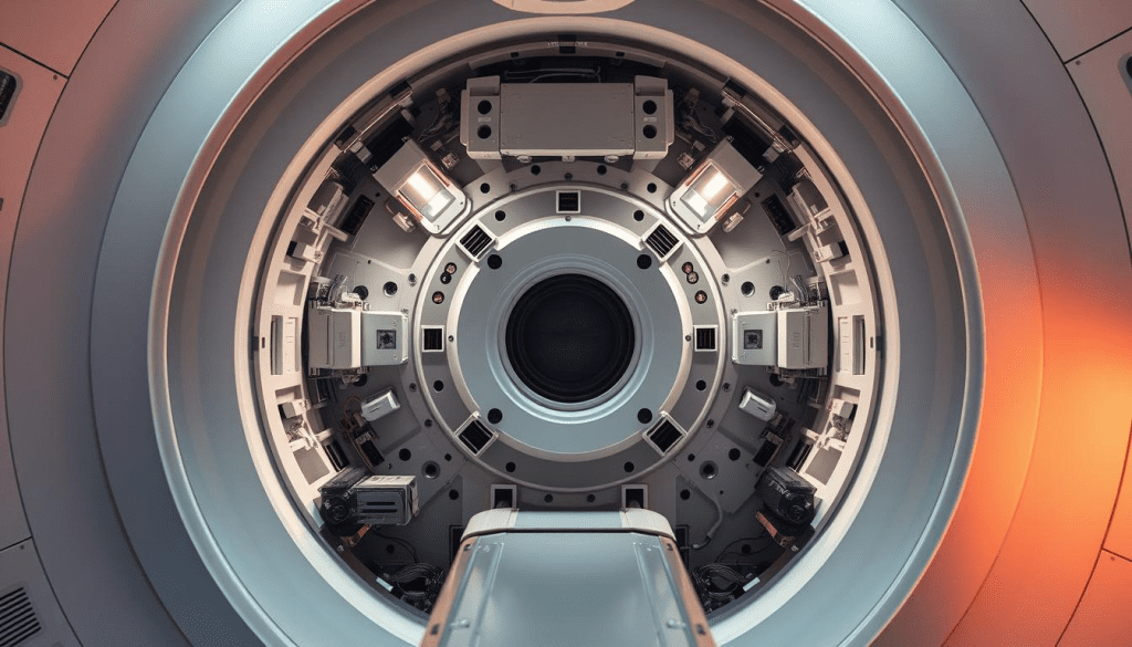
A modern gamma camera has key parts that work together. They help detect and process gamma radiation. The main parts are:
- A collimator, which directs gamma rays onto the detector
- A scintillation crystal that converts gamma rays into visible light
- Photomultiplier tubes that amplify the light signal
- Sophisticated electronics for signal processing and image formation
The collimator is key for the camera’s sharpness. It’s made of dense materials like lead. It has many small holes to let through gamma rays and block others.
| Component | Function |
| Collimator | Directs gamma rays onto the detector |
| Scintillation Crystal | Converts gamma rays into visible light |
| Photomultiplier Tubes | Amplify the light signal for processing |
| Sophisticated Electronics | Process signals and form diagnostic images |
Image Processing and Reconstruction Techniques
After detecting gamma rays, the camera turns them into electrical signals. Then, advanced techniques are used to make detailed images. These methods fix issues like scatter radiation and how much the rays are weakened.
Advanced algorithms, like iterative methods, are used to make images clearer and more accurate. These algorithms use info about the camera and how gamma rays work. This helps make high-quality images for diagnosis.
Knowing how a gamma camera works shows its complexity and importance in nuclear medicine. It also shows why we need to keep improving gamma camera technology. This will help us get even better at diagnosing diseases.
How Gamma Cameras Capture Physiological Processes
Gamma cameras are key in capturing how our bodies work. They help doctors diagnose and treat diseases. We use these diagnostic imaging tools to see inside our bodies.
From Gamma Photons to Digital Images
The journey starts with gamma photons from radiopharmaceuticals in our bodies. These photons turn into digital images through scintillation crystals and photomultiplier tubes. A study on gamma camera imaging shows this process is vital for clear images.
Spatial Resolution and Image Quality Factors
Image quality depends a lot on spatial resolution. Several things affect this, like the collimator design and the scintillation crystal type. Also, how advanced the image processing algorithms are matters a lot.
| Factor | Impact on Spatial Resolution | Effect on Image Quality |
| Collimator Design | Directly affects the ability to resolve adjacent structures | Improves image clarity and diagnostic accuracy |
| Scintillation Crystal Type | Influences the conversion efficiency of gamma photons | Enhances image sensitivity and resolution |
| Image Processing Algorithms | Impacts the reconstruction and enhancement of images | Improves image quality and diagnostic confidence |
Energy Discrimination in Modern Systems
Today’s gamma cameras can tell different energy levels of gamma photons apart. This is great when using radiopharmaceuticals in imaging. It lets us focus on specific body processes.
By blocking out unwanted energy, we get better images and more accurate diagnoses. As gamma camera tech gets better, so will our ability to see inside our bodies clearly.
Common Clinical Applications of Gamma Camera Imaging
Gamma cameras have changed how we diagnose diseases. They give us important insights into how our bodies work. We use them to find and track many diseases.
Cardiac Imaging and Heart Disease Detection
Gamma cameras are key in heart disease diagnosis. They help us see how well the heart works and find problems. This helps doctors decide the best treatment.
Myocardial perfusion imaging is very useful. It shows how well the heart gets blood, both when it’s working hard and when it’s not. This is key for managing heart disease.
Oncological Applications in Cancer Diagnosis
In cancer care, gamma cameras are essential. They help find and track cancer. Bone scans are used to see if cancer has spread to bones. They also help find the first place cancer might go.
Bone Scans for Skeletal Disorders
Bone scans are important for many bone problems. They help find cancer in bones and check for pain or infection. They’re very good at finding bone issues early.
Thyroid Function Assessment
Gamma cameras also help with thyroid issues. They use special dyes to see thyroid problems. This helps doctors treat thyroid diseases better.
Gamma cameras help us care for many conditions. This includes heart disease, cancer, bone problems, and thyroid issues. They give us detailed information to help patients.
SPECT Imaging: Three-Dimensional Diagnostic Capabilities
SPECT imaging is a big step forward in nuclear medicine. It gives us detailed 3D views of the body’s inner workings. This helps us understand complex health issues better. A gamma camera moves around the patient, taking pictures from different sides. These pictures are then put together to form a complete 3D image.
Principles of Single Photon Emission Computed Tomography
SPECT imaging works by catching gamma rays from a special medicine given to the patient. As the camera moves, it picks up these rays from all sides. This lets us make detailed images of the body’s cross-sections.
Key components of SPECT imaging include:
- Gamma camera with one or more detector heads
- Collimators to direct gamma rays
- Computer system for image reconstruction
3D Reconstruction and Visualization Techniques
To make 3D images, SPECT uses special algorithms to fix up the 2D pictures. Then, advanced tools help doctors understand these 3D images. This gives them important clues about the patient’s health.
Clinical Advantages of SPECT Over Planar Imaging
SPECT imaging is better than 2D pictures in many ways. It’s more accurate and can see complex body parts in 3D. This is really helpful for finding and treating diseases like cancer, heart problems, and brain disorders.
| Imaging Modality | Diagnostic Capability | Clinical Application |
| Planar Imaging | 2D representation | General diagnostic purposes |
| SPECT Imaging | 3D representation | Detailed assessment of complex conditions |
SPECT imaging gives us clearer and more precise views of the body. It’s a key tool in today’s medicine for diagnosing and treating diseases.
Patient Experience During a Gamma Camera Procedure
Gamma camera imaging can seem scary, but knowing what happens can help. We make sure our patients know what to expect. This makes the experience smoother for everyone.
Preparation and Radiopharmaceutical Administration
Before starting, patients might need to prepare. This could mean fasting or avoiding certain medicines. Our team gives clear instructions for each step.
Then, patients get a special medicine that helps the camera see what’s inside. This medicine is given through an IV. It lights up the area we want to see, and the camera captures the images.
The Imaging Process: What Patients Can Expect
During the scan, patients need to stay very quiet for a bit. The camera moves around them to get all the pictures. It’s usually not painful and can take from 30 minutes to a few hours.
For some scans, patients might get Single Photon Emission Computed Tomography (SPECT). This means the camera takes 3D pictures by moving around the body. We make sure patients are comfortable the whole time.
Safety Considerations and Radiation Exposure
Keeping patients safe is our main goal. We use just the right amount of radiation to get good pictures. We follow strict rules to keep everyone safe, including our staff.
| Safety Measure | Description | Benefit |
| Radiopharmaceutical Dosage | Carefully calculated to minimize radiation exposure | Reduces risk to the patient |
| Shielding | Use of lead aprons and other shielding methods | Protects against unnecessary radiation |
| Staff Training | Comprehensive training on safety protocols | Ensures adherence to safety standards |
Knowing about gamma camera procedures can make patients feel better. Our team works hard to keep everyone safe and comfortable during imaging.
Recent Technological Advancements in Gamma Camera Systems
Gamma camera technology has seen big improvements, making medical tests more accurate and efficient. These changes include better detectors, hybrid imaging systems, and advanced software for processing images.
Improvements in Detector Technology and Spatial Resolution
Detector technology has greatly improved in gamma cameras. This has led to clearer images and better accuracy in diagnoses. Modern detectors are more sensitive, capturing detailed images of the body’s functions.
The introduction of CZT detectors has been a game-changer. They are more sensitive and offer better image quality than older NaI detectors. This means doctors can make more accurate diagnoses.
| Detector Type | Sensitivity | Energy Resolution |
| Sodium Iodide (NaI) | Moderate | 9-10% |
| Cadmium Zinc Telluride (CZT) | High | 5-6% |
Hybrid Imaging Systems: SPECT/CT and SPECT/MRI
Hybrid imaging systems have made a big impact. They combine SPECT with CT or MRI, giving doctors both function and anatomy in one go. This makes diagnoses more accurate and complete.
SPECT/CT systems are great for finding and understanding tumors. They use CT’s detailed images to pinpoint where problems are in the body.
“The integration of SPECT with CT has transformed the field of nuclear medicine, providing unparalleled diagnostic capabilities.” – A Nuclear Medicine Specialist
Software Innovations for Enhanced Image Processing
Software has also been a key factor in improving gamma cameras. New algorithms enhance image quality, reduce noise, and improve diagnosis. Tools like iterative reconstruction and advanced filtering are now essential.
Advanced software can also turn 2D SPECT images into 3D models. This is very helpful for planning surgeries and understanding disease extent.
As gamma camera tech keeps getting better, we can look forward to even more precise and useful medical images. The ongoing improvements in detectors, hybrid systems, and software will help patients get better care and results.
Comparing Gamma Cameras with Other Diagnostic Imaging Modalities
Choosing the right diagnostic imaging tool is key. Gamma cameras have special benefits compared to others. Each tool has its own strengths and best uses.
Gamma Camera vs. PET Scanning
Gamma cameras and PET scanning are both important in nuclear medicine imaging. But they do different things. Gamma cameras catch single photons from a radiopharmaceutical, showing how things work. PET scanning catches pairs of photons, giving clearer images.
PET scanning is more sensitive and detailed, great for cancer. But gamma cameras are versatile and used for many things because of the wide range of radiopharmaceuticals.
Functional vs. Anatomical Imaging Techniques
Functional imaging techniques, like gamma cameras, look at how the body works. They show how organs and systems function, not just their shape.
On the other hand, CT and MRI scans show the body’s inside structures well. They’re good for anatomy but not as good for function as nuclear medicine.
Using both functional and anatomical imaging together gives a full picture. For example, SPECT/CT combines gamma camera data with CT scan details.
Selecting the Appropriate Imaging Modality for Different Conditions
The right diagnostic imaging tools depend on the condition. Gamma cameras are great for heart tests and some cancer diagnoses because they show function.
For detailed anatomy, like structural issues or injuries, MRI or CT scans are better. PET scanning is key in oncology for cancer staging and treatment tracking.
Choosing an imaging modality means thinking about the clinical question, the patient’s situation, and each technology’s strengths. This way, healthcare providers can make the best choices for patient care.
Conclusion: The Future of Gamma Camera Technology in Modern Medicine
Gamma camera technology is set to be even more important in medicine’s future. New advancements in nuclear medicine imaging will make gamma cameras even better. This will help them be used in more ways in healthcare.
New tools and techniques in diagnostic imaging will lead to better patient care. Gamma camera imaging will keep being key in understanding and treating diseases. It shows how the body works, helping doctors make accurate diagnoses.
Gamma camera tech will get even better, with clearer images and quicker scans. These improvements will help doctors make more precise diagnoses. This means patients will get better care and treatment plans tailored just for them.
FAQ
What is a gamma camera and how does it work?
What are radiopharmaceuticals and how are they used in gamma camera imaging?
What are the core components of a modern gamma camera?
How do gamma cameras capture physiological processes?
What are the common clinical applications of gamma camera imaging?
What is SPECT imaging and how does it differ from planar imaging?
What can patients expect during a gamma camera procedure?
How does gamma camera technology compare to other diagnostic imaging modalities like PET scanning?
What are the recent technological advancements in gamma camera systems?
What is the future of gamma camera technology in modern medicine?
References
- Nuclear Medicine Instrumentation. (n.d.). In StatPearls. https://www.ncbi.nlm.nih.gov/books/NBK597384/


