Last Updated on November 27, 2025 by Bilal Hasdemir

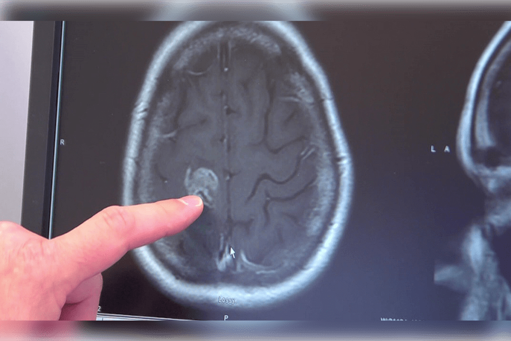
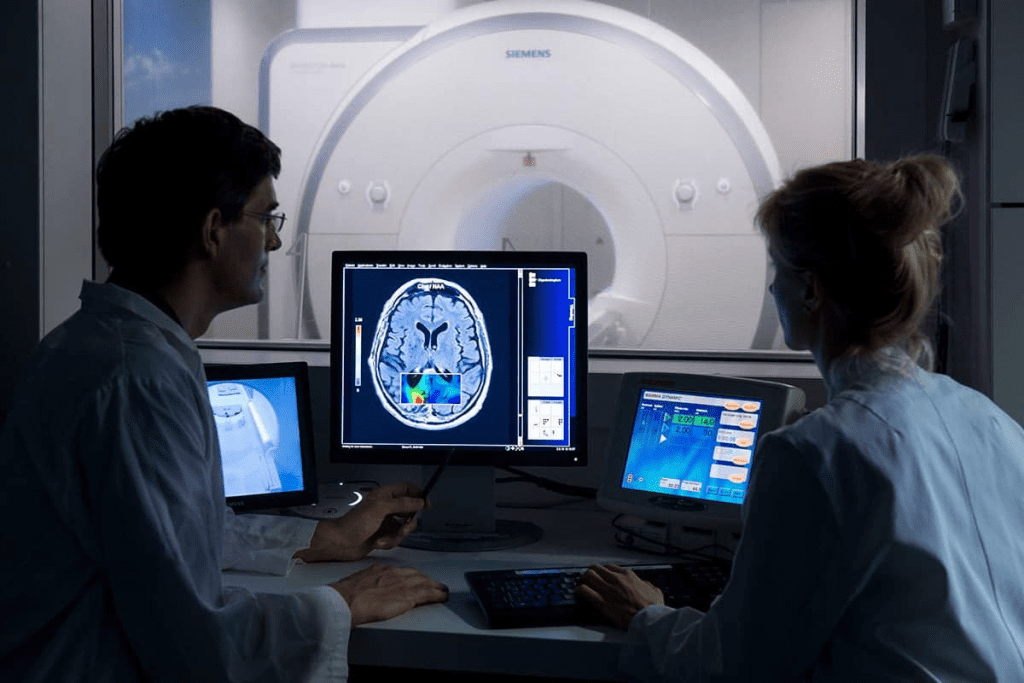
At Liv Hospital, we understand the concerns surrounding brain tumor detection. CT scans are often used to find brain tumors. But, they can miss some tumors. Advanced imaging modalities like MRI and bone scintigraphy are key in finding and managing brain tumors.
CT scans use X-rays to show detailed brain images. But, they might not show the whole tumor. Bone scintigraphy, especially with technetium-99m, helps show how active and spread the tumor is.
We know how important it is to diagnose and treat tumors correctly. By using CT scans with other imaging, we can find more tumors and plan better treatments.
Key Takeaways
- CT scans have limitations in detecting brain tumors.
- MRI and bone scintigraphy are valuable alternatives.
- Technetium-99m bone scintigraphy provides insights into tumor activity.
- Combining imaging modalities improves detection and treatment.
- Accurate diagnosis is crucial for effective brain tumor management.
The Fundamentals of CT Scanning in Neuroimaging

CT scanning has changed neuroimaging by making quick and effective diagnoses of brain issues. It gives us detailed images of the brain. These images help us understand and treat many neurological problems.
How CT Technology Creates Brain Images
CT scans use X-rays to make brain images. They rotate an X-ray source and detectors around the head. This captures data to create detailed images.
The quality of these images depends on the technology and scanning protocols. Advanced CT scanners can make clearer, more detailed images. This helps doctors make more accurate diagnoses.
Types of CT Protocols Used for Brain Assessment
There are different CT protocols for brain assessments. Non-contrast CT is used in emergencies, like finding acute hemorrhages. Contrast-enhanced CT uses a contrast agent to show tumors or other brain issues.
- Non-contrast CT for acute hemorrhage detection
- Contrast-enhanced CT for tumor detection
- CT angiography for vascular assessment
Standard Applications in Neurological Diagnosis
CT scanning is key in neurological diagnosis. It’s great for quick checks in emergencies, like head injuries or strokes. It also helps track neurological diseases and treatment success.
Understanding CT scanning and its uses in neuroimaging is important. It helps us see how it aids in diagnosing and treating brain issues. The technology keeps getting better, improving patient care.
Can a CT Scan Miss a Brain Tumor? Examining Detection Limitations

It’s important to know the limits of CT scans in finding brain tumors. CT scans are useful but not perfect. Several things can make it hard to spot tumors, like the tumor’s size, where it is, how dense it is, and when the scan is done.
Size Thresholds for Reliable Detection
One big problem with CT scans is finding small tumors. Tumors under 5 mm are hard to see, especially if they don’t show up well with contrast or are in tricky spots.
Using contrast agents can help find more small tumors. But, even with contrast, very tiny tumors might still be missed.
Location Challenges: Skull Base and Posterior Fossa
Where a tumor is can also affect if it’s seen on a CT scan. Tumors near the skull base or in the posterior fossa are tough to spot because of bone artifacts.
“The skull base and posterior fossa are regions where CT scan limitations are most pronounced due to beam hardening artifacts and partial volume averaging.”
This makes it harder to find tumors in these areas. It might lead to missing tumors if not checked with other imaging methods.
Density and Contrast Complications
Tumors that look the same as the brain on CT scans are hard to see. Contrast agents help by making tumors stand out. This is because they show up differently from the brain.
| Tumor Type | Density | Contrast Enhancement |
| Gliomas | Variable | Often enhances |
| Metastases | Usually hypodense | Typically enhances |
| Meningiomas | Often isodense or hyperdense | Usually enhances strongly |
Timing Factors in Tumor Visualization
When the CT scan is done compared to when contrast is given can also matter. The right timing is key to seeing how tumors react to contrast.
In summary, CT scans are good for finding brain tumors but have their limits. These include size, location, density, and timing. Knowing these limits helps doctors decide when more tests are needed.
Types of Brain Tumors Most Frequently Missed on CT Imaging
Even with the latest CT technology, some brain tumors are hard to spot. We’ll look at the types of tumors that often slip through the cracks on CT scans. We’ll also talk about how other imaging methods can help find them.
Small Metastatic Lesions
Small metastatic lesions are a big problem for CT scans. They’re too tiny for CT to see clearly. Early detection is key because catching them early can make a big difference in treatment success.
Isodense Tumors Without Enhancement
Isodense tumors are hard to spot because they blend in with the brain. If they don’t show up with contrast, they’re almost invisible. Advanced imaging is needed to find these tumors accurately.
Early-Stage Gliomas and Infiltrative Tumors
Early-stage gliomas and infiltrative tumors are tricky to find on CT scans. They might not show up because they’re so subtle. MRI is better for spotting these tumors because it shows soft tissue details better.
Brainstem and Cerebellar Tumors
Tumors in the brainstem and cerebellum are hard to see on CT scans. This is because of artifacts from the bone around them. MRI is better for these areas because it doesn’t get messed up by bone artifacts, giving a clearer view.
In summary, while CT scans are useful, some brain tumors are missed. Knowing these limitations helps choose the right imaging method for accurate diagnosis.
MRI: The Superior Alternative for Brain Tumor Detection
MRI is now the top choice for finding brain tumors. It beats CT scans in accuracy. This is because MRI has special tech and features.
Technological Advantages Over CT Scanning
MRI is better than CT scans in many ways. It makes detailed images without harmful radiation. This makes patients safer and lets doctors take more pictures without worry.
Key technological advantages include:
- Superior soft tissue contrast
- Multiplanar imaging capabilities
- Ability to perform functional imaging techniques
Enhanced Soft Tissue Differentiation
MRI is great at showing soft tissues clearly. This helps doctors tell tumors apart and see their edges.
The clarity MRI offers helps doctors make better diagnoses. They can plan treatments more accurately. CT scans can’t always do this as well.
Specialized MRI Sequences for Tumor Characterization
MRI has special sequences for studying tumors. These include diffusion-weighted imaging and magnetic resonance spectroscopy.
These methods give insights into tumor biology. They help doctors understand the tumor better for treatment.
Limitations and Contraindications
Even though MRI is great, it has its limits. Some people can’t have MRI because of metal implants or pacemakers. But, many implants now work with MRI.
Also, MRI might cost more and be harder to get than CT scans in some places. But, for finding brain tumors, MRI’s benefits are worth it.
Advanced Nuclear Medicine Techniques in Brain Tumor Assessment
Nuclear medicine has changed how we assess brain tumors. It helps us understand tumor characteristics better. This leads to more accurate diagnoses and effective treatments.
PET Scanning for Metabolic Activity
PET scanning is a key tool for brain tumor assessment. It shows how active tumors are metabolically. PET scanning with fluorodeoxyglucose (FDG) spots high glucose levels, often in cancerous tumors.
PET scanning brings many benefits, including:
- Enhanced diagnostic accuracy
- Improved tumor grading
- Better treatment planning
- Effective monitoring of treatment response
SPECT Imaging Applications
SPECT imaging is another valuable tool in brain tumor assessment. It reveals details about blood flow and metabolism in tumors. SPECT imaging with certain radiopharmaceuticals can spot specific tumor traits.
SPECT imaging is used in various ways, including:
- Differentiating tumor recurrence from radiation necrosis
- Assessing tumor vascularity
- Guiding biopsy procedures
Integration with Conventional Imaging
Combining PET and SPECT with CT and MRI has transformed brain tumor assessment. This approach gives a full view of tumor characteristics. It includes anatomical, metabolic, and functional details.
This combination enhances our ability to:
- Improve diagnostic accuracy
- Enhance treatment planning
- Monitor treatment response more effectively
A leading nuclear medicine expert notes, “The fusion of PET and MRI has opened new avenues in brain tumor assessment. It provides both functional and anatomical information in a single examination.” This shows the growing role of multimodal imaging in neuro-oncology.
“The combination of PET and MRI offers a powerful tool for assessing brain tumors, providing valuable insights into tumor biology and behavior.”
Three-Phase Bone Scintigraphy: Principles and Technology
Three-phase bone scintigraphy is a complex test that uses technetium-99m to check bone health. It’s key in diagnosing and treating bone problems, especially those linked to brain tumors.
Technetium-99m Bone Scan Methodology
Technetium-99m bone scans work by using a special compound that bonds with bone. Technetium-99m diphosphonates are often used because they stick to bone, showing where bone is being built up.
The Three Distinct Phases of Bone Scanning
A three-phase bone scan has three parts: the arterial or angiographic phase, the soft tissue or blood pool phase, and the delayed or bone phase. The first phase shows how the tracer moves through blood vessels. The second phase shows where the tracer goes in soft tissues. The third phase, taken hours later, shows bone activity, highlighting areas of bone growth.
This method helps doctors diagnose and treat bone issues, like cancer spread and bone tumors. Using it with CT and MRI scans makes it even more useful for diagnosis.
3-Phase Bone Scan Interpretation in Neurological Applications
Interpreting 3-phase bone scans is key in neurological fields. It gives us insights into bone-related issues. We use it to check bone health and find problems linked to the brain.
Normal vs. Abnormal Uptake Patterns
Knowing the difference in uptake patterns is crucial. Normal scans show even uptake, except in high turnover areas like joints. Abnormal scans show either too much or too little activity. Too much activity might mean fractures, infections, or tumors. Too little could mean poor blood flow or dead bone.
To spot these issues, we look at the scan with the patient’s history in mind. We also compare it with other scans like CT or MRI. This helps us make better treatment plans.
Differentiating Infection from Neoplastic Processes
Distinguishing infections from tumors is a big challenge. Both can show up as increased activity on scans. We look at the pattern and intensity of uptake, along with the patient’s symptoms. Infections tend to spread out, while tumors are more focused.
- Wide uptake in early phases might point to an infection.
- Later, focused uptake could mean a tumor.
- It’s important to match the scan with the patient’s story and other scans.
Skull-Based Tumor Detection Capabilities
Three-phase bone scans are great for finding tumors in the skull. They’re especially good when the tumor affects the bone. This makes them very sensitive for such tumors.
Signs of a skull tumor on a scan include:
- Specific spots of high activity in the skull.
- Uneven activity patterns.
- Activity that matches a known tumor location.
Metastatic Disease Identification in the Skeletal System
Three-phase bone scans are also good for spotting metastatic disease. Metastases show up as multiple spots of high activity in the bones. This pattern is often specific.
The benefits of using these scans for metastases include:
- They’re very good at finding bone tumors.
- They can check the whole skeleton at once.
- They’re useful for tracking how well treatment is working.
By understanding 3-phase bone scans, we can better diagnose and treat skeletal and neurological problems.
Clinical Applications of Radionuclide Bone Scans in Neuro-Oncology
Radionuclide bone scans are key in neuro-oncology. They help find bone metastases. These scans spot changes in bone metabolism, showing if disease has spread.
Detecting Skull and Vertebral Metastases from Primary Brain Tumors
Radionuclide bone scans are vital for finding skull and vertebral metastases from brain tumors. These metastases can greatly affect patient outcomes and treatment plans. The scans are good at catching early signs of metastatic lesions.
- Early detection of metastatic disease
- Monitoring of treatment response
- Identification of skeletal complications
Monitoring Treatment Response in Osseous Involvement
These scans are also great for tracking how metastases react to treatment. They watch bone metabolism changes over time. This helps doctors see if treatments are working.
For example, if radionuclide uptake goes down, it means treatment is working. But if it goes up, it might mean the disease is getting worse.
Triple-Phase Bone Scan vs. Single-Phase Techniques
Triple-phase bone scans have more benefits than single-phase ones. They show different bone activity phases. This helps doctors tell apart different disease types.
| Phase | Description | Clinical Utility |
| Flow Phase | Initial blood flow to the area | Assesses perfusion |
| Blood Pool Phase | Distribution of tracer in the blood pool | Evaluates inflammation or tumor activity |
| Delayed Phase | Uptake of tracer into bone | Reflects bone metabolism |
Integration with CT and MRI Findings
Combining radionuclide bone scans with CT and MRI scans improves diagnosis. This mix gives doctors a full picture of the disease. It’s especially helpful in tricky cases where detailed diagnosis is needed.
Multimodal Imaging Approach: Creating Optimal Diagnostic Protocols
Using many imaging methods is key for accurate diagnosis and treatment planning in brain tumor patients. By combining different techniques, we get a full picture of the tumor’s details, location, and how it affects the brain.
First-Line Imaging Selection Criteria
We look at several things when choosing the first imaging method. These include the patient’s symptoms, medical history, and the suspected tumor type. CT scans are often first because they’re quick and good at finding bleeding or calcifications. But MRI is usually better for seeing soft tissues and understanding the tumor’s spread.
Sequential and Complementary Imaging Strategies
After the first scan, we might use more scans to get a better look at the tumor. For example, if a CT scan is first, we might follow it with an MRI for more details. PET or SPECT scans can also help by showing how active the tumor is or what it’s made of.
Cost-Effectiveness and Radiation Considerations
We balance the need for accurate diagnosis with cost and radiation concerns. CT scans use radiation but are often needed first. MRI doesn’t use radiation but can be more expensive. We decide based on what’s best for each patient.
Patient-Specific Factors in Imaging Selection
Each patient’s needs are unique when it comes to imaging. For instance, some patients can’t have MRI because of metal implants. Others might need special care with contrast agents due to kidney issues. We adjust our approach to fit each patient’s situation.
Conclusion: Ensuring Accurate Brain Tumor Detection Through Comprehensive Imaging
Getting brain tumors right is key for good treatment and better health. We’ve looked at different ways to see tumors, like CT scans, MRI, and nuclear medicine. Each method has its own good points and areas where it falls short.
Using many imaging types together makes finding tumors more accurate. MRI is great for seeing soft tissues, while CT scans are fast. The triple phase bone scan helps spot bone metastases. Mixing these methods helps us find and understand brain tumors better.
We stress the importance of using many imaging types in brain cancer care. This way, doctors can plan treatments that really fit each patient. Using all these imaging methods is crucial for improving care and results in diagnosing brain tumors.
FAQ
What are the limitations of CT scans in detecting brain tumors?
CT scans have some big limitations when it comes to finding brain tumors. They can miss small tumors or those in tricky spots. Also, they might not work well with tumors that look the same as the brain tissue. This can lead to missing some tumors, especially the small ones.
How does MRI compare to CT scans in brain tumor detection?
MRI is better than CT scans for finding brain tumors. It shows more detail in soft tissues and has special sequences for tumors. MRI is great for spotting small tumors, tumors that look the same as the brain, and early gliomas.
What is three-phase bone scintigraphy, and how is it used in neuro-oncology?
Three-phase bone scintigraphy is a test that uses a special kind of medicine to find bone problems. In brain cancer, it helps find tumors in the skull and spine. It also checks how well treatments are working and tells the difference between infections and tumors.
How is a 3-phase bone scan interpreted in neurological applications?
When we look at a 3-phase bone scan, we check for normal or abnormal signs. We also try to tell if it’s an infection or a tumor. The scan shows how blood flows, pools, and changes in bone metabolism, helping us find tumors and metastases.
What are the advantages of a multimodal imaging approach in neuro-oncology?
Using different imaging methods like CT, MRI, PET, and SPECT together helps a lot. It gives a full picture of brain tumors. This way, doctors can make better decisions and see how treatments are working.
How do patient-specific factors influence imaging selection in neuro-oncology?
Every patient is different, and their medical history, symptoms, and tumor details matter a lot. Choosing the right imaging tests depends on these factors. This way, patients get the best and safest tests for their needs.
What is the role of PET and SPECT imaging in brain tumor assessment?
PET and SPECT scans show how tumors work and what they’re made of. PET scans look at how tumors use energy, while SPECT scans check blood flow and other functions. This helps doctors understand tumors better.
Can a CT scan miss a small brain tumor?
Yes, CT scans might not catch small brain tumors, especially if they’re the same color as the brain. MRI is usually better at finding these tiny tumors.
How do CT and MRI findings integrate with radionuclide bone scans in neuro-oncology?
Combining CT and MRI with bone scans gives a complete view of tumors. It helps find metastases in the skull and spine, track treatment success, and guide treatment plans.
What are the benefits of using technetium-99m in bone scintigraphy?
Technetium-99m is a top choice for bone scans because it’s safe and works well. It has the right half-life and energy for clear images. Patients usually do well with it.
References
- Lu, Y. M., Yu, X. Q., & Sun, X. H. (2016). The most suitable guidelines for performing bone scans in prostate cancer patients for detection of bone metastases. Procedia Environmental Sciences, 31, 37-43. https://www.sciencedirect.com/science/article/pii/S1879522615003620
- Chong, A., Song, C., & Shin, H. (2014). Application of bone scans for prostate cancer staging. Journal of Cancer Research and Therapeutics, 10(3), 605-610. https://www.ncbi.nlm.nih.gov/pmc/articles/PMC4137016/


