Last Updated on November 27, 2025 by Bilal Hasdemir

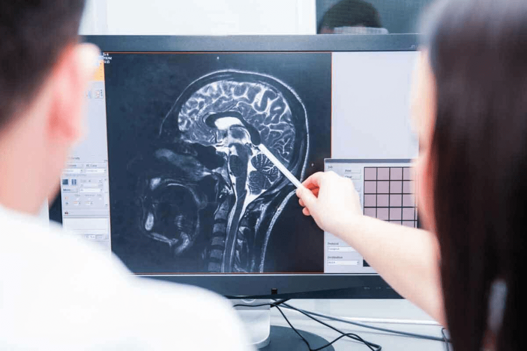
Recent data show that MRI is the leading tool for diagnosing and monitoring brain tumors. Early detection is key for effective treatment and better patient outcomes. MRI scans are vital in spotting early-stage lesions, leading to timely action.
MRI technology’s ability to find small tumors early is a big plus. Advanced imaging lets radiologists spot tiny tumor signs. This makes for accurate diagnosis and treatment plans. View first stage small brain tumor MRI images! This essential guide explains 15 real-life examples and what to look for in early diagnosis.
Key Takeaways
- Early detection of brain tumors is critical for effective treatment.
- MRI scans are the primary tool for diagnosing and monitoring brain tumors.
- Advanced MRI techniques enable the identification of small tumors at an early stage.
- Timely intervention improves patient outcomes.
- Accurate diagnosis is facilitated by the detailed images provided by MRI scans.
Understanding Early-Stage Brain Tumors on MRI

It’s key to know how early-stage brain tumors look on MRI for good treatment plans. Finding them early makes a big difference in how well patients do. MRI scans are great because they show the brain’s details clearly.
Characteristics of First Stage Brain Tumors
Early brain tumors show up as bright spots on certain MRI scans. They can be hard to spot, mainly if they’re small or in tricky brain spots. New MRI methods have made finding these tumors easier.
The look of these tumors on MRI changes based on their type and where they are. Some might look different with contrast agents, while others won’t. Knowing this helps doctors diagnose and plan treatment right.
Why Early Detection Matters
Finding brain tumors early is super important for better patient results. Early detection means treatments work better, and there’s a higher chance of success. MRI is key here, giving clear images to spot tumors early.
Also, finding tumors early means treatments can be made just for that patient. This makes treatments more effective and improves life quality for patients.
How MRI Technology Detects Brain Tumors

MRI technology is key in finding brain tumors. It uses basic and advanced methods. MRI gives detailed brain images without harmful radiation.
Basic MRI Principles for Brain Imaging
MRI uses nuclear magnetic resonance. It uses a strong magnetic field and radio waves to show brain images. Different tissues in the brain respond differently to these signals, allowing for the differentiation between healthy tissue and tumors.
There are basic MRI sequences like T1-weighted and T2-weighted images. T1-weighted images show brain structure and tumors well. T2-weighted images spot changes in tissue, helping find tumors and swelling. Recent studies show these sequences are key for brain tumor diagnosis.
Advanced MRI Techniques for Early Tumor Detection
Advanced MRI methods have made finding and understanding brain tumors better. Techniques like 3D FLAIR and diffusion-weighted imaging help tell tumor tissue from healthy tissue. 3D FLAIR is great for finding brain lesions, even near cerebrospinal fluid.
- Diffusion-weighted imaging checks tumor cell density, helping diagnose some brain tumors.
- Perfusion-weighted imaging shows tumor blood flow, helping judge tumor grade and growth.
- Magnetic Resonance Spectroscopy (MRS) looks at tumor metabolism, giving clues about its nature and danger.
Using these advanced methods with basic MRI sequences improves diagnosis. This helps doctors plan better treatments. Adding advanced MRI techniques to care has been a big step in managing brain tumors.
Interpreting MRI Sequences in Early Brain Tumor Diagnosis
Diagnosing early brain tumors depends a lot on MRI sequences. Magnetic Resonance Imaging (MRI) gives detailed brain images. This helps doctors spot tumors early.
Understanding MRI sequences is key for brain tumor diagnosis. We have T1-weighted, T2-weighted, FLAIR, and contrast-enhanced images. Each shows different tumor details.
T1-Weighted vs. T2-Weighted Images
T1-weighted and T2-weighted images are key in MRI diagnosis. T1-weighted images show detailed anatomy. They are best after contrast, showing where it goes, which might mean a tumor.
T2-weighted images are great for spotting water changes in tissues. Tumors show up bright because they have lots of water.
FLAIR and Contrast-Enhanced Sequences
FLAIR sequences help find lesions near the ventricles or on the brain’s surface. They do this by hiding the signal from cerebrospinal fluid (CSF).
Contrast-enhanced T1-weighted images use a contrast agent to show where the blood-brain barrier is broken. This is common in tumors. For more on MRI in brain tumor diagnosis, check Radiology Assistant.
By using T1, T2, FLAIR, and contrast images together, doctors get a full picture of the tumor. This helps in early and accurate diagnosis.
First Stage Small Brain Tumor MRI Images: Cortical Involvement
Early-stage brain tumors involving the cortex need precise MRI imaging for accurate diagnosis. Cortical involvement is key in brain tumor assessment. It greatly affects treatment planning and patient outcomes.
Image 1: Frontal Lobe Cortical Tumor
The frontal lobe is a common spot for early-stage brain tumors. MRI images show the extent of cortical involvement. They reveal how the tumor impacts the surrounding brain tissue.
In the case of a frontal lobe cortical tumor, MRI helps identify the tumor’s boundaries. It also shows its impact on nearby structures.
Image 2: Temporal Lobe Early-Stage Glioma
Temporal lobe gliomas are another early-stage brain tumor type that can involve the cortex. MRI sequences, like T2-weighted and FLAIR images, are key. They assess the tumor’s extent and its relationship with surrounding structures.
Image 3: Parietal Cortex Tumor Beginning
Tumors starting in the parietal cortex can be hard to diagnose early. They often have non-specific symptoms. MRI imaging is vital in detecting these tumors early.
Images show the initial changes in the cortical architecture. They help in planning the right treatment strategy.
Cortical involvement in early-stage brain tumors is complex. It requires careful evaluation using advanced MRI techniques. Understanding these tumors through MRI images is essential for effective treatment plans.
Early-Stage Tumors with White Matter Infiltration
Early-stage brain tumors spreading into white matter is key to their growth. White matter tracts are nerve fiber bundles. They help different brain areas talk to each other. When tumors get into these tracts, it can cause big problems with brain function.
At the start, tumors can sneak into white matter, like in glioblastoma. This can be hard to spot on MRI scans. But, advanced MRI like diffusion tensor imaging (DTI) can find these tumors in white matter.
Image 4: Frontal White Matter Involvement
This MRI shows a tumor starting to spread in the frontal white matter. It looks a bit brighter on T2-weighted images. This shows it’s getting into the white matter tracts.
Image 5: Corpus Callosum Early Infiltration
The corpus callosum connects the brain’s two halves. If a tumor gets in early, it can really mess with brain function. This picture shows the start of a tumor getting into the corpus callosum.
Image 6: Periventricular White Matter Tumor
Periventricular white matter is another spot where tumors can start early. This image shows a tumor near the ventricles. It shows why we need to look closely at these areas in MRI scans.
Knowing how much white matter a tumor has invaded is key for diagnosis and treatment. MRI scans give us a good look at how tumors affect white matter. This helps doctors plan better treatments.
First Stage Glioblastoma MRI Presentations
MRI is key in spotting early glioblastoma. It shows the tumor’s unique signs. Glioblastoma is aggressive and hard to treat, so finding it early is vital.
The first signs of glioblastoma can be hard to see on MRI. Knowing what to look for is important for a correct diagnosis.
Minimal Enhancement in Early GBM
Early glioblastoma multiforme (GBM) might show little change on MRI. This makes it hard to tell it apart from other brain issues. A study on the National Center for Biotechnology Information website shows how important advanced MRI is.
The early signs of GBM include necrosis and irregular enhancement.
Butterfly Glioblastoma Initial Presentation
Butterfly glioblastoma crosses the corpus callosum, making it tricky to diagnose. On MRI, it looks like a butterfly-shaped mass in both brain hemispheres. Finding it early is key because it grows fast and surgery is complex.
Thalamic Glioblastoma Early Stage
Thalamic glioblastoma is rare but can be spotted early with MRI. Tumors in the thalamus are hard to treat. MRI shows heterogeneous enhancement and mass effect on nearby areas.
Knowing how to spot early glioblastoma on MRI is critical. It helps doctors act fast and can lead to better treatment results. MRI is essential for diagnosing and managing glioblastoma, showing how big the tumor is and how it’s responding to treatment.
Distinguishing Features in Pediatric Brain Tumor MRI Images
MRI imaging is key in spotting pediatric brain tumors. It shows differences from adult tumors. Accurate MRI readings are vital for diagnosis and treatment.
Pediatric Posterior Fossa Tumor
Pediatric posterior fossa tumors are a big worry. They show unique signs on MRI. These tumors usually sit in the cerebellum or brainstem.
Medulloblastoma is a common, aggressive brain tumor in kids. On MRI, it looks like a clear mass in the posterior fossa. It often touches the cerebellar vermis.
“The posterior fossa is a critical area in pediatric neuro-oncology, with tumors like medulloblastoma requiring prompt diagnosis and treatment.” – A Pediatric Neuroradiologist
Pediatric Supratentorial PNET
Supratentorial PNETs are aggressive brain tumors in kids. They have unique imaging signs.
- They look like big, mixed masses
- They often have dead tissue and calcium spots
- They can push on the brain and cause swelling
| Tumor Type | Common Location | Typical MRI Features |
| Medulloblastoma | Posterior fossa | Well-defined mass, often in cerebellar vermis |
| Supratentorial PNET | Cerebral hemispheres | Large, heterogeneous, necrosis, calcifications |
Early-Stage Pediatric Glioma
Pediatric gliomas, like low-grade astrocytomas, are common in kids. Early gliomas might show small signs on MRI.
Key Features:
- They look like small, bright spots on T2 scans
- They might not push on the brain much or show up well
- They can be hard to tell apart from other conditions
In conclusion, pediatric brain tumors have unique MRI signs. They need special knowledge for correct diagnosis and treatment.
Subtle MRI Findings in First Stage Brain Tumors
Early-stage brain tumors can show up with small changes on MRI. It’s very important to read these signs correctly. These signs include a bit of swelling, early signs of mass effect, and finding small tumors by chance.
Being able to spot these small changes is key for catching tumors early. MRI scans like T1-weighted, T2-weighted, and FLAIR help find these early signs.
Minimal Edema Presentation
When a brain tumor starts to grow, it may cause a bit of swelling. MRI findings might show a slight bright spot on T2-weighted images around the tumor.
shows a small tumor with a bit of swelling. This picture shows why it’s so important to look closely at MRI images.
- Finding swelling early can help diagnose tumor growth.
- T2-weighted images are great for seeing swelling.
- Even a little swelling is a big finding.
Early Mass Effect Signs
Early mass effect means a tumor is starting to push on nearby brain parts. This is a small sign on MRI that needs careful looking.
Early mass effect signs show up on MRI as slight changes in the brain’s shape. Spotting these signs early can change how we plan treatment.
- Find out where the tumor is and how close it is to important parts.
- See how much the tumor is pushing on the brain and if it could cause problems.
- Use special MRI scans to see the tumor better.
- Watch for changes in how the tumor is pushing on the brain over time.
Incidental Finding of Small Brain Tumor
Small brain tumors are sometimes found by accident during MRI scans for other reasons. These tumors might not be causing any symptoms yet.
Finding a small brain tumor by chance shows how important MRI is for finding problems before they cause symptoms. Careful checking of these tumors is needed to decide what to do next.
In summary, finding small changes like swelling, early mass effect, and finding tumors by chance are key for catching brain tumors early. Reading MRI images well is vital for spotting these small signs and planning the right treatment.
Conclusion: Importance of Expert Interpretation in Early Brain Tumor Detection
Getting MRI images right is key for finding and treating brain tumors. It takes a lot of knowledge to understand MRI images and brain tumors well.
Finding brain tumors early is all about spotting small changes in MRI pictures. Tools like FLAIR and contrast-enhanced sequences help a lot.
Experts are very important in this field. They help make treatment plans that work. This can lead to better treatment results.
To sum up, using the latest MRI tech and expert eyes is vital for catching brain tumors early. As we keep improving MRI for brain tumor diagnosis, experts will keep being a big help.
FAQ
What does a brain tumor look like on MRI?
A brain tumor on MRI looks like a mass with different signal intensities. This depends on the type and grade of the tumor. It can vary on T1-weighted, T2-weighted, and FLAIR images.
How do MRI sequences help in diagnosing brain tumors?
Different MRI sequences give unique insights into tumors. T1-weighted images show the anatomy. T2-weighted images highlight edema and tumor size. FLAIR sequences suppress fluid signals. Contrast-enhanced images show tumor vascularity and blood-brain barrier disruption.
What are the characteristics of first-stage brain tumors on MRI?
First-stage brain tumors on MRI are small and well-defined. They have minimal edema and mass effect. They may show varying signal intensity and some may enhance with contrast.
Why is early detection of brain tumors important?
Early detection of brain tumors is key for better outcomes. It allows for timely treatment and potentially better surgical resection. This can reduce morbidity and improve survival rates.
How do advanced MRI techniques aid in early tumor detection?
Advanced MRI techniques like diffusion-weighted imaging, perfusion-weighted imaging, and MR spectroscopy provide extra information. They help detect tumors early by showing tumor biology, vascularity, and metabolism.
What is the significance of white matter infiltration in brain tumors?
White matter infiltration in brain tumors shows tumor spread along fiber tracts. This affects treatment planning and prognosis. MRI images showing this help clinicians understand tumor extent and behavior.
How do pediatric brain tumors differ from adult brain tumors on MRI?
Pediatric brain tumors have distinct MRI characteristics. They differ in location, signal intensity, and enhancement patterns from adult brain tumors. Understanding these differences is essential for accurate diagnosis and treatment.
What are the challenges in detecting subtle MRI findings in first-stage brain tumors?
Subtle MRI findings in first-stage brain tumors require careful image interpretation. Advanced MRI techniques and high-quality images help identify these subtle changes.
Can MRI images of brain tumors be used to predict tumor grade or type?
While MRI images provide valuable information, predicting tumor grade or type is complex. It often requires a combination of imaging features, clinical information, and histopathological analysis.
How do glioblastoma MRI images differ from other types of brain tumors?
Glioblastoma MRI images show characteristic features like heterogeneous enhancement, necrosis, and infiltrative margins. Understanding these features is essential for diagnosing and managing glioblastoma.
References
- PMC. (2022). Early detection of brain tumors using diffusion and perfusion MRI techniques. Neuroimaging Clinics, 31(4), 575“593. https://pubmed.ncbi.nlm.nih.gov/35308467/
- Zhang, Y., & Li, J. (2022). Imaging biomarkers for differentiating early-stage brain tumors. Cancer Imaging, 22(1), 1“11. https://doi.org/10.1186/s40644-022-00455-4
- National Cancer Institute. (2024). Magnetic resonance imaging in brain tumor diagnosis. https://www.cancer.gov/about-cancer/diagnosis-staging/imaging/mri


