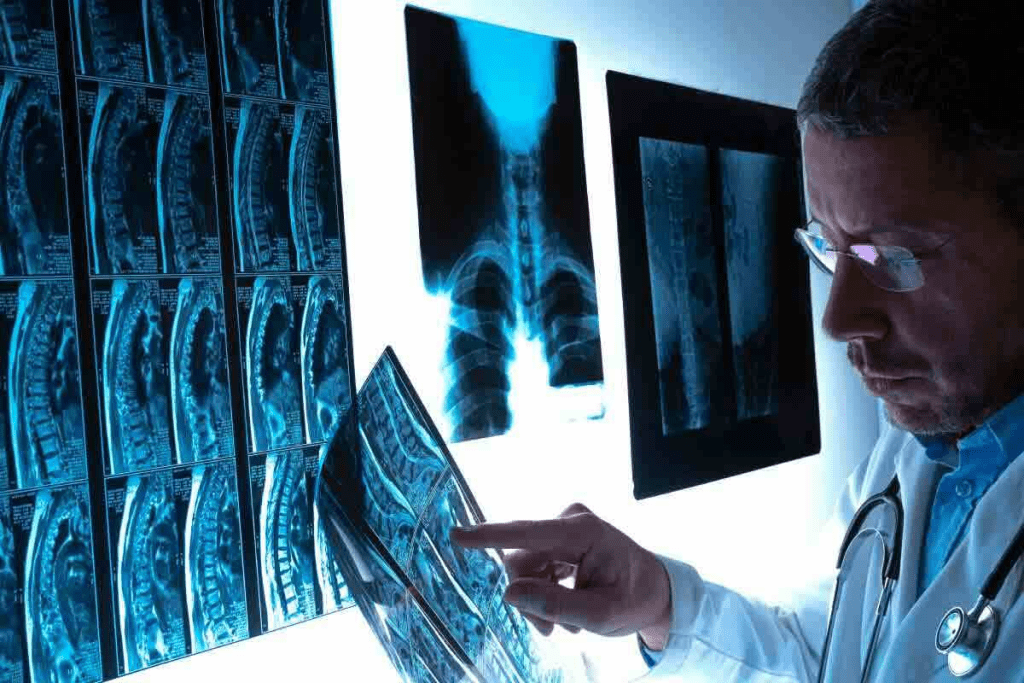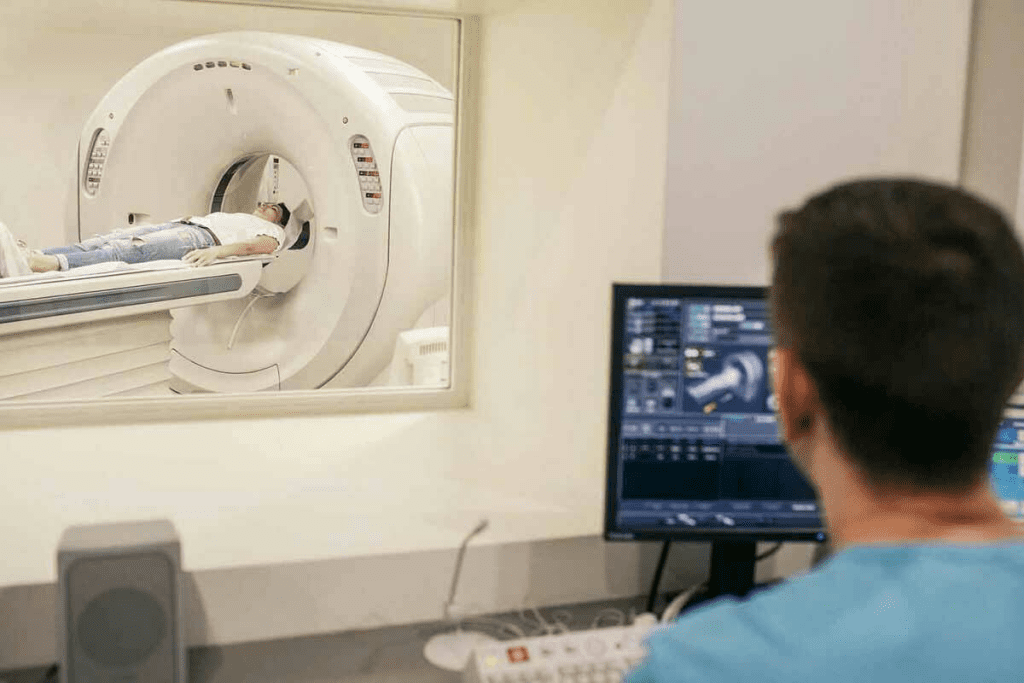
We use technetium-99m labelled compounds in a 3 phase bone scan radiology to check bone health. This method is key for spotting bone damage or disease. The 3 phase bone scan involves three imaging phases: the first phase assesses blood flow to the bone, the second phase evaluates soft tissue involvement, and the third phase detects bone metabolism activity. This detailed imaging helps in diagnosing fractures, infections, tumors, and other bone conditions with high accuracy.
A bone scan happens in a hospital’s nuclear medicine unit. It can take up to four hours. This includes the time for the tracer to move and the scan itself. The scan uses a radioactive tracer to find areas of high bone turnover.
It’s important to understand these scans for correct diagnosis and treatment. The scan has three phases: dynamic flow, blood pool, and delayed. Each phase gives important info about the bone’s health.
Key Takeaways
- Technetium-99m labelled compounds are used in 3 phase bone scan radiology.
- The procedure evaluates osseous vascularity and osteoblastic activity.
- A bone scan can take up to four hours to complete.
- The scan helps in identifying areas of increased bone turnover.
- Understanding scan interpretation is crucial for diagnosis and treatment.
The Fundamentals of Nuclear Medicine Bone Imaging

It’s important to know how nuclear medicine bone imaging works. This is key for understanding three-phase bone scintigraphy results. It helps us see how bones work and find problems.
Bone scintigraphy uses a special dye to check bone health. It looks at the whole skeleton. This test is great for finding diseases and hidden fractures.
What Defines a Three Phase Bone Scintigraphy
A three-phase bone scintigraphy has three parts. The dynamic flow phase checks blood flow. The blood pool phase looks at soft tissue. The delayed phase shows bone activity. Each part gives different clues about health.
The first part, the dynamic flow phase, checks blood flow right after the dye is given. The blood pool phase comes next, showing where the dye goes in soft tissues. The delayed phase, 2-4 hours later, shows where bones are most active.
Evolution of Bone Scan Techniques in Clinical Practice
Bone scan methods have gotten better over time. New cameras and dyes make scans more accurate. Studies show these improvements help doctors more in many areas.
New tech like SPECT (Single Photon Emission Computed Tomography) and SPECT/CT make scans even better. They give clear pictures of bones and how they work. This helps doctors find and understand bone problems better.
In short, knowing about nuclear medicine bone imaging is crucial. It helps doctors use three-phase bone scintigraphy to its fullest. As tech keeps improving, bone scans will keep being a key tool in healthcare.
Key Fact #1: How Technetium-99m Functions in Bone Scanning

Technetium-99m is key in bone scanning because of its special properties. We use Technetium-99m labeled compounds like methylene diphosphonate (MDP) or hydroxydiphosphonate (HDP) in bone scintigraphy. These help us see bone metabolism and find different pathologies.
Chemical Properties of Tc-99m MDP and Related Compounds
The chemical makeup of Tc-99m MDP and similar compounds is perfect for bone imaging. Tc-99m MDP sticks well to bone, especially where bone turnover is high. This is because it binds to hydroxyapatite in bone. The chemical structure of these compounds also lets them be labeled with Technetium-99m efficiently, making a stable radiopharmaceutical.
The table below summarizes the key chemical properties of Tc-99m MDP:
| Chemical Property | Description |
| Molecular Structure | Contains phosphonate groups for bone binding |
| Labeling Efficiency | High affinity for Technetium-99m |
| Biodistribution | Primarily accumulates in bone and excreted through kidneys |
Physiological Basis of Tracer Uptake in Bone Tissue
The uptake of Tc-99m MDP in bone tissue comes from bone remodeling. Areas with more bone turnover, like those with disease, take up more of the tracer. This is because the radiopharmaceutical attaches to new bone, making it great for finding bone diseases.
Knowing how tracer uptake works is key to understanding bone scan results. We can spot normal and abnormal uptake patterns. This helps us diagnose things like fractures, infections, and metastatic bone disease.
Key Fact #2: The Three Critical Phases of 3 Phase Bone Scan Radiology
The three-phase bone scan is key in nuclear medicine. It gives a full view of bone health by looking at different bone functions and problems in three stages.
Dynamic Flow Phase: Vascular Assessment
The dynamic flow phase is the first part of the scan. It quickly takes pictures of the area after the radiopharmaceutical is given. This phase checks blood flow to bones and soft tissues.
Vascular assessment here spots issues like infections or injuries. It shows if there’s too much or too little blood flow.
Blood Pool Phase: Soft Tissue Evaluation
The blood pool phase comes after the dynamic flow phase. It takes pictures a few minutes later. This phase looks at soft tissue’s blood activity, showing inflammation or infection.
During this phase, soft tissue evaluation is key. It finds issues like cellulitis or abscesses that might not be bone-related but affect the scan’s results.
Delayed Phase: Bone Metabolism Imaging
The delayed phase is the most important part. It happens 2-4 hours after the radiopharmaceutical is given. By then, the bone has taken up the tracer, showing bone metabolism and activity.
Bone metabolism imaging in this phase is vital. It helps diagnose bone disorders like fractures, infections, or tumors.
| Phase | Timing | Primary Assessment |
| Dynamic Flow | Immediate | Vascular perfusion |
| Blood Pool | A few minutes post-injection | Soft tissue activity |
| Delayed | 2-4 hours post-injection | Bone metabolism |
In conclusion, the three-phase bone scan is a powerful tool for diagnosing bone health. It looks at different aspects of bone function and disease in three stages. This helps doctors understand bone conditions better and choose the right treatments.
Key Fact #3: Essential Equipment and Technical Considerations
Getting good results from skeletal scintigraphy depends on the right equipment and tech. The quality of 3 phase bone scan radiology relies on several things. These include the technology used and the scanning techniques.
Gamma Camera Technology in Skeletal Scintigraphy
The gamma camera is key in skeletal scintigraphy. It captures internal radiation patterns and turns them into images. Gamma camera technology has improved a lot, making bone scans better and more precise.
The gamma camera can spot gamma rays from the Tc tracer. This lets us see bone metabolism in detail. It’s vital for diagnosing and tracking bone issues.
Quality Control and Labeling Efficiency in 99mTc Bone Scans
Quality control is crucial for accurate 3 phase bone scans. It checks if the Tc tracer is working right. Labeling efficiency means the technetium-99m is well attached to compounds like methylene diphosphonate (MDP). Good labeling is key for clear images.
| Quality Control Measure | Description | Importance |
| Labeling Efficiency | Successful binding of Tc to MDP | High |
| Gamma Camera Calibration | Ensuring accurate detection of gamma rays | High |
| Tracer Preparation | Correct preparation of Tc tracer | High |
Keeping quality control high, like labeling efficiency and gamma camera calibration, is vital. It helps make top-notch bone scans. These scans are crucial for accurate diagnosis and treatment plans.
Key Fact #4: Diagnostic Patterns in 3 Phase Bone Scan Interpretation
In 3 phase bone scan interpretation, we look at patterns to figure out bone disorders. These patterns help us diagnose and manage bone system issues.
Normal Distribution Patterns Across All Phases
A normal 3 phase bone scan shows a specific pattern. In the first phase, we see symmetrical blood flow. The second phase shows mild soft tissue activity. The third phase highlights bone activity with some soft tissue activity left.
These patterns are key for spotting any oddities. Symmetrical uptake in bones is a sign of a healthy scan.
Pathological Uptake Signatures
Abnormal conditions show up as odd uptake patterns in the scan. For example, osteomyelitis shows increased activity in all phases, showing inflammation and bone reaction.
- Acute osteomyelitis has high uptake in all phases, showing inflammation.
- Chronic osteomyelitis has less uptake in early phases but keeps activity in the delayed phase.
- Fractures and bone metastases have unique patterns, helping us diagnose them.
Quantitative Analysis Methods
Quantitative analysis makes 3 phase bone scans more meaningful. Region of interest (ROI) analysis lets us measure activity in specific spots. This gives us a clearer view of bone activity.
- ROI analysis involves drawing around areas of interest on the scan images.
- By calculating uptake ratios, we can compare activity between spots.
- These numbers help track disease changes and treatment effects.
By mixing visual checks with numbers, we get a better grasp of 3 phase bone scan patterns.
Key Fact #5: Clinical Applications of Radionuclide Bone Scans
Radionuclide bone scans are key in medical care. They help diagnose and manage bone issues. We use them to check bone health, find trauma, and look at cancer.
Trauma and Occult Fracture Detection
Radionuclide bone scans are great for finding trauma and hidden fractures. They’re useful when X-rays don’t show anything. These scans can spot fractures X-rays miss, helping doctors treat patients right.
- Early detection of stress fractures in athletes
- Identification of occult fractures in elderly patients
- Assessment of fracture healing and non-union
Oncological Evaluation and Metastatic Surveillance
These scans are vital for cancer care. They help find bone metastases and check how well treatments work. Because they’re so sensitive, they’re key for cancer patients.
Infection vs. Inflammation Differentiation
Radionuclide bone scans help tell infection from inflammation. Both can cause similar symptoms but need different treatments. By looking at how the tracer spreads, we can tell them apart and choose the right treatment.
Metabolic and Degenerative Bone Disease Assessment
These scans are also good for checking bone diseases like Paget’s and osteomalacia. They help doctors diagnose and track these conditions. They give us important info for managing these diseases.
In short, radionuclide bone scans are very useful in medicine. They help find trauma, check for cancer, and tell infection from inflammation. They’re a crucial part of nuclear medicine today.
Key Fact #6: Comparative Advantages of Triple Phase Bone Scanning
Triple phase bone scanning is a key tool in nuclear medicine. It has many benefits in finding bone problems. We’ll look at its sensitivity, specificity, how it works with other tests, and its cost.
Sensitivity vs. Specificity in Bone Pathology Detection
Triple phase bone scanning is great at finding bone issues early. It’s especially good at spotting osteomyelitis or bone metastases. Though it might not be perfect, it’s a good first step to check for problems.
Sensitivity is key in finding issues first. The triple phase scan gives insights into bone health and problems.
Complementary Role with Advanced Imaging Modalities
Bone scans work well with MRI and CT scans. These tests show the body’s structure, while bone scans show how bones are working. Together, they help solve complex cases or see how far a disease has spread.
| Imaging Modality | Primary Use | Complementary Information |
| Triple Phase Bone Scan | Functional information about bone metabolism | Sensitivity in detecting bone pathology |
| MRI | Detailed soft tissue imaging | Anatomical detail, extent of disease |
| CT Scan | Detailed bone and soft tissue imaging | Anatomical detail, structural changes |
Cost-Effectiveness and Radiation Considerations
Choosing a diagnostic tool means thinking about cost and radiation. Bone scans are cheaper than some tests but still use some radiation. We must think about the benefits of the scan against the risk of radiation.
Knowing the benefits of triple phase bone scanning helps us use it better. This improves patient care and results.
Key Fact #7: Challenges and Pitfalls in Technetium-99m Scintigraphy
Reading technetium-99m scintigraphy results can be tricky. This is because of technical and patient-related issues. Despite its value in showing bone health, its accuracy can be affected by many things.
Technical Artifacts and Their Recognition
Technical problems can mess up the quality of technetium-99m scintigraphy scans. These issues might come from faulty equipment, wrong setup, or problems with the injection. For example, an injection mistake can make it hard to see real problems.
Knowing about these issues is key to understanding the scans right.
Some common technical problems include:
- Motion artifacts from patient movement during the scan
- Attenuation artifacts from dense objects or body parts
- Injection artifacts from wrong tracer use
Patient Factors Affecting Scan Quality
Patient factors also affect how good technetium-99m scintigraphy scans are. Things like how hydrated the patient is, their kidney function, and body shape can change how the tracer spreads. For instance, not drinking enough water can make soft tissues show up too much, hiding or faking problems.
To improve scan quality, getting the patient ready is important. Making sure they drink enough water and using the right amount of tracer helps. Knowing about these patient-specific things helps doctors understand the scans better.
Interpretation Challenges in Complex Cases
It’s hard to understand technetium-99m scintigraphy in complicated cases. When there are many problems at once, like cancer spreading or infections in different places, it’s tough to tell them apart. Doctors need to look at the patient’s history and other images to get a clear picture.
| Challenge | Description | Mitigation Strategy |
| Technical Artifacts | Equipment malfunctions or injection issues | Regular equipment maintenance and proper injection technique |
| Patient Factors | Hydration status, renal function, body habitus | Adequate patient preparation and adjusted tracer dosage |
| Complex Cases | Multiple pathologies or overlapping conditions | Correlation with clinical history and other imaging modalities |
Conclusion: The Evolving Role of 3 Phase Bone Scans in Modern Radiology
3 phase bone scans are key in modern radiology, giving important insights into bone health. The evolving role of these scans is thanks to new technology and techniques. This makes them better at finding bone problems.
These scans are essential for diagnosing and treating patients, especially when other tests don’t help. The improvement in Technetium-99m scintigraphy keeps them useful in today’s medicine. They help in many different medical situations.
In summary, the growth and use of 3 phase bone scans show their vital role in today’s radiology. They help doctors check bone health and make better care plans for patients.
FAQ
What is a 3 phase bone scan?
A 3 phase bone scan is a way to check bone health. It uses special compounds to see if bones are healthy or not. This helps doctors find and treat bone problems.
How does technetium-99m function in bone scanning?
Technetium-99m finds areas where bone is changing a lot. It helps doctors see where problems might be. This is because it sticks to bone tissue well.
What are the three critical phases of 3 phase bone scan radiology?
The three phases are the flow, blood pool, and delayed phases. Each phase shows different things about bones. They help doctors understand bone health better.
What is the significance of the delayed phase in 3 phase bone scan radiology?
The delayed phase shows how bones are working. It helps find problems like fractures or infections. This is very important for diagnosis.
How does gamma camera technology contribute to 3 phase bone scan radiology?
Gamma cameras detect the radiation from technetium-99m. This makes clear images of bones. These images help doctors diagnose bone issues.
What are some common clinical applications of radionuclide bone scans?
Bone scans are used for many things. They help find fractures, check for cancer, and see if bones are infected. They also help with bone diseases.
How does 3 phase bone scan radiology compare to other imaging modalities?
Bone scans are very good at finding bone problems. They work well with other tests to understand bones better. This helps doctors make accurate diagnoses.
What are some challenges and pitfalls in technetium-99m scintigraphy?
There are a few issues with bone scans. Things like technical problems and how the patient feels can affect the scan. Also, some cases are hard to interpret.
How can technical artifacts be mitigated in 3 phase bone scan radiology?
To avoid technical problems, quality checks are important. Using the right technology and making sure compounds are labeled correctly helps too.
What is the role of quantitative analysis in 3 phase bone scan interpretation?
Quantitative analysis helps measure how much technetium-99m is in bones. This gives doctors important information about bone health. It helps them diagnose bone conditions.
How does 3 phase bone scan radiology contribute to the diagnosis of bone conditions?
Bone scans give a full picture of bone health. They check blood flow, soft tissues, and bone activity. This makes them a key tool for diagnosing bone problems.
References
- Palestro, C. J., Love, C. (2006). Three-phase bone scintigraphy in musculoskeletal infection. Radiologic Clinics of North America, 44(4), 855-872. https://pubmed.ncbi.nlm.nih.gov/16893769/


