Last Updated on November 27, 2025 by Bilal Hasdemir

At Liv Hospital, we’re dedicated to top-notch healthcare that puts patients first. We use the PSMA PET scan to find cancer. This scan looks for the Prostate-Specific Membrane Antigen (PSMA) protein, which prostate cancer cells often have too much of.
This advanced test has changed how we find and understand prostate cancer. Studies show that PSMA PET/CT can detect recurrent or metastatic prostate cancer with a sensitivity ranging from 65% to 85% depending on PSA levels and disease burden, and specificity exceeding 95%.
Key Takeaways
- PSMA PET scan is a highly sensitive imaging technique for detecting prostate cancer.
- PSMA PET/CT has shown high specificity (>95%) and variable sensitivity (65“85%) in detecting prostate cancer metastases, particularly in biochemical recurrence.
- PSMA PET/CT has shown up to 92% accuracy in detecting prostate cancer metastases.
- This diagnostic tool enables more effective treatment plans for prostate cancer patients.
- Liv Hospital is committed to delivering world-class healthcare with a patient-centered approach.
Understanding PSMA PET Scan Technology

PSMA PET scan technology is key for finding and managing prostate cancer. It has changed how we look at cancer, making it easier to spot and understand prostate cancer.
What is a PSMA PET Scan?
A PSMA PET scan is a high-tech way to see cancer. It uses Positron Emission Tomography (PET) and a special tracer for Prostate-Specific Membrane Antigen (PSMA). PSMA PET/CT scans are great at finding cancer cells early or when they come back.
This scan uses a tiny amount of a radioactive tracer that sticks to PSMA. The PET scanner then finds this radiation, showing where the cancer is.
How PSMA PET/CT Imaging Works
PSMA PET/CT imaging combines PET’s function with CT’s structure. This mix gives a full picture of cancer, showing how it works and where it is.
First, a PSMA-targeting tracer is injected. Then, the PET finds this tracer, while the CT scan shows the body’s structure. Together, they help doctors find and check cancer spots.
Types of PSMA Tracers Used in Imaging
There are many PSMA tracers for PET/CT scans, each with its own benefits. The right tracer can make the scan more accurate.
| Tracer | Characteristics | Advantages |
| 68Ga-PSMA-11 | High affinity for PSMA, rapid clearance | High sensitivity for detecting prostate cancer lesions |
| 18F-PSMA-1007 | Longer half-life, lower urinary excretion | Better detection of lesions near the bladder |
| 18F-DCFPyL | High PSMA affinity, favorable biodistribution | Improved image quality and lesion detection |
PSMA tracers have made diagnosing prostate cancer more accurate. This helps doctors plan better treatments. As research goes on, we’ll see even better ways to use PSMA PET/CT scans.
The Science Behind PSMA Expression in Cancer Cells
Understanding PSMA and Its Role in Prostate Cancer
Prostate-Specific Membrane Antigen (PSMA) is a protein found in prostate cancer cells. It’s a key target for both finding and treating the disease. This is because it’s present in high amounts in prostate cancer.
The Importance of PSMA in Diagnosis
PSMA PET scans have changed how we diagnose prostate cancer. They offer a precise way to spot cancer cells. This has made diagnosing and planning treatments more accurate.
Therapeutic Applications of PSMA
PSMA is also being looked at for treatment. Scientists are working on therapies that target PSMA-positive cells. These aim to kill cancer cells without harming healthy ones.
In short, PSMA is vital for diagnosing and treating prostate cancer. Research into PSMA-targeted treatments is promising. As we learn more about PSMA, we’ll see better ways to fight prostate cancer.
PSMA PET Scan Procedure: What to Expect

A PSMA PET scan is a special test for finding and tracking prostate cancer. Knowing what happens can make you feel less worried and more ready.
Patient Preparation Requirements
Before your PSMA PET scan, you need to get ready. Here are some things to do:
- Tell your doctor about any medicines, allergies, or health issues
- Stay away from certain foods or drinks that might mess with the scan
- Get to the scan place at least 30 minutes early for paperwork and getting ready
Getting ready is important for good results. By following your doctor’s advice, you make the process easier and more effective.
Step-by-Step Imaging Process
The PSMA PET scan process has a few steps:
- Getting the PSMA tracer: A tiny bit of radioactive stuff is put into a vein in your arm.
- Waiting: The tracer spreads and sticks to cancer cells, which takes about 60 minutes.
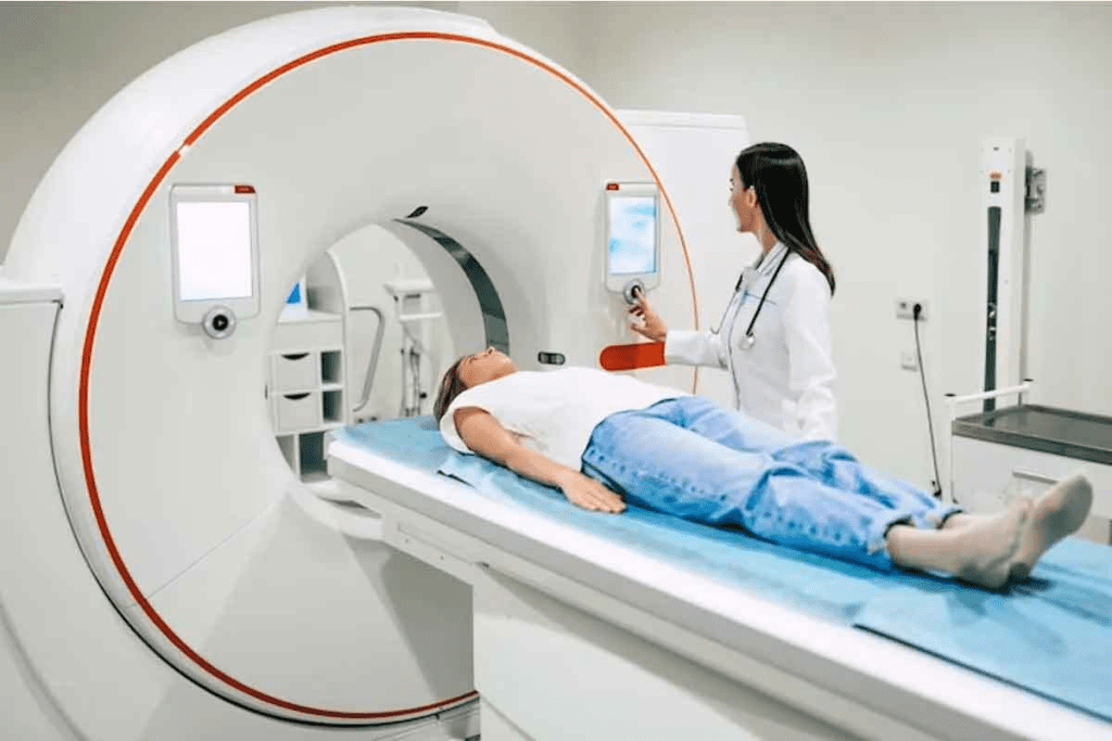
- Scanning: You lie on a table that moves into a PET/CT scanner. It takes pictures of your body.
Duration and Post-Scan Protocols
The whole PSMA PET scan, from start to finish, takes a few hours. After it’s done, you can go back to your usual day. But, it’s key to follow any instructions from your doctor, like:
| Post-Scan Instruction | Purpose |
| Drinking lots of water | To get rid of the radioactive stuff |
| Staying away from others | To keep them from getting too much radiation |
| Seeing your doctor again | To talk about the scan results and what’s next |
Knowing what happens during a PSMA PET scan can help you feel more comfortable. If you have any questions or worries, always talk to your doctor.
Detecting Prostate Cancer: Primary Applications
PSMA PET scans are key in finding and managing prostate cancer. They focus on the prostate-specific membrane antigen. This makes them very good at spotting cancer that has spread.
These scans have changed how doctors fight cancer. They help doctors find cancer more accurately. This leads to better treatment plans.
As technology gets better, PSMA PET scans will play an even bigger role. They might become a mainstay in diagnosing and treating prostate cancer.
Prostate Cancer: Normal vs. Abnormal PSMA PET Scan Results
It’s important to know the difference between normal and abnormal PSMA PET scan results. These scans are key in finding prostate cancer. They give clear images that help doctors plan the best treatment.
Characteristics of Normal Scans
A normal PSMA PET scan shows little to no tracer uptake in the prostate or nearby tissues. Some areas like the salivary glands, liver, and spleen might show some uptake. This is because PSMA is naturally found in these organs. But, the level and pattern should be as expected.
Identifying Abnormal Uptake Patterns
Abnormal scans show more tracer uptake in places that shouldn’t have it. In prostate cancer, this might be in the prostate, seminal vesicles, or in lymph nodes or bones. The way and how much it’s taken up can tell us a lot about the cancer.
| Uptake Pattern | Possible Interpretation |
| Focal uptake in prostate | Primary prostate cancer |
| Uptake in lymph nodes | Lymph node metastasis |
| Uptake in bones | Bone metastasis |
Interpreting Intensity Levels
The PSMA tracer uptake is measured by the Standardized Uptake Value (SUV). Higher SUV values mean more aggressive disease. We use these values along with the patient’s history to decide the best treatment.
Understanding PSMA PET scan results helps us diagnose and treat prostate cancer better. This leads to better outcomes for patients.
Comparative Accuracy: PSMA PET vs. Conventional Imaging
PSMA PET scans are more accurate than traditional imaging for prostate cancer. They can spot cancer cells more precisely. This is key for planning treatment.
Sensitivity and Specificity Rates
Research shows PSMA PET/CT scans are more accurate than CT and bone scans, with sensitivity ranging from 65% to 85% and specificity exceeding 95% in biochemical recurrence.
“The high sensitivity and specificity of PSMA PET/CT make it a valuable tool in the diagnosis and staging of prostate cancer,” as noted in recent clinical studies. This enhanced accuracy helps in making informed decisions regarding treatment options.
Advantages Over CT and Bone Scans
PSMA PET scans have several benefits over CT and bone scans. They can spot cancer cells early, even when they’re tiny. This is great for finding metastatic disease that other scans can’t see.
- Improved detection of small lesions
- Enhanced accuracy in identifying metastatic disease
- Better assessment of treatment response
So, PSMA PET scans are becoming the go-to for prostate cancer diagnosis and staging.
Detection of Small Lesions
PSMA PET scans are great at finding small lesions that other scans miss. This is important for early detection and treatment of prostate cancer. It allows for more focused and effective therapy.
Finding small lesions is a big challenge in cancer diagnosis. PSMA PET scans solve this by showing detailed images of even the smallest cancerous areas.
In conclusion, PSMA PET scans are more accurate than traditional imaging for prostate cancer. Their high sensitivity and specificity, along with their ability to spot small lesions, make them essential for diagnosing and treating prostate cancer.
Does PSMA PET Scan Detect Other Cancers?
PSMA PET scans are mainly for finding prostate cancer. But, they can also spot other cancers that have PSMA. This is good news for diagnosing and treating different types of cancer.
Non-Prostate Cancers with PSMA Expression
PSMA is not just for prostate cancer. Research has found it in other cancers too. For example, some kidney cancer, thyroid cancer, and lung cancer have PSMA. This opens up new ways to use PSMA PET scans for these cancers.
| Cancer Type | Frequency of PSMA Expression |
| Kidney Cancer | 15“25% (clear cell renal cell carcinoma) |
| Thyroid Cancer | 5“10% (papillary and anaplastic subtypes) |
| Lung Cancer | 3“8% (non-small cell lung cancer) |
Incidental Findings in Clinical Practice
In real-world use, PSMA PET scans sometimes find cancers not related to prostate. These finds are very important. They can help doctors find cancers they didn’t know about before. For example, a study in the Journal of Nuclear Medicine found neuroendocrine tumors and other non-prostate malignancies with PSMA PET scans.
“The use of PSMA PET/CT has expanded beyond prostate cancer, revealing its potential in detecting other cancers. This has significant implications for patient management and treatment strategies.”
Nuclear Medicine Specialist
Research on PSMA in Various Tumor Types
Research is looking into PSMA in many tumor types. They want to know how PSMA PET scans can help with different cancers. Studies are checking PSMA in breast cancer, colon cancer, and more. This research is key to using PSMA PET scans for more cancers.
As research goes on, we’ll learn more about PSMA PET scans. They might be used for many cancers soon. This could make diagnosing and treating cancer better for many patients.
Clinical Impact on Treatment Planning
PSMA PET scan results are very important for planning treatments. They give doctors the information they need to make better choices for patients. This leads to more personalized and effective care for prostate cancer patients.
How PSMA PET Results Influence Therapy Decisions
PSMA PET scans help doctors decide the best treatment for prostate cancer. They show where and how much cancer is present. This helps doctors choose the right treatment plan.
“The high sensitivity and specificity of PSMA PET/CT allow for accurate detection of prostate cancer recurrence, facilitating targeted therapy and improving patient outcomes.”
Source: Journal of Clinical Oncology
Radiation Treatment Field Adjustments
PSMA PET scans help with planning radiation treatments. They show where the cancer is, so doctors can target it better. This reduces harm to healthy tissues and makes treatments more effective.
Surgical Planning Modifications
PSMA PET scans also affect surgical plans for prostate cancer. They give surgeons detailed information on the disease. This helps them choose the best surgery and avoid unnecessary steps.
- Improved accuracy in identifying cancerous tissues
- Enhanced ability to plan targeted therapies
- Better patient outcomes through more informed treatment decisions
In conclusion, PSMA PET scan results greatly impact treatment planning for prostate cancer. They influence therapy choices, radiation plans, and surgery. As the field grows, PSMA PET imaging will continue to be key in managing prostate cancer.
Limitations and Challenges of PSMA PET Imaging
PSMA PET imaging has changed how we diagnose prostate cancer. Yet, it has its own set of challenges. Knowing these challenges is key to making accurate diagnoses and planning effective treatments.
False Positive and False Negative Results
PSMA PET imaging can sometimes show false positives and negatives. False positives can cause unnecessary worry and extra tests. False negatives can lead to late diagnosis and treatment.
Many things can cause these errors. For example, some non-cancerous conditions can show up as cancer on scans. Also, some cancers might not show enough PSMA, leading to false negatives.
| Cause | Effect | Clinical Implication |
| Physiological uptake in normal tissues | False positive results | Unnecessary additional testing or anxiety |
| Low PSMA expression in cancer | False negative results | Delayed diagnosis or treatment |
| Technical issues during scanning | Both false positives and negatives | Compromised image quality and accuracy |
Physiological Uptake in Normal Tissues
PSMA PET imaging isn’t just for prostate cancer. Some normal tissues and benign conditions can also show PSMA uptake. For example, the salivary glands, liver, and spleen can have physiological uptake.
Technical and Interpretive Challenges
The quality of PSMA PET imaging depends on the technology and the experts reading the scans. Technical issues, like image quality and tracer distribution, can impact the scan’s accuracy. Also, it can be hard to tell the difference between benign and malignant uptake.
To improve, we need to keep working on our techniques. We should aim for better image quality and train more experts. By facing and solving these challenges, we can make PSMA PET imaging even more useful for prostate cancer care.
Future Directions in PSMA PET Technology
PSMA PET technology is growing, promising better care for prostate cancer patients. We’re exploring new ways to improve diagnosis and treatment. This includes better tracers and advanced imaging techniques.
Emerging Tracers and Techniques
New PSMA tracers are being made to work better. For example, 18F-PSMA-1007 and 18F-DCFPyL might offer clearer images and better detection rates. These advancements could change how we see and treat prostate cancer.
Some new techniques include:
- Creating tracers that last longer for easier imaging
- Making tracers more specific to reduce false positives
- Using PSMA PET with other imaging methods for better accuracy
| Tracer | Characteristics | Potential Benefits |
| F-PSMA-1007 | High affinity for PSMA, improved stability | Better image quality, higher detection rates |
| Ga-PSMA-11 | Established tracer with good PSMA affinity | Proven track record, widely available |
Integration with Artificial Intelligence
AI is being added to PSMA PET imaging. AI can make image analysis faster and more accurate. It can help in several ways, like:
- Automating the detection and measurement of lesions
- Segmenting images for clearer tumor outlines
- Creating predictive models for treatment outcomes
“The fusion of AI with PSMA PET imaging has the power to change how we use diagnostic info. It could lead to more tailored and effective treatments.”
Theranostic Applications
Theranostics combines therapy and diagnostics, and PSMA PET is at the forefront. It uses PSMA-targeting ligands with therapeutic radionuclides. This allows for targeted treatment of prostate cancer cells while protecting healthy tissue.
Theranostic applications offer several benefits, including:
- Personalized treatment plans based on tumor characteristics
- More effective treatment with fewer side effects
- The ability to monitor treatment response in real-time
The future of PSMA PET technology is bright. With new tracers, AI, and theranostics, we can better diagnose and treat prostate cancer.
Patient Considerations: Insurance Coverage and Accessibility
When looking into prostate cancer diagnosis, insurance coverage and accessibility are key. The cost of medical imaging, like PSMA PET scans, can be overwhelming.
Current Reimbursement Status
The cost of PSMA PET scans varies a lot. In the U.S., Medicare and some private insurers now cover them for some cases. But how much they cover can vary a lot.
It can be hard to figure out what’s covered. Patients should talk to their doctors and insurance about their plans. Also, remember that insurance rules can change with new research and tech.
Geographic Availability
Where you live also affects access to PSMA PET scans. Big hospitals and cancer centers are more likely to offer them. But people in rural areas might find it harder to get to these places.
Telemedicine and remote consultations help. They let patients talk to experts from anywhere. This doesn’t replace in-person scans, but it helps with first talks and follow-ups.
Healthcare is working to make PSMA PET scans more available. This includes teaming up between hospitals and teaching doctors and patients about this tech.
We’re pushing to make new prostate cancer diagnosis tools available to everyone. Knowing about insurance and where to get scans helps patients make smart choices about their health.
Conclusion
We’ve looked into how PSMA PET scans change the game for prostate cancer diagnosis and treatment. These scans use special technology to find cancer in the prostate and might even spot other cancers. This is a big deal for doctors and patients alike.
PSMA PET scans are now key in fighting prostate cancer. They’re much better at finding cancer than older methods. This means doctors can plan treatments more accurately, helping patients get better faster.
New advancements in PSMA PET technology are on the horizon. Things like artificial intelligence and new tracers could make these scans even better. More people will get to use them, thanks to better insurance coverage.
To wrap it up, PSMA PET scans are a huge leap forward in finding and treating prostate cancer. They give doctors the info they need to make better treatment plans. As we keep improving, we’ll see even more benefits for patients.
FAQ
What is a PSMA PET scan, and how does it work?
A PSMA PET scan is a test that uses a PET scanner to find cancer cells. It injects a radioactive tracer that sticks to cancer cells. This lets the scanner see where the cancer is.
What is PSMA, and why is it important in prostate cancer diagnosis?
PSMA is a protein found on prostate cancer cells. It’s key for finding cancer with PSMA PET scans. This helps doctors see how much cancer is there and plan treatment.
How does a PSMA PET scan differ from conventional imaging techniques?
PSMA PET scans are better at finding prostate cancer than CT and bone scans. They spot smaller cancers and give more accurate info about the disease.
Can a PSMA PET scan detect other cancers besides prostate cancer?
Yes, PSMA PET scans can find other cancers like kidney and thyroid cancer. But they’re mainly used for prostate cancer.
What are the advantages of using PSMA PET scans in prostate cancer diagnosis?
PSMA PET scans are more accurate and can find small cancers. They help doctors plan better treatments. This can lead to better patient outcomes.
Are there any limitations or challenges associated with PSMA PET scans?
Yes, there are challenges like false results and finding cancer in normal tissues. Also, not everyone has access to these scans, and insurance coverage varies.
How do PSMA PET scan results influence treatment planning?
PSMA PET scan results help doctors plan treatments. They show how much cancer there is. This helps decide on surgery, radiation, or other treatments.
What is the future of PSMA PET technology?
The future of PSMA PET technology looks bright. New tracers and AI are being explored. These could make scans even better for finding and managing prostate cancer.
Is a PSMA PET scan covered by insurance?
Insurance coverage for PSMA PET scans varies. Some plans cover it for certain reasons, while others don’t.
How can I access a PSMA PET scan?
Getting a PSMA PET scan depends on where you live and if it’s available. Talk to your doctor to see if it’s an option for you.
References
- Sutherland, M., et al. (2021). Utilization of computerized tomography and magnetic resonance imaging in cervical spine trauma. Journal of Clinical Imaging Science, 11(1), 1“7. https://pmc.ncbi.nlm.nih.gov/articles/PMC8318846/
- Hect, J. L., et al. (2023). Relationship of cervical soft tissue injury and surgical decision-making. Global Spine Journal, 13(5), 1123“1130. https://pmc.ncbi.nlm.nih.gov/articles/PMC10339037/


