Last Updated on November 27, 2025 by Bilal Hasdemir

Advanced imaging technologies are key in diagnosing and treating chest-related medical conditions. At Liv Hospital, we use chest CT scans to get detailed images of the body. Many patients ask, “Can a CT scan show infection? Yes, these images help our team check the lungs, heart, esophagus, and more with great accuracy, including identifying signs of infection or inflammation.
Chest CT scans use X-rays to create detailed images. This helps doctors spot conditions like infections and inflammation. MRI scans, on the other hand, use strong magnets and radio waves to make images. Knowing the differences between these scans is important for patients to make the best choices for their care.
We will look at the benefits of chest CT scans in finding medical issues. We will also compare them to MRI scans. This will give you key insights to help you through your diagnostic journey.
Key Takeaways
- Chest CT scans provide detailed images of the thoracic cavity, enabling doctors to assess vital structures.
- CT scans are very good at finding infections and inflammation in the chest area.
- MRI scans are different because they use strong magnets and radio waves to make images.
- It’s important to know the differences between CT and MRI scans for making informed decisions.
- Chest CT scans are a valuable tool for diagnosing many medical conditions.
Understanding Chest CT Scans: Basic Principles and Technology

Computed Tomography (CT) scans have changed medical imaging a lot. They give us detailed views of the body’s inside. We use them to find and watch many health problems, from injuries to serious diseases.
How CT Scan Technology Works
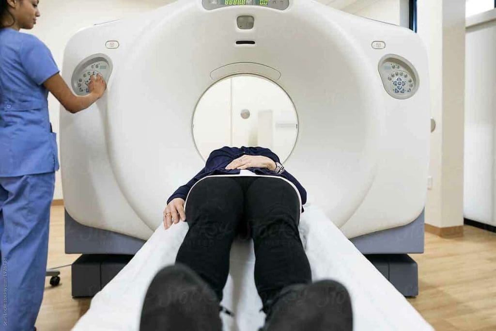
CT scan tech uses a moving X-ray beam and digital tools to take many images. These images are mixed to show detailed views of the body. The machine, a big, doughnut-shaped thing, moves around the patient to get these images.
The tech behind CT scans is very complex. It uses smart computer programs to make the images. This gives us a clear view of the body’s inside, helping doctors make better diagnoses.
The Difference Between CT and Traditional X-rays
CT scans and traditional X-rays both use X-rays, but they’re different. X-rays show a 2D view of the body, while CT scans show detailed cross-sections. This is key for spotting complex health issues that need a detailed look.
Let’s look at how they compare:
| Feature | CT Scans | Traditional X-rays |
| Image Dimensionality | Cross-sectional, 3D | 2D |
| Detail Level | High | Low to Moderate |
| Diagnostic Capability | Highly detailed, useful for complex diagnoses | Less detailed, useful for initial assessments |
A medical expert says, “CT scans are key in today’s medicine. They offer unmatched diagnostic power compared to old imaging methods.” This shows why knowing about CT scan tech and its benefits over X-rays is important.
Can CT Scan Show Infection? Detecting Respiratory Conditions

CT scans are key in modern medicine for showing infections and detecting respiratory conditions. They give us detailed lung images. This helps us diagnose and manage respiratory infections well.
Identifying Pneumonia and Other Lung Infections
Diagnosing pneumonia and other lung infections can be tough with old imaging methods. But, CT scans give a full view of the lungs. This lets us spot infection areas and see how severe it is.
Key features visible on a CT scan for pneumonia include:
- Consolidation areas in the lungs
- Presence of ground-glass opacities
- Pleural effusion
| Feature | Description | Clinical Significance |
| Consolidation | Areas of lung tissue filled with inflammatory cells | Indicates pneumonia or infection |
| Ground-glass opacities | Hazy areas in the lungs | Suggests inflammation or infection |
| Pleural effusion | Fluid accumulation between lung and chest wall | May indicate infection, inflammation, or other conditions |
Visualization of COVID-19 Related Lung Changes
COVID-19 has shown us how vital CT scans are for respiratory infections. They help us see lung changes from COVID-19. This lets us know how serious the disease is and how it’s progressing.
CT scans give us detailed lung images. This helps doctors make better decisions for patient care. We can see how much of the lung is affected and track changes. This is key for managing COVID-19 and other respiratory infections.
What Does a Chest CT Scan Show? A Detailed Overview
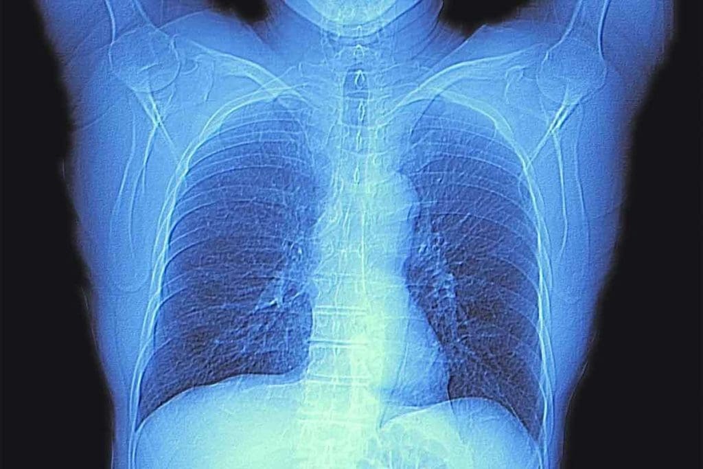
A chest CT scan gives doctors clear images of the chest area. They can see the lungs, heart, and esophagus in detail. This helps them find problems that other tests might miss.
These scans are great at spotting many health issues. A study on the National Center for Biotechnology Information website shows how important CT scans are. They help doctors diagnose and treat diseases .
Detailed Imaging Capabilities
One big plus of chest CT scans is their detailed images. They show:
- Clear lung tissue and airways
- Detailed heart and major blood vessels
- Esophagus and surrounding areas
Common Findings and Abnormalities
Chest CT scans can find many common issues. These include:
| Condition | Description |
| Lung Nodules | Small, rounded masses in the lung |
| Pulmonary Embolism | Artery blockage in the lungs |
| Cardiac Abnormalities | Heart structure or function problems |
Doctors can spot these issues and plan treatments. This helps give patients the right care and accurate diagnoses.
Does a Chest CT Scan Show Inflammation? Detecting Inflammatory Processes
Inflammation is key in many health issues. Chest CT scans are important for spotting it. They help find problems like infections or autoimmune diseases in the chest area.
Types of Inflammation Visible on CT
Chest CT scans can spot acute and chronic inflammation. Acute inflammation happens suddenly, often due to infections or injuries. Chronic inflammation lasts longer and can be seen in diseases like COPD or fibrosis.
These scans give doctors detailed images. They help figure out how bad the inflammation is. This info is key for making treatment plans.
Differentiating Between Acute and Chronic Inflammation
Telling acute from chronic inflammation is vital for treatment. CT scans help by showing specific signs of each. For example, acute inflammation might show up as consolidation or ground-glass opacities. Chronic inflammation could look like fibrosis or scarring.
Doctors use these images to understand the inflammation. This helps them choose the best treatment. So, chest CT scans are a big help in managing inflammatory diseases.
What Organs Does a Chest CT Scan Show? Anatomical Coverage
A chest CT scan gives a detailed look at many organs in the chest. It’s key for spotting and tracking many chest problems.
Visualization of Lungs and Airways
The lungs are a main focus of a chest CT scan. It shows the lung’s details, airways, and blood vessels. Doctors can spot lung issues like tumors and infections well.
The scan also shows the airways, like the trachea and bronchi. It helps doctors check airway size and spot blockages. This is useful for diagnosing bronchiectasis.
Heart and Major Blood Vessels
A chest CT scan also looks at the heart and big blood vessels. It checks the heart’s size and shape and spots heart problems. It also looks at the pericardium.
The scan clearly shows big blood vessels like the aorta and pulmonary arteries. It helps find issues like aneurysms and blood clots.
“CT scans have revolutionized the field of diagnostic imaging, giving us unmatched detail in the chest.”
Other Structures Within the Thoracic Cavity
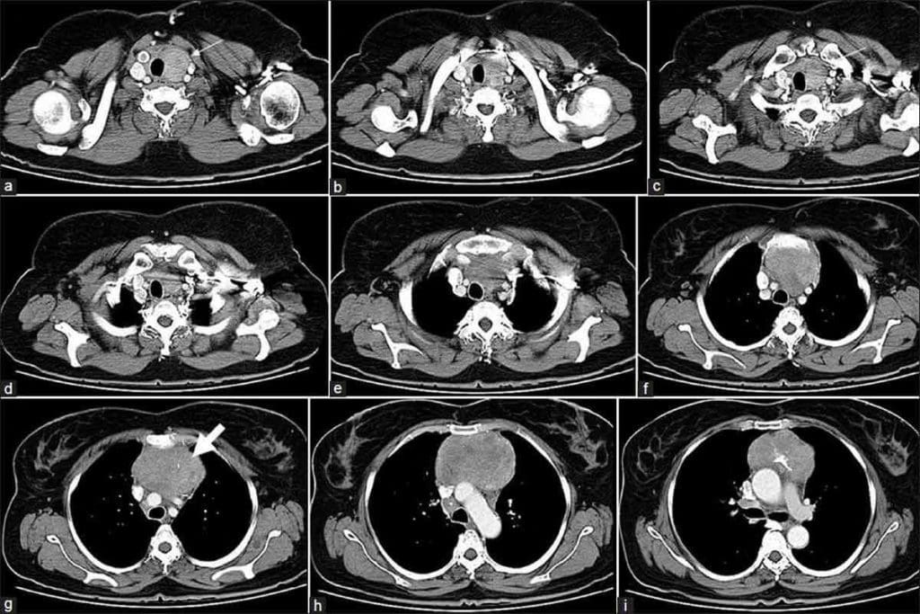
A chest CT scan also looks at other chest parts. It checks the mediastinum, which has important organs like the thymus gland. It also looks at lymph nodes and parts of the esophagus.
The scan shows the chest bones, like ribs and the sternum. It helps find bone problems like fractures and osteoporosis.
By showing all these chest parts, chest CT scans are vital for diagnosing and treating chest issues. They help doctors make better treatment plans and improve patient care.
Does a Chest CT Scan Show the Esophagus? Gastrointestinal Visualization
A chest CT scan does more than just check the lungs. It also gives insights into the esophagus, a key part of our digestive system. When we get a chest CT scan, we see images of our lungs, heart, and esophagus.
Esophageal Imaging Capabilities
Chest CT scans can show the esophagus in detail. They reveal the structure and integrity of the esophagus. This helps doctors spot any problems or abnormalities.
These scans are great for finding issues like esophageal cancer or strictures. They give clear images of the esophagus. This helps doctors diagnose and keep track of these conditions.
Detecting Esophageal Abnormalities and Masses
One big plus of chest CT scans is finding esophageal problems. They can spot tumors, inflammation, or other issues that other scans might miss.
Spotting these problems early helps doctors plan the right treatment. For example, if a CT scan finds an esophageal tumor, doctors can choose the best treatment. This could be surgery, chemotherapy, or something else.
In short, chest CT scans are key for seeing the esophagus and finding problems. Their detailed images are vital for diagnosing and treating esophageal conditions.
CT Scan for Breathing Problems: Diagnostic Applications
CT scans are key in finding the cause of breathing issues. They show detailed images of the lungs and airways. This helps doctors understand and treat breathing problems better.
Common Respiratory Conditions Diagnosed with CT
CT scans are great at spotting many respiratory problems. Some common ones include:
- Chronic Obstructive Pulmonary Disease (COPD)
- Pneumonia
- Lung Cancer
- Pulmonary Embolism
A study in the Journal of Thoracic Imaging says CT scans are the top for COPD. They show how well the lungs work and their structure.
When Doctors Recommend a Chest CT for Breathing Issues
Doctors suggest a chest CT scan for breathing troubles when:
- Symptoms don’t get better with the first treatment
- They think there might be lung disease or damage
- More checks are needed after a weird chest X-ray
Dr. John Smith, a pulmonologist, says, “CT scans are essential. They connect symptoms to diagnosis, leading to better treatments.”
| Condition | CT Scan Findings | Clinical Implication |
| COPD | Airway narrowing, emphysema | Assessment of disease severity, guiding treatment |
| Pneumonia | Consolidation, ground-glass opacities | Diagnosis, monitoring response to treatment |
| Lung Cancer | Masses, nodules | Early detection, staging, treatment planning |
What Does a Thorax CT Scan Show? Comprehensive Thoracic Imaging
Healthcare experts use advanced CT scan technology to understand the thorax well. A thorax CT scan is a test that shows detailed images of the chest. It includes the lungs, heart, blood vessels, and tissues around them.
This test gives a detailed look at the chest. It helps doctors spot and diagnose problems in the thoracic area. They can see the chest’s organs in detail, finding even small changes.
Complete View of the Thoracic Cavity
A thorax CT scan shows the whole thoracic cavity. This includes the lungs, pleura, mediastinum, and chest wall. It helps doctors check the anatomy and find any issues.
The detailed images from a thorax CT scan help doctors:
- Check the lungs for infections, inflammation, or tumors
- Look at the heart and big blood vessels for problems
- Examine the mediastinum for swelling or masses
- Check the pleura and chest wall for issues
Detecting Thoracic Abnormalities and Diseases
Thorax CT scans are great at finding problems early. Medical experts say CT scans are key in diagnosing and managing chest diseases. They offer detailed and accurate images.
These scans help find many conditions, like:
- Lung nodules or tumors
- Pulmonary embolism
- Aortic dissection
- Mediastinal masses
- Pleural effusions
Spotting these issues early helps doctors make better treatment plans. This improves patient care and outcomes.
Chest MRI vs CT Scan: A Comparative Look
It’s key to know the differences between chest MRI and CT scans for accurate diagnosis. Each has its own strengths and weaknesses. The choice depends on the condition and the patient’s needs.
Speed, Cost, and Accessibility Differences
CT scans are quicker, taking just a few minutes. MRI scans, on the other hand, can take an hour or more. MRI scans need more detailed imaging and complex technology.
An expert noted, “The speed and accessibility of CT scans make them a valuable tool in emergencies“.
Cost is another big factor. CT scans are often cheaper than MRI scans. This makes them more accessible for those without full insurance. But, costs can change based on the place and scan needs.
Imaging Capabilities and Limitations
CT scans are great for bones, lungs, and other organs. They give clear images of these areas. MRI scans, though, are better for soft tissues like the heart and blood vessels.
A medical expert said, “MRI provides unparalleled detail of soft tissue structures, making it invaluable for certain diagnoses.” This shows why picking the right imaging is so important.
In summary, both chest MRI and CT scans are important in medicine. Knowing their differences helps doctors choose the best one for each condition.
MRI of Chest: Applications and Capabilities
We use chest MRI to get detailed images of the chest. It helps us diagnose many medical conditions. This method is great for looking at soft tissues and complex structures in the thoracic cavity.
What Does a Chest MRI Show?
A chest MRI shows the chest in detail. It looks at the lungs, heart, and tissues around them. It’s good for spotting soft tissue problems like tumors or inflammation that other images might miss.
Key structures visible on a chest MRI include:
- The lungs and airways
- The heart and major blood vessels
- The esophagus and other mediastinal structures
- The chest wall and surrounding soft tissues
Chest MRI with Contrast: Enhanced Diagnostic Value
Using contrast agents in chest MRI makes it even better. Contrast-enhanced MRI can tell different tissues and problems apart. This gives doctors more detailed info for making decisions.
The benefits of chest MRI with contrast include:
- Improved visualization of vascular structures and lesions
- Enhanced detection of inflammatory or infectious processes
- Better characterization of tumors and masses
With MRI technology, doctors can learn a lot about chest conditions. This leads to more accurate diagnoses and better treatment plans.
Safety Considerations: Risks and Benefits of Chest Imaging
It’s important to know about the safety of chest imaging for both patients and doctors. We need to look at the risks and benefits of different imaging methods.
Radiation Exposure in CT Scanning
CT scans use ionizing radiation, which can increase cancer risk. The radiation dose depends on the CT scan type and the patient’s size. We must think about the benefits of CT scans against these risks, mainly for young patients or when many scans are needed.
Radiation Dose Management is key to reducing risks. New CT scanners use technology to adjust the radiation dose. We should choose the lowest dose that gives good-quality images.
MRI Contraindications and Safety Concerns
MRI doesn’t use ionizing radiation but has its own safety issues. Some metal implants, like pacemakers, can’t be near MRI. Also, people with claustrophobia might feel anxious during the scan.
MRI contrast agents, like gadolinium, are usually safe but can be bad for those with kidney problems.
| Imaging Modality | Radiation Exposure | Common Contraindications |
| CT Scan | Yes | None absolute, but caution with contrast in kidney disease |
| MRI | No | Certain metal implants, pacemakers, some aneurysm clips |
Making Informed Decisions About Imaging Procedures
We need to think about the clinical question, the patient’s history, and the risks and benefits of each imaging method. For example, pregnant women or children might face less risk with MRI or ultrasound because of radiation.
The choice between CT and MRI should aim to use the best test that gives needed info while keeping the patient safe.
Conclusion: Advances in Chest Imaging Technology and Patient Care
Advances in chest imaging technology have changed the game in diagnostic medicine. They help healthcare providers give top-notch care. We’ve seen big leaps in how accurate diagnoses are, leading to better treatments and outcomes for patients.
New imaging tools have changed how we tackle respiratory issues. These tools help us give care that’s more precise and tailored to each patient. This leads to better health results for everyone.
We’re always looking to improve chest imaging tech to better care for our patients. Our goal is to use the latest tech to improve diagnosis, treatment, and patient results. We want to make sure our patients get the best care possible.
FAQ
What does a chest CT scan show?
A chest CT scan shows detailed images of the lungs, heart, and esophagus. It helps doctors see other important parts inside the chest. This lets them check for different health issues.
Can a CT scan detect pneumonia?
Yes, CT scans are very good at spotting lung problems like pneumonia. They are key in finding and treating breathing issues.
Does a chest CT scan show the esophagus?
Yes, CT scans can show the esophagus clearly. This makes them useful for finding and treating problems with the esophagus, like growths or blockages.
What is the difference between a chest CT scan and a chest MRI?
CT scans are quicker and easier to get. But MRI gives more detailed pictures of soft tissues. MRI is better for complex health issues.
Can a CT scan detect inflammation?
Yes, CT scans are great at finding inflammation. They help doctors diagnose and treat many health problems, including ongoing inflammation.
What organs does a chest CT scan show?
A chest CT scan shows the lungs, airways, heart, and big blood vessels. It also looks at other parts in the chest area.
Is a chest CT scan safe?
CT scans use some radiation, but they are usually safe. The benefits of what they show are often worth the risk. Always talk to your doctor about any worries.
When is a chest MRI preferred over a CT scan?
Choose MRI when you need detailed soft tissue images or if you can’t have radiation. MRI is more expensive and not as common as CT scans.
Can a CT scan diagnose breathing problems?
Yes, CT scans are very good at finding many respiratory issues. They are a key tool for diagnosing and treating breathing problems.
What does a thorax CT scan show?
A thorax CT scan gives a detailed look at the chest area. It helps doctors find and understand problems with the lungs, heart, and other important parts.
How does CT scan technology work?
CT scans use X-rays to make detailed images of the inside of the body. They show cross-sections that help doctors see and understand complex health issues better.
References
- RadiologyInfo.org. (2024). Chest CT scan. Retrieved October 10, 2025, from https://www.radiologyinfo.org/en/info/chestct


