Last Updated on November 27, 2025 by Bilal Hasdemir
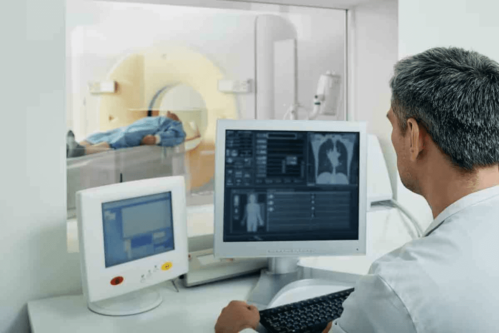
At LivHospital, we know how scary it can be when you have unexplained pain or weird symptoms. That’s why we use advanced tools like CT scans to give you clear answers.
CT scans are key in checking for abdominal pain and finding inflammation and cancer. They give detailed pictures that help our team spot signs of inflammation. This lets us make the right diagnosis.
We use the latest CT scanning methods to distinguish inflammation from cancer. This way, we can give our patients the best care possible. Knowing how CT scans work helps us treat you right.
Key Takeaways
- CT scans are a valuable diagnostic tool for assessing abdominal pain.
- Advanced CT scanning techniques help differentiate between inflammation and cancer.
- Liv Hospital utilizes cutting-edge technology to provide accurate diagnoses.
- Comprehensive care is provided to patients with abdominal issues.
- CT scans help identify indirect signs of inflammation.
Understanding CT Scan Technology

CT scan technology is key in modern medicine. It helps diagnose inflammation and cancer. CT scans give us detailed images of the body, helping doctors treat many conditions.
Basic Principles of CT Imaging
CT scans use X-rays to show the body’s inside. An X-ray source and detectors move around the body. They capture data from many angles.
This data is turned into images by computers. A study on the National Center for Biotechnology Information shows how this tech has improved.
CT imaging works by how X-rays pass through tissues. Bone absorbs more X-rays than soft tissue. This helps create clear images of different body parts.
Types of CT Scans Used in Diagnostic Medicine
There are many CT scans for different needs. Here are a few:
- Non-contrast CT scans: Spot acute hemorrhages, stones, and injuries.
- Contrast-enhanced CT scans: Use a contrast agent to show blood vessels or tumors.
- High-resolution CT scans: Show small details, like lung bronchioles.
| Type of CT Scan | Primary Use | Key Features |
| Non-contrast CT | Detecting acute hemorrhages, stones | No contrast agent used |
| Contrast-enhanced CT | Highlighting blood vessels, tumors | Contrast agent used to enhance visibility |
| High-resolution CT | Detailed imaging of small structures | High detail, useful for lung imaging |
Knowing about different CT scans helps doctors better care for patients. The right scan depends on the patient’s needs and the doctor’s question.
The Role of CT Scans in Evaluating Abdominal Pain
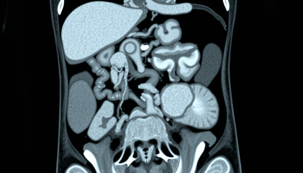
When people have abdominal pain, CT scans are key in finding the cause. Pain can come from many things like inflammation, infection, or cancer. It’s hard to figure out without seeing inside the body.
CT scans give us detailed pictures of the abdomen. They help us find where the pain is coming from. This guides us on how to treat it. CT scans are very useful in both emergency and regular check-ups.
Common Clinical Indications for Abdominal CT
There are many reasons to get a CT scan of the abdomen. These include:
- Acute abdominal pain of unknown origin
- Suspected appendicitis or diverticulitis
- Trauma to the abdomen
- Abdominal masses or tumors
- Infections or abscesses
These issues need quick and accurate diagnosis. CT scans can help. By using a CT scan first, doctors can quickly decide on the best care.
CT Scan as First-Line Diagnostic Tool
Often, we start with a CT scan for abdominal pain. CT scans show everything inside the abdomen. They are fast and accurate, which is important in emergencies.
CT scans help us tell apart different causes of pain. This is key for the right treatment.
Using CT scans first can lead to better care. It shortens the time to find and treat the problem. It also means fewer tests, making care smoother.
Inflammation CT Scan: How Inflammatory Processes Appear
CT scans show us how inflammation affects tissues. We look for specific signs of inflammation when we check a CT scan.
Indirect Signs of Inflammation on CT Images
Inflammation on CT scans is spotted through indirect signs. These include swelling, fluid buildup, and thickened organ walls. These signs suggest inflammation is happening.
For example, in appendicitis, a CT scan might show a thickened appendix wall and fat stranding. In pancreatitis, it can reveal an enlarged pancreas and fluid around it.
Tissue Changes That Indicate Inflammatory Processes
Inflammation causes tissue changes seen on CT scans. These changes include fat tissue swelling, abscesses, or inflammatory masses.
Using advanced CT techniques, like contrast-enhanced CT, makes these changes clearer. Contrast agents help show inflammation by highlighting increased blood flow in affected areas.
By looking at these changes and signs, doctors can diagnose and understand how severe the inflammation is. This helps them make the right treatment plans.
Common Inflammatory Conditions Detected by CT Scans
We use CT scans to find many inflammatory disorders, mainly in the abdomen and chest. These scans give us key details. They help us spot and treat different inflammatory conditions.
Abdominal Inflammatory Disorders
CT scans are great at finding problems in the belly. They can spot appendicitis, diverticulitis, and pancreatitis well. For example, they can show an inflamed appendix and any complications like abscesses.
Diverticulitis, which is inflammation in the colon, is also diagnosed with CT scans. They show how bad the inflammation is and help decide treatment. Pancreatitis, or inflammation of the pancreas, is also checked with CT scans. They help see how severe it is and if there are any other problems.
Respiratory and Thoracic Inflammation
In the chest, CT scans are key for finding respiratory and thoracic inflammation. Pneumonia and pleurisy are common issues that CT scans can spot. Pneumonia looks like solid areas on CT scans, and pleurisy shows up as inflammation of the pleura with fluid.
CT scans also help see how big these problems are and if there are any complications. For pneumonia, they can find abscesses or empyema. For pleurisy, they show how much fluid there is and if it needs to be drained.
By accurately diagnosing and assessing these conditions, we can give better treatment. The detailed info from CT scans is vital for managing belly and chest inflammation.
How Cancer Appears on CT Imaging
Cancer on CT scans shows clear signs that doctors look for. Knowing these signs is key for correct diagnosis and treatment.
Characteristic Features of Malignant Masses
Malignant masses on CT scans have specific signs. They often have irregular borders, showing they’ve spread into nearby tissues. They also have heterogeneous density, meaning different tissue types within the mass.
Necrosis in the tumor, seen as low-density areas, is common in big tumors. The size and shape of the mass matter too. Larger, irregular shapes usually mean cancer.
Density and Enhancement Patterns of Tumors
The density and how tumors enhance on CT scans are very telling. Contrast enhancement is key, as cancer tumors often show heterogeneous enhancement. This means different parts of the tumor react differently to contrast.
Some tumors have calcifications, which are important clues. For example, lung or ovarian cancers have specific calcification patterns. These patterns help doctors figure out the cancer type. For more on CT scans in cancer diagnosis, see this research article.
Doctors use these signs to decide if a tumor is likely cancerous. CT imaging has changed oncology, making diagnosis and treatment planning more precise.
CT Scan Abdomen Cancer: Detection Capabilities
Abdominal CT scans are key in finding and managing abdominal cancers early. They give us detailed images of the belly area. This helps us spot and understand different cancers.
What Cancers Can an Abdominal CT Scan Detect
These scans can find many cancers, like liver, pancreatic, kidney, and colon cancers. They show these organs clearly. This lets us see tumors and how big they are.
Liver Cancer: CT scans can spot liver tumors, like hepatocellular carcinoma. They show how big and spread out these tumors are. CT scans are very good at finding big liver tumors.
Pancreatic Cancer: Finding pancreatic tumors is tough because of where the pancreas is. But, CT scans are often the first choice for looking for these tumors. Adding contrast makes tumors easier to see.
Sensitivity and Specificity for Different Cancer Types
How well CT scans work depends on the cancer type and size. They’re very good at finding big tumors in the liver and kidney. But, they might miss small pancreatic tumors.
| Cancer Type | Sensitivity (%) | Specificity (%) |
| Liver Cancer | 85-90 | 90-95 |
| Pancreatic Cancer | 75-85 | 85-90 |
| Kidney Cancer | 90-95 | 95-98 |
| Colon Cancer | 80-90 | 90-95 |
The table shows how well CT scans work for different cancers. Knowing this helps doctors understand CT scan results better. It helps them make better decisions for patients.
Differentiating Between Inflammation and Cancer on CT
Understanding the difference between inflammation and cancer on CT scans is complex. CT scans can spot signs of both, but sometimes it’s hard to tell them apart. This requires a close look and sometimes more tests.
Key Distinguishing Features
There are key signs that help tell inflammation from cancer on CT scans. Inflammation usually has diffuse or ill-defined margins. Cancer, on the other hand, has more defined borders and may spread into nearby tissues.
Another clue is how the area looks after getting contrast. Inflammation often enhances early and washes out quickly. Cancer, though, enhances more over time.
Challenging Cases and Diagnostic Dilemmas
But, some cases are tricky to figure out. For example, chronic inflammation can look like cancer. Some tumors also show atypical enhancement.
A radiology expert says, “The mix-up in imaging signs between inflammation and cancer can cause problems. It shows we need a team effort to figure it out.”
“The radiologist must consider the clinical context, laboratory findings, and sometimes additional imaging modalities to arrive at an accurate diagnosis.”
In tough cases, more tests like biopsies or PET scans might be needed to make a sure diagnosis.
By knowing the main differences and the tricky parts, radiologists can get better at telling inflammation from cancer on CT scans. This helps improve patient care.
The Role of Contrast in CT Scan Accuracy
Contrast-enhanced CT scans are key in modern medicine. They give us clear images for accurate diagnoses and treatment plans. This is true for many medical conditions.
Contrast agents make CT scans more accurate. They highlight differences in body tissues and structures. A top radiologist says, “Contrast agents are essential for precise imaging.”
How Contrast Enhances Visualization
Contrast agents change how X-rays interact with body tissues. They make certain areas more visible on CT scans. This helps doctors spot problems like tumors or inflammation.
In the abdomen, contrast agents are very helpful. They show the differences between organs and structures. This makes it easier for radiologists to find and understand lesions.
Contrast CT Scan Abdomen Cancer Detection Improvements
For finding abdominal cancers, contrast-enhanced CT scans are very effective. They help doctors see tumours and healthy tissues apart. This makes staging and treatment planning more accurate.
Research shows contrast agents help find abdominal cancers better. They work well for small tumors or those in hard-to-see places. This lets doctors act quickly and precisely.
A leading oncologist says, “Contrast-enhanced CT scans have changed how we diagnose and treat abdominal cancer. They help us find cancers early and plan better treatments.”
Advanced CT Techniques for Better Differentiation
CT technology has improved a lot. Now, we have advanced techniques to tell the difference between inflammation and cancer. These new methods help us make better diagnoses and choose the right treatments.
Dual-Energy CT Applications
Dual-energy CT (DECT) scans at two energy levels. It gives us more info about tissue types. DECT helps spot certain lesions better and makes contrast agents clearer. This makes it easier to tell if something is inflamed or cancerous.
DECT has many benefits:
- It helps tell different materials apart
- It spots iodine and other contrast agents better
- It’s great for looking at kidney stones
- It might mean we need fewer extra tests
Perfusion CT Imaging
Perfusion CT imaging looks at blood flow in tissues. It’s key for seeing how vascular lesions are. Areas with lots of blood flow might show inflammation or tumors. Perfusion CT gives us numbers to back up these findings.
Perfusion CT’s advantages are:
- It gives us numbers on blood flow
- It helps us understand how vascular lesions are
- It can track how well treatments are working
Specialized Imaging Protocols
Special imaging protocols are made for certain needs. They use the right contrast, timing, and tech. This way, we get more useful info about the lesion. It makes us more sure about what we see.
Some examples include:
- Dynamic contrast-enhanced CT for blood flow checks
- High-resolution CT for small details
- Low-dose CT to cut down on radiation
Using these advanced CT methods, we can better tell inflammation from cancer. This leads to more accurate diagnoses and better treatments.
Multidisciplinary Approach to CT Interpretation
Getting a CT scan right needs teamwork. We think that working together is key for correct diagnosis and good treatment plans.
Collaboration Between Radiologists and Specialists
Reading CT scans is a team effort. Radiologists are important, but they don’t do it alone. They team up with surgeons, oncologists, and gastroenterologists to match the scan with the patient’s story and health history.
For example, when someone has belly pain, the radiologist and a gastroenterologist work together. They look at the CT scan and the patient’s symptoms and lab results. This teamwork helps find the real cause of the pain, like inflammation or infection.
Integrating Clinical Information with Imaging Findings
Combining a patient’s medical history with the scan is vital. Radiologists who know the patient’s background can give more accurate readings.
For instance, a cancer patient’s CT scan is checked against their treatment and current health. This helps the radiologist understand the scan better.
| Clinical Information | Imaging Findings | Interpretation |
| History of cancer | Mass detected on CT scan | Possible tumor recurrence |
| Abdominal pain | Inflammation on CT scan | Possible appendicitis |
| Trauma patient | Injury detected on CT scan | Possible internal bleeding |
Biopsy Correlation with CT Findings
Biopsy results can confirm what the CT scan suggests. Sometimes, the CT scan points to a diagnosis, but the biopsy says something else.
For example, a patient with a suspicious mass might get a biopsy. The biopsy can tell if the mass is cancer or not. This helps confirm or rule out the CT scan’s findings.
By working together on CT scans, we can make diagnoses more accurate. This teamwork leads to better treatment plans and care for patients.
Conclusion: The Future of CT Imaging in Inflammation and Cancer Diagnosis
CT technology is getting better, helping us find and understand inflammation and cancer better. This means we can give patients more accurate diagnoses and care. CT scans are key in spotting problems in the belly, finding inflammation, and spotting tumors.
By using new CT methods like dual-energy CT and perfusion CT, we can tell the difference between inflammation and cancer more clearly. This is a big step forward.
At Liv Hospital, we use the latest in medical imaging to help our patients. Our team works together to make sure patients get the right diagnosis and treatment. For more on how imaging helps in diagnosing diseases, check out research studies on multimodal imaging.
The future of CT imaging looks bright for tackling complex diseases and finding new treatments. As technology keeps improving, we expect to see even better at diagnosing and treating patients.
FAQ
Can a CT scan detect inflammation in the abdomen?
Yes, a CT scan can spot inflammation in the abdomen. It does this by looking for signs like changes in tissue and how things enhance.
How does a CT scan differentiate between inflammation and cancer?
A CT scan tells the difference between inflammation and cancer by looking at specific signs. It checks for tumor density and how they enhance, as well as certain inflammatory signs.
What cancers can an abdominal CT scan detect?
An abdominal CT scan can find many cancers. It looks at organs like the liver, pancreas, and kidneys. It spots cancers by their unique features.
Does a CT scan with contrast improve cancer detection in the abdomen?
Yes, contrast in a CT scan makes finding cancer better. It helps see tumors and their features more clearly.
Can a CT scan show abdominal pain causes?
Yes, a CT scan can find what’s causing abdominal pain. It looks at the details of the abdomen to see if it’s inflammation or cancer.
What is the role of advanced CT techniques in diagnosing inflammation and cancer?
Advanced CT techniques, like dual-energy CT and perfusion CT, are key. They give more detailed and accurate info for diagnosing inflammation and cancer.
How do radiologists differentiate between inflammatory and malignant conditions on a CT scan?
Radiologists look at specific signs on a CT scan to tell the difference. They check for enhancement patterns and tissue changes. They also consider what the patient’s symptoms are.
Can CT scans detect all types of abdominal cancers?
CT scans are good at finding many abdominal cancers. But, how well they work can depend on the cancer type and other things.
What are the benefits of a multidisciplinary approach to CT scan interpretation?
A team approach to reading CT scans is very helpful. It includes radiologists, doctors, and specialists. This way, they can use all the information they have to make a better diagnosis.
Will a CT scan always show inflammation or cancer?
A CT scan can often show inflammation and cancer. But, how accurate it is can depend on many things. This includes the type of condition, the quality of the scan, and the skill of the radiologist.
Reference
- Johnson, T. R., & Nakayama, Y. (2016). MRI vs CT for thoracic imaging. Journal of Thoracic Imaging, 31(4), 221-233. https://journals.lww.com/thoracicimaging/abstract/2016/07000/imaging_in_advanced_non_small_cell_lung_cancer__a.6.aspx


