Last Updated on November 27, 2025 by Bilal Hasdemir

Choosing between a PET CT and an MRI is key to diagnosing and treating medical conditions. At Liv Hospital, we focus on top-notch healthcare and put patients first. This helps you make smart choices for better health outcomes.
It’s important to know the differences between PET CT vs MRI. PET CT scans are great for spotting cancer early and tracking its growth. On the other hand, MRI gives clear pictures of soft tissues and organs. This makes MRI perfect for brain and muscle problems.
Key Takeaways
- PET CT scans are ideal for tracking disease progression and detecting cancers early.
- MRI is best for detailed images of soft tissues and organs.
- Choosing between PET CT and MRI depends on the specific medical condition.
- PET CT involves low levels of radiation, while MRI does not use radiation.
- MRI is more common and accessible in most diagnostic centers.
Understanding Medical Imaging: PET, CT, and MRI Basics

Knowing how PET CT and MRI scans work is key for both patients and doctors. These scans are vital in today’s healthcare world. They help find and treat many health issues. PET CT and MRI scans give us special views of the body.
What is PET CT Scanning?
PET CT scans mix Positron Emission Tomography (PET) and Computed Tomography (CT). They show how the body works and its structure. PET CT scans are great for finding and tracking cancer.
What is MRI Scanning?
MRI scans use a strong magnetic field and radio waves to see inside the body. They don’t use harmful radiation, making them safer for some patients. MRI is known for its clear images of soft tissues. This is why it’s good for checking the brain, muscles, and spine.
The Role of Advanced Imaging in Modern Medicine
PET CT and MRI scans are very important in today’s medicine. They help doctors make accurate diagnoses and plan treatments. The right scan depends on the patient’s needs and the health issue.
- Cancer diagnosis and staging
- Neurological disorder assessment
- Cardiovascular disease evaluation
- Musculoskeletal condition diagnosis
Understanding PET CT and MRI scans helps everyone make better choices. This is true for both patients and doctors.
Fundamental Technology Differences Between PET CT vs MRI

PET CT and MRI scans use different technologies. This affects how they are used in hospitals. Each has its own way of imaging, showing different things.
How PET CT Works: Radioactive Tracers and Metabolic Activity
PET CT scans mix Positron Emission Tomography (PET) and Computed Tomography (CT). They show how active cells are and what they look like. The PET part uses radioactive tracers to find active areas, like tumors.
The tracers send out positrons. These meet electrons and make gamma rays. The PET scanner catches these rays. Then, it combines this with CT data for a detailed view.
How MRI Works: Magnetic Fields and Radio Waves
MRI uses nuclear magnetic resonance to make images. It has a strong magnetic field and radio waves. These help align hydrogen nuclei in the body.
When the radio waves disturb this alignment, signals are sent out. These signals help create detailed images. MRI is great for soft tissues, like the brain and muscles.
Key Technological Distinctions
PET CT and MR scans areperformed in different ways. PET CT uses tracers and X-rays for metabolic and anatomical details. MRI uses magnetic fields and radio waves for detailed anatomy and function.
This difference affects how they are used. PET CT is good for cancer, heart, and brain studies. MRI is better for detailed body scans and checking how body parts work.
Knowing these differences helps doctors choose the right scan for each patient.
Comparing the Machines: PET Scan Machine vs MRI Machine
Understanding the differences between PET scan machines and MRI machines is key. These differences affect the scanning process, patient experience, and what facilities need.
Physical Characteristics and Setup
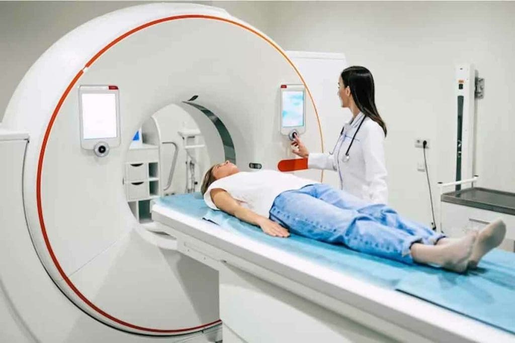
PET CT scanners combine PET and CT technologies in one machine. This allows for both functional and anatomical imaging at once. PET CT machines have a larger gantry for the CT part. They also need a radioactive tracer given to the patient before scanning.
MRI machines use magnetic fields and radio waves to produce images. They have a big cylindrical magnet for the scan. MRI machines come in different strengths, measured in Tesla (T). Higher strengths mean more detail but can cause claustrophobia.
Patient Experience During Each Scan
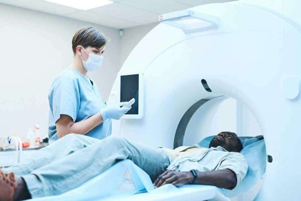
PET CT scans require patients to stay very quiet on a table moving through the scanner. This usually takes 30 minutes to an hour. The tracer given through an IV line means patients must wait for it to spread before scanning.
MRI scans also need patients to lie very quietly on a table, moving into the MRI machine. Scans can last from 15 to 90 minutes, based on the needed detail. Patients must remove all metal and might feel claustrophobic due to the machine’s enclosed space.
Facility Requirements for Each Technology
PET CT and MRI machines need different facilities. PET CT places require special handling and storage of radioactive materials. This means they need shielding and safety rules. MRI facilities must shield the room to block magnetic and radiofrequency interference, using a Faraday cage.
| Characteristics | PET CT Machine | MRI Machine |
| Primary Use | Functional and anatomical imaging | Detailed anatomical imaging, soft tissue contrast |
| Technology | Combines PET and CT technologies | Uses magnetic fields and radio waves |
| Patient Preparation | Requires radioactive tracer administration | Requires removal of metal objects, may need contrast agent |
| Scan Duration | Typically 30 minutes to 1 hour | 15 to 90 minutes, depending on complexity |
| Facility Requirements | Specialized shielding for radioactive materials | Faraday cage to prevent magnetic interference |
Radiation Exposure and Safety Considerations
PET CT scans use radiation, unlike MRI scans. It’s important to know the risks of radiation and how to reduce them.
Radiation in PET CT: Risks and Precautions
PET CT scans use radioactive tracers to see how the body works. While one scan is safe, many can raise cancer risk. We keep the tracer dose low to protect you.
Precautions for PET CT scans include:
- Careful patient selection and dosing
- Use of alternative imaging methods when possible
- Strict adherence to radiation safety guidelines
MRI: A Radiation-Free Alternative
MRI scans don’t use radiation, making them safer for those needing many scans. MRI uses a strong magnetic field and radio waves to see inside the body. But MRI has its own safety rules, like removing metal before scanning.
Patient Preparation for Each Procedure
Getting ready for PET CT and MRI scans is key to safety and quality images. For PET CT, patients often fast and avoid certain meds. For MRI, removing metal items is essential to avoid injury.
Knowing the safety steps and what to do before PET CT and MRI scans helps patients feel more at ease.
Image Resolution and Detail: What Each Scan Reveals
PET CT and MRI scans have different strengths in image quality and detail. They meet different needs in medical imaging. This makes them great together in helping patients.
Functional and Metabolic Information
PET CT scans are top-notch for functional and metabolic information. They use special tracers to show how tissues work. This is key in finding and understanding cancer.
By combining PET and CT, doctors get both how tissues work and their structure in one scan. This makes diagnosis more accurate.
Anatomical Precision and Soft Tissue Contrast
MRI scans are known for their anatomical precision and soft tissue contrast. They use magnetic fields and radio waves to show soft tissues clearly. This is super helpful for the brain, spine, and joints.
MRI’s high-quality images help doctors see tissue structure and find small problems. These might not show up on other scans.
Comparative Visualization Capabilities
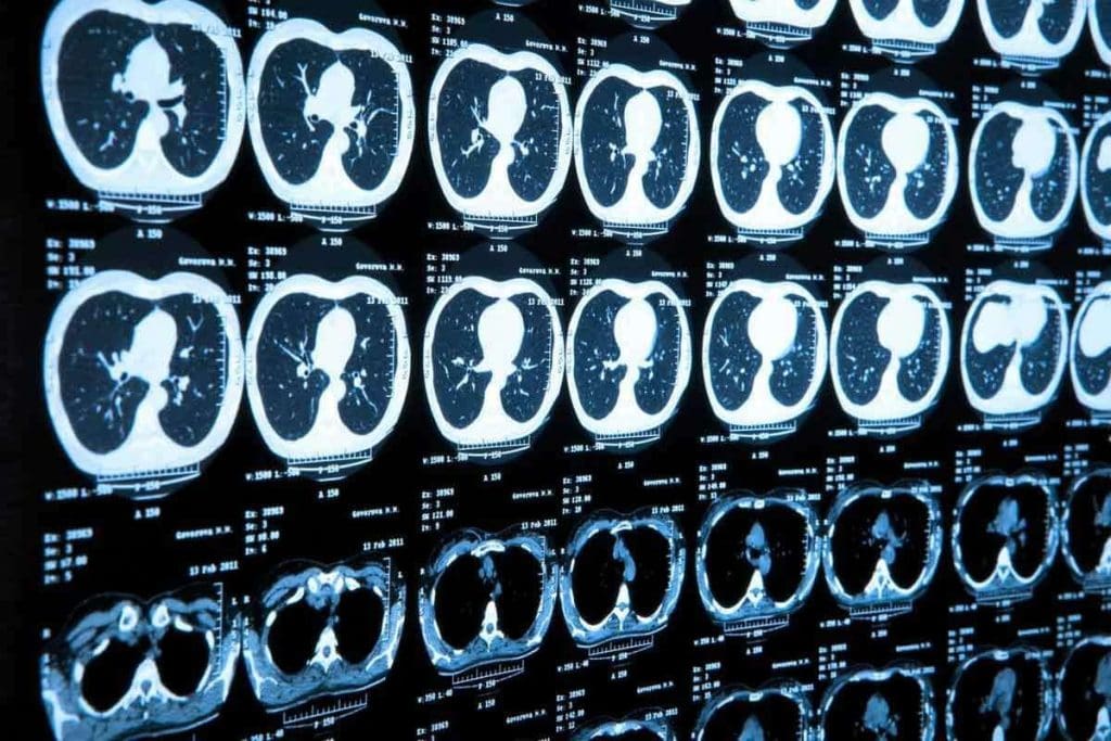
PET CT and MRI have their own best points. PET CT is great at showing metabolic changes. It’s often used for cancer staging and tracking treatment.
| Characteristics | PET CT | MRI |
| Primary Use | Cancer staging, metabolic activity | Soft tissue imaging, structural analysis |
| Image Detail | Functional and anatomical information | High-resolution soft tissue images |
| Diagnostic Strengths | Metabolic activity, cancer detection | Soft tissue contrast, structural integrity |
The table shows that PET CT and MRI do different things well. Knowing this helps doctors choose the best scan for each patient.
Using both PET CT and MRI helps doctors make better choices. This leads to better care for patients.
Clinical Applications: When to Choose PET CT vs MRI
Choosing between PET CT and MRI scans depends on what information you need. Each has its own strengths for different medical uses.
Optimal Uses for PET CT Scans
PET CT scans are great for cancer care. They help find and track cancer, see how treatments work, and spot when cancer comes back. They mix PET’s metabolic info with CT’s detailed images.
PET CT is great for:
- Finding and checking cancer types
- Seeing if treatments are working
- Finding cancer again
Ideal Scenarios for MRI Scans
MRI scans are top picks for brain and muscle imaging. They show soft tissues well and give clear views of the brain and spine. MRI is also good for looking at joint and muscle problems.
MRI shines in:
- Spotting neurological issues like multiple sclerosis and stroke
- Checking muscle and bone injuries
- Looking at soft tissue tumors and infections
Conditions Requiring Both Imaging Methods
Some health issues need both PET CT and MRI scans. For example, in some cancers, PET CT checks metabolic activity. MRI gives detailed body structure info.
Knowing when to use PET CT or MRI helps doctors choose the best imaging for each patient.
PET Scan Versus MRI for Cancer Detection and Monitoring
PET scans and MRI are key in fighting cancer. They do different jobs. PET scans spot metabolic activity and check how treatments work. MRI gives detailed views of tumors and the tissues around them.
Early Cancer Detection Capabilities
Finding cancer early is key to beating it. PET scans are great at this because they show metabolic changes in cells. This helps find cancer before it grows or spreads.
“PET scans can spot cancerous tissues early, even when they’re small,” says Dr. John Smith, a top oncologist. This early detection is critical for starting treatment on time.
Tumor Characterization and Staging
MRI is top-notch for detailed images of the body. It’s essential for tumor characterization and staging. MRI shows the tumor’s size, where it is, and how it affects nearby tissues. This helps doctors plan surgeries or radiation therapy.
MRI’s ability to see soft tissues is unmatched. It’s vital for figuring out how far cancer has spread. This helps in accurate cancer staging.
Monitoring Treatment Response and Recurrence
PET scans are great for checking how well a tumor responds to treatment. They look at metabolic changes to see if treatment is working.
- PET scans show if a tumor is active or if treatment is working.
- MRI tracks changes in tumor size and shape over time.
- Both are key to catching cancer recurrence early.
In summary, both PET scans and MRI are essential for cancer detection and tracking. Each has its own strengths. The choice between them depends on the patient’s needs and the situation.
Neurological and Cardiac Applications
Diagnosing and treating neurological and cardiac issues rely on advanced imaging. Techniques like PET CT and MRI give deep insights into the brain and heart. This helps doctors make better decisions.
Brain Imaging: Functional vs. Structural Analysis
MRI is the best choice for brain scans because it shows detailed images. It’s great for finding things like tumors and injuries. But, PET CT scans show how the brain works, which is key for diseases like Alzheimer’s.
For example, MRI can show brain tumors in detail. PET CT can then check how active the tumor is. This helps doctors understand how serious it is and how it’s responding to treatment.
Cardiac Assessment Differences
PET CT and MRI are both good for heart checks. MRI is great for looking at the heart’s structure and how it works. It can spot scars and see if parts of the heart are working.
PET CT, though, is better at showing how the heart uses energy. It’s good for finding out if the heart is getting enough blood. This helps doctors decide if they need to fix any heart problems.
- PET CT Advantages: Shows how the heart uses energy, checks if parts of the heart are working.
- MRI Advantages: Gives clear images of the heart, checks how well it’s working, and if there are scars.
Comparative Effectiveness for Specific Conditions
Choosing between PET CT and MRI depends on the condition. For some issues, like brain tumors or heart disease, both might be used together. This gives a full picture of the problem.
In summary, PET CT and MRI are key for diagnosing and treating brain and heart problems. Knowing what each can do helps doctors pick the best test for each patient.
Hybrid Imaging: The Evolution of Combined PET-MRI Systems
Hybrid imaging, like PET-MRI, is a big leap in medical tech. It mixes PET’s metabolic info with MRI’s detailed images. This gives a full view of how things work and what they look like.
Technology Behind Integrated Systems
Making PET-MRI systems is a complex task. It needs top-notch hardware and software. This includes PET detectors that work well in MRI’s strong magnetic field.
One big challenge is keeping PET detectors working in theMRIs field. Manufacturers have come up with special designs and materials to solve this problem.
Clinical Benefits of Combined Imaging
PET-MRI brings big benefits to doctors and patients. It gives both function and anatomy in one scan. This makes diagnosing diseases more accurate.
- Improved diagnostic accuracy for complex neurological and oncological conditions
- Enhanced characterization of tumors and their metabolic activity
- Reduced need for multiple imaging sessions, improving patient comfort and reducing overall healthcare costs
As hybrid imaging tech gets better, so will our ability to diagnose and treat. PET and MRI together are a big step towards better medical imaging.
Conclusion: Making Informed Decisions About Medical Imaging
Exploring the differences between PET CT and MRI scans shows that each has its own strengths. This knowledge helps healthcare providers and patients choose the best imaging for their needs. It’s about picking the right tool for the job.
The choice between PET CT and MRI depends on several factors. These include the condition being diagnosed and the need for certain types of information. PET CT is great for showing how active cells are, which is key in cancer diagnosis. MRI, on the other hand, is better for seeing soft tissues and is often used for brain and muscle issues.
By understanding these differences, we can make better choices in medical imaging. This leads to more accurate diagnoses and effective treatments. As imaging technology grows, staying up-to-date with its capabilities is vital for the best care.
FAQ
What is the main difference between a PET scan and an MRI?
A PET scan looks at how cells in the body work. It uses special tracers to see this. An MRI, on the other hand, shows detailed pictures of the body’s inside. It uses magnetic fields and radio waves for this.
Is a PET scan or an MRI better for cancer detection?
Both PET scans and MRIs are good for finding cancer. But they look at different things. PET scans find cancer by how active the cells are. MRI shows detailed pictures of tumors and the area around them. The right choice depends on the cancer type and what the doctor needs to see.
What are the advantages of PET CT over MRI?
PET CT combines PET’s cell activity view with CT’s detailed pictures. This is great for checking cancer spread and how well treatments work. MRI is better for soft tissue details and some injuries.
Are PET scans or MRI scans more sensitive for detecting neurological conditions?
MRI is better for seeing structural problems in the brain, like injuries. PET scans are good for looking at brain cell activity. This is helpful for diseases like Alzheimer’s.
How do PET CT and MRI compare in terms of radiation exposure?
PET CT scans use radiation from the tracer aand the T part. MRI doesn’t use radiation. So, MRI is safer for people who need many scans or are worried about radiation.
Can PET CT and MRI be used together?
Yes, doctors can use PET CT and MRI together or separately. Some places have hybrid PET-MRI systems. These systems combine the best of both in one scan, which can help doctors more.
What is the difference between a PET scan machine and an MRI machine?
PET scan machines find gamma rays from tracers in the body. MRI machines use magnetic fields and radio waves to make images. These machines are very different in design and how they work.
How do I prepare for a PET CT scan versus an MRI?
For a PET CT scan, you might need to fast and avoid some medicines. For an MRI, you’ll need to remove metal and tell your doctor about any implants or if you’re claustrophobic.
Are there specific conditions where one is preferred over the other?
Yes, some conditions are better seen with PET CT or MRI. PET CT is good for cancer because it shows cell activity. MRI is better for soft tissue, like in the brain or muscles.
What is the role of hybrid PET-MRI systems in medical imaging?
Hybrid PET-MRI systems are a big step forward. They let doctors see both metabolic activity and detailed anatomy at the same time. This can make diagnoses better, reduce the need for more scans, and help patients more.
References
- Ratner, E., & Pellerito, J. S. (2020). Hybrid imaging in oncology: PET/CT, PET/MRI, and SPECT/CT. Radiologic Clinics of North America, 58(3), 553-573. https://pubmed.ncbi.nlm.nih.gov/32215821/
- Catana, C. (2019). PET/MRI for neurological applications. Journal of Nuclear Medicine, 60(3), 351-357. https://pubmed.ncbi.nlm.nih.gov/30641009/


