Last Updated on November 27, 2025 by Bilal Hasdemir

PET vs CT Scan for Cancer: Understanding the Difference
Choosing the right imaging test is a crucial step in accurate cancer diagnosis. At Liv Hospital, we prioritize patient care and use the most advanced diagnostic tools to ensure precise results and effective treatment planning.
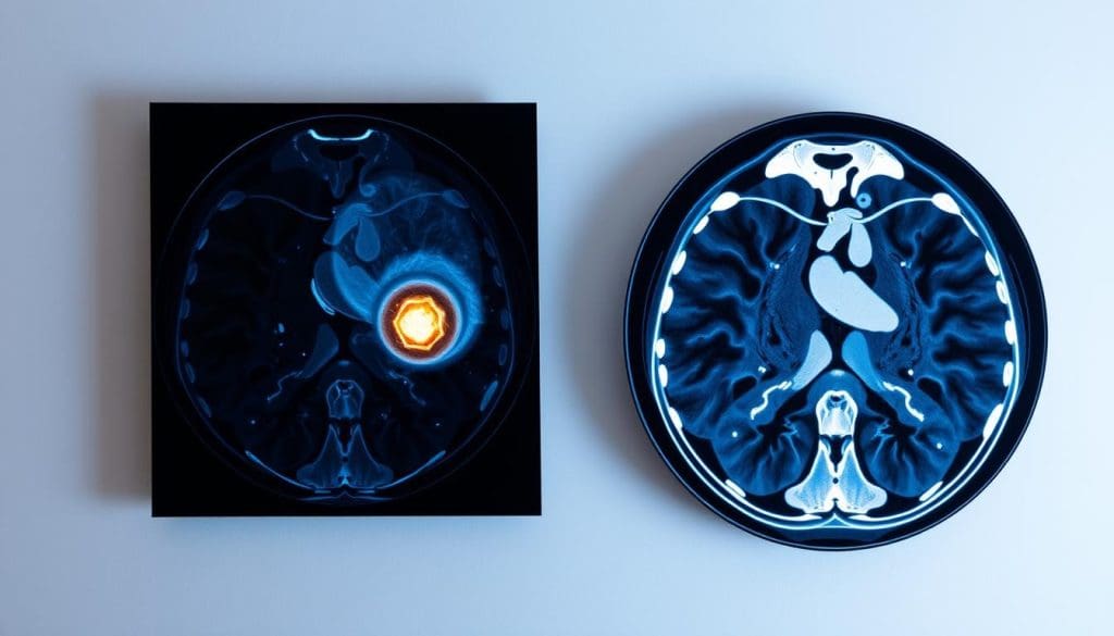
When comparing PET vs CT scan for cancer, each plays a unique role. A PET scan identifies areas of high cellular activity and metabolic changes, helping detect cancer early and monitor how well treatment is working. In contrast, a CT scan provides detailed images of internal structures, allowing doctors to see the exact size, shape, and location of tumors.
By combining both scans, specialists can gain a complete picture of a patient’s condition ” structure and function together. At Liv Hospital, we aim to make every patient’s cancer care journey clearer and more informed by explaining these key differences with care and compassion.
Key Takeaways
- Understand the fundamental differences between PET and CT scans for cancer diagnosis.
- Learn how PET scans detect cellular activity and metabolic changes.
- Discover the role of CT scans in providing detailed anatomical images.
- Explore the benefits of using PET and CT scans together in cancer care.
- Find out how Liv Hospital’s patient-centered approach ensures effective diagnosis and treatment.
The Critical Role of Imaging in Cancer Management

Imaging technologies are key in the battle against cancer. They help doctors diagnose and treat the disease. Imaging is crucial for managing cancer, allowing for accurate diagnosis and treatment.
Why Accurate Diagnostic Imaging Matters
Getting a cancer diagnosis accurately is vital. It helps doctors find tumors, know how far they’ve spread, and check if treatments are working. Accurate imaging is key to planning the best treatment.
It also means fewer surgeries or biopsies are needed. This improves patient care and saves money for healthcare.
Evolution of Cancer Imaging Technologies
Imaging tech has grown a lot, helping doctors better plan treatments. PET and CT scans give important information on tumors. These advancements have made diagnosis and treatment planning more accurate.
As imaging tech keeps improving, it remains essential for managing cancer. It helps from the start of diagnosis to ongoing care.
What is a PET Scan? Metabolic Imaging Explained

Positron Emission Tomography (PET) scans are a key tool in fighting cancer. They show how cells work by looking at their metabolic activity. This is different from other scans that just look at structure.
The Science Behind Positron Emission Tomography
PET scans use radioactive tracers that are injected into the body. These tracers go to areas where cells are very active, like in cancer. They then send out gamma rays that the scanner picks up.
This helps create detailed images of how the body’s cells are working.
Radioactive Tracers and Cellular Activity Detection
The main tracer used is Fluorodeoxyglucose (FDG), a special sugar molecule. Cancer cells take up more of this sugar than normal cells. So, they show up clearly on the scan.
This makes it easier to find cancer and see how active it is.
What to Expect During a PET Scan
When you have a PET scan, you’ll lie on a table that moves into the scanner. It’s painless and takes about 30 to 60 minutes. You might need to fast beforehand and avoid hard activities.
The scan is important for finding and tracking cancer. It helps doctors understand how well treatments are working.
What is a CT Scan? Anatomical Mapping in Detail
Computed Tomography (CT) scans have changed medical imaging a lot. They give detailed views of the body’s inside parts. This tech is key to finding and treating many health issues, like cancer.
How Computed Tomography Creates Structural Images
CT scans mix X-ray tech and computer power to show the body’s inside parts clearly. An X-ray tube spins around the body, taking pictures from many angles. A computer then puts these images together to show a detailed, three-dimensional view.
X-ray technology is key here. It helps show different tissue types. This makes high-quality images that spot problems like tumors or injuries.
X-ray Technology and Cross-Sectional Imaging
The X-ray tech in CT scans is top-notch. It makes fast, accurate images. Unlike regular X-rays, CT scans show cross-sections. This helps doctors see inside the body well, which is very useful for finding cancer.
CT scans’ cross-sectional view is great for checking complex areas. Places like the abdomen or pelvis, where many organs are close together, are easier to see.
The Patient Experience During CT Scanning
When you get a CT scan, you lie on a table that moves into the scanner. It’s quick, lasting just a few minutes. You might need to hold your breath for a bit to get clear pictures.
Getting a CT scan can make some people nervous. Our medical team is here to help. We make sure you’re comfortable and answer any questions you have.
PET vs CT Scan for Cancer: Fundamental Difference #1 – Detection Mechanism
PET and CT scans differ in how they find cancer. PET scans look at how cells work, while CT scans show the body’s structure. Knowing this helps us see what each scan is good at.
Metabolic Activity vs Anatomical Structure
PET scans find cancer by spotting cells that work too hard. CT scans, on the other hand, show the body’s inside parts. Together, they help doctors understand cancer better.
How PET Reveals “Hot Spots” of Cancer Activity
PET scans use special tracers that find cancer cells. These “hot spots” show where cancer is and how bad it is. This helps doctors find cancer early and see if treatments are working.
CT’s Precision in Identifying Structural Abnormalities
CT scans use X-rays to show the body’s inside details. They help find tumors or other problems. CT scans are great for planning biopsies and other procedures.
Knowing how PET and CT scans work helps doctors pick the best one for each patient. This leads to better diagnoses and treatment plans.
Key Difference #2: Sensitivity and Early Detection Capabilities
When it comes to cancer detection, how well imaging works is key. Finding tumors early is important for better treatment and outcomes. So, knowing how PET and CT scans work is vital.
PET’s Advantage in Detecting Small or Hidden Tumors
PET scans are great at finding small or hidden tumors. They do this by showing where cancer cells are active. This is because they use special tracers to spot tumors that other scans might miss. This helps find cancers early, which can lead to better treatment results.
When CT Scans May Miss Early-Stage Cancers
CT scans are good at showing body structures, but might miss early cancers. This is because they use X-rays to see inside the body. They might not catch the metabolic changes that early cancer causes.
Comparative Studies on Detection Sensitivity
Many studies have looked at how well PET and CT scans find cancer. These studies show that PET scans are better at spotting small tumors. For example, in lymphoma or lung cancer, PET scans find cancer sooner. This makes a big difference in how well patients do.
It’s important to remember that PET scans are good at showing where cancer is active. But CT scans are better at showing the body’s structure. Using both together, as in PET-CT scans, gives a full picture of the cancer.
Key Difference #3: Accuracy in Cancer Staging and Spread
Getting cancer staging right is key to finding the best treatment. Staging cancer means checking how far it has spread in the body. This is important for knowing how well a patient will do and what treatments to use.
Evaluating Metastasis: PET vs CT Effectiveness
PET scans are great at spotting cancer spread because they show where cancer cells are active. This is because cancer cells use more energy than normal cells. So, PET scans can find cancer in places CT scans might miss.
PET scans use special tracers that light up cancer cells. This makes them easy to see, even in small lymph nodes or other hidden spots.
Lymph Node Assessment Differences
PET scans are better than CT scans for checking lymph nodes. CT scans look at how big the lymph nodes are. But PET scans can find cancer in nodes that are of normal size.
| Characteristics | PET Scan | CT Scan |
| Detection Method | Metabolic activity | Anatomical structure |
| Lymph Node Assessment | Detects cancerous activity regardless of size | Rely on size criteria |
| Sensitivity to Metastasis | High sensitivity | Limited by anatomical changes |
Impact on Treatment Planning and Prognosis
How well cancer is staged affects treatment plans and patient outlook. PET scans give a clearer picture of cancer spread. This helps doctors choose the best treatments, which can lead to better results for patients.
Effective treatment planning depends on knowing how far cancer has spread. This helps decide if surgery or radiation is needed, or if chemotherapy is better.
Key Difference #4: Radiation Exposure and Safety Considerations
Radiation exposure is a big worry in cancer imaging. PET and CT scans have different levels and types of radiation. Both are key in managing cancer, but they expose patients to different amounts of radiation.
Comparing Radiation Doses Between Modalities
PET scans use radioactive tracers that emit positrons and gamma rays. The dose from a PET scan can be between 4 to 7 millisieverts (mSv). This depends on the tracer and the patient’s health.
CT scans, on the other hand, use X-rays to see inside the body. The dose from a CT scan can range from 2 to 10 mSv or more. This varies with the scan type and body part being checked.
| Imaging Modality | Typical Radiation Dose (mSv) |
| PET Scan | 4-7 |
| CT Scan | 2-10 or more |
The table shows that both PET and CT scans use radiation. But the doses can change a lot based on the procedure and patient.
Risk-Benefit Analysis for Cancer Patients
For cancer patients, the benefits of accurate diagnosis often outweigh the risks of radiation. It’s important to weigh the risks and benefits. This includes looking at the patient’s age, health, and whether there are other imaging options.
“The decision to use PET or CT scans should be based on a careful evaluation of the benefits and risks for each patient. This should consider their unique situation and the needs of their cancer diagnosis.”
” Expert in Nuclear Medicine
Safety Protocols and Minimizing Exposure
Healthcare providers follow strict safety rules to lower radiation exposure. They use the least amount of radioactive tracers needed for PET scans. They also adjust CT scan settings to reduce dose while keeping image quality good. They also think about the patient’s specific needs to choose the best imaging method.
It’s vital to keep radiation exposure low for patient safety. Advances in imaging tech are making PET and CT scans safer for patients.
Key Difference #5: The Combined Power of PET-CT Fusion Imaging
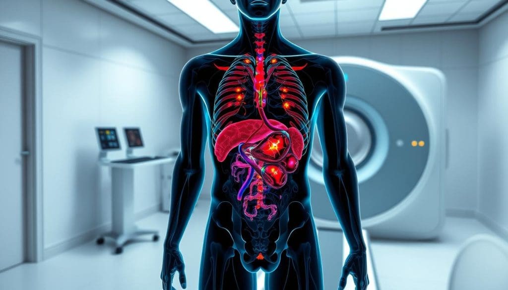
PET-CT fusion imaging is a powerful tool in the fight against cancer. It combines metabolic and anatomical information. This gives a deeper understanding of the disease.
How Integrated PET-CT Technology Works
PET-CT fusion imaging uses software to align PET and CT images. This lets doctors see the tumor’s location, size, and activity. It provides a detailed and accurate picture.
Enhanced Diagnostic Accuracy with Fusion Imaging
PET and CT scans together improve diagnostic accuracy. They help doctors stage cancer better and find metastases. This is key for effective treatment plans.
Studies show PET-CT fusion imaging boosts diagnostic confidence. A study in the Journal of Nuclear Medicine found it was more accurate than PET or CT alone.
Clinical Evidence Supporting Combined Approaches
There’s strong evidence for PET-CT fusion imaging in cancer. Studies have shown its benefits in lung, colorectal, and lymphoma cancers. The table below highlights some key findings.
| Cancer Type | Benefit of PET-CT Fusion Imaging | Reference |
| Lung Cancer | Improved staging accuracy | Study A |
| Colorectal Cancer | Enhanced detection of metastases | Study B |
| Lymphoma | Better assessment of treatment response | Study C |
In conclusion, PET-CT fusion imaging is a big step forward in cancer diagnosis. It combines PET and CT scans for a better understanding of the disease. This improves accuracy and helps in making treatment decisions.
Key Differences #6 and #7: Clinical Applications and Patient-Specific Factors
PET and CT scans have different uses in medicine. The choice between them depends on the type of cancer and what the patient needs. It’s important to understand how these scans are used and how they fit each patient’s needs.
Cancer Types and Their Optimal Imaging Methods
Each cancer type needs a specific imaging method. PET scans are great for managing lymphomas, melanomas, and some lung cancers. They show how active the cancer cells are. On the other hand, CT scans are better for lung, liver, and pancreas cancers. They give detailed pictures of the body’s structures.
- Lymphoma: PET scans are often used for staging and assessing treatment response.
- Lung Cancer: Both PET and CT scans are used, with PET being more sensitive for detecting metastasis.
- Pancreatic Cancer: CT scans are typically used for initial diagnosis and staging.
Cost, Availability, and Accessibility Considerations
The cost and availability of PET and CT scans affect their use. PET scans are more expensive because of the radioactive tracer. They are also less common than CT scanners, making them harder to find in some places.
| Imaging Modality | Cost | Availability |
| PET Scan | Higher | Less Common |
| CT Scan | Lower | More Common |
Patient Factors Influencing Scan Selection
Many factors can affect which scan a patient gets. For example, patients with kidney disease may be at risk from the contrast dye used in CT scans. In these cases, PET scans might be safer.
Liv Hospital’s Patient-Centered Imaging Approach
At Liv Hospital, we focus on the patient’s needs when choosing between PET and CT scans. Our team looks at the cancer type, the patient’s health, and their preferences. This way, we ensure the best imaging solution for each patient.
We combine cutting-edge technology with caring service. Our goal is to provide top-notch imaging that helps our patients get the best results.
Conclusion: Navigating Cancer Imaging Decisions
It’s important to know the difference between PET and CT scans when dealing with cancer. This knowledge helps doctors make better choices for their patients. It ensures the best care possible.
At Liv Hospital, we focus on our patients first. We keep improving our diagnostic tools. Our team works with patients to find the best imaging method for them. This could be PET, CT, or both.
Choosing the right imaging is key to fighting cancer. It leads to better diagnoses and treatments. We aim to give patients the information and support they need. This helps them through their cancer journey.
FAQ
What is the main difference between a PET scan and a CT scan for cancer diagnosis?
PET scans look for metabolic activity, showing “hot spots” of cancer. CT scans, on the other hand, give detailed images of the body’s structure.
Which scan is more sensitive for detecting small or hidden tumors?
PET scans are better at finding small or hidden tumors. This makes them great for catching cancer early.
How do PET and CT scans differ in evaluating metastasis and lymph node involvement?
PET scans are better for spotting metastasis. CT scans, though, give clear images of lymph nodes. The right choice depends on the cancer type and situation.
What are the radiation exposure risks associated with PET and CT scans?
Both scans use radiation. But the amounts are different. We follow strict rules to keep radiation low and protect patients.
What is PET-CT fusion imaging, and how does it enhance diagnostic accuracy?
PET-CT fusion combines metabolic info from PET scans with CT’s detailed images. This gives a full view of tumor biology and structure.
How do healthcare professionals choose between PET and CT scans for cancer patients?
The choice depends on many such, such like cancer type and patient health. At Liv Hospital, we focus on what’s best for each patient.
Are PET scans or CT scans more suitable for monitoring treatment response?
Both scans can track treatment response. PET scans focus on metabolic activity, while CT scans show anatomical details.
Can PET and CT scans be used together for cancer diagnosis and treatment?
Yes, using both scans together offers a deeper understanding of tumors. This improves diagnosis and treatment planning.
How do PET and CT scans compare in terms of cost and availability?
Costs and availability vary by location and provider. At Liv Hospital, we aim to offer these scans while keeping care of high quality.
What is the role of PET-CT fusion imaging in cancer management?
PET-CT fusion imaging is key in cancer care. It helps in accurate diagnosis, staging, and planning. This leads to better patient care and treatment.
References
- Delbeke, D., & Martin, E. C. (2018). Positron emission tomography/computed tomography in the evaluation of cancer patients. Seminars in Nuclear Medicine, 48(6), 480-495. https://www.ncbi.nlm.nih.gov/pmc/articles/PMC6524530/
- Pfannenberg, C., Aschoff, P., & Both, M. (2020). PET/CT in oncological imaging: current standards and future directions. Radiology and Oncology, 54(1), 1-10. https://www.ncbi.nlm.nih.gov/pmc/articles/PMC7074027/
- National Cancer Institute. (2023). Positron emission tomography (PET) scans. https://www.cancer.gov/about-cancer/diagnosis-staging/diagnosis/pet-scan-fact-sheet


