Last Updated on November 26, 2025 by Bilal Hasdemir
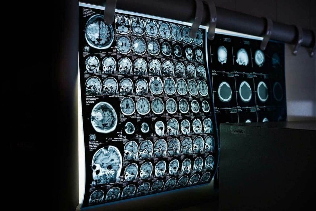
When doctors check for bone fractures, they often use X-rays and MRI. At Liv Hospital, our team uses both to give the best care. X-rays are usually the first choice for finding broken bones. But MRI gives a clearer view, which is great for soft tissue injuries.
We use advanced MRI technology to see inside the body. This is super helpful for soft tissues and small injuries that X-rays might miss. MRI is a key tool for us, helping us give the right treatment and care.
Key Takeaways
- MRI technology provides detailed images of soft tissues and internal body structures.
- X-ray is typically used as the first line of defense for detecting bone fractures.
- MRI is very good at finding small injuries that X-rays can’t see.
- Liv Hospital’s expert team uses both X-ray and MRI for complete checks.
- Advanced MRI technology helps us make better treatment plans for patients.
The Fundamental Differences Between MRI and X-ray Technology
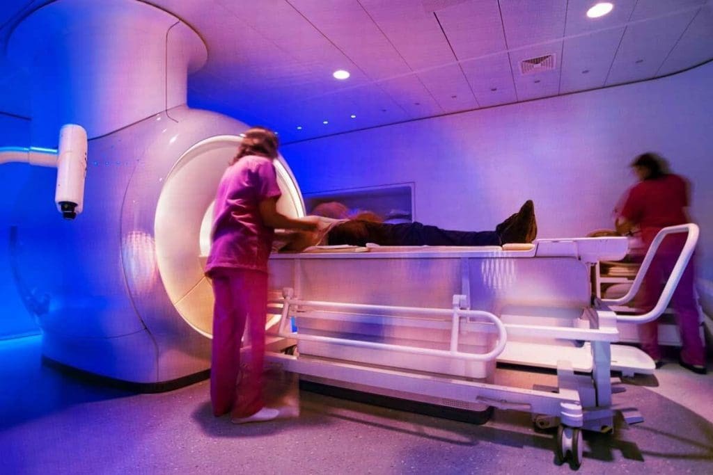
Knowing the differences between MRI and X-ray is key for good diagnosis and treatment. At Liv Hospital, we use the latest imaging tech to care for our patients.
Medical imaging has changed how we find and treat health issues. X-rays and MRI are two main tools used. They work differently and have their own strengths.
How X-rays Create Images Using Radiation
X-rays use radiation to show what’s inside our bodies. They’re great for seeing bones and finding breaks. The radiation goes through the body, and denser parts like bone show up white.
Key benefits of X-ray technology include:
- Quick and relatively inexpensive
- Wide availability
- Effective for acute bone imaging
How MRI Creates Images Using Magnetic Fields
MRI uses a strong magnetic field and radio waves to show body details. It’s safer than X-rays because it doesn’t use radiation. MRI is best for soft tissues like tendons and organs.
For more info on choosing imaging, check out this resource on X-ray, CT, and MRI.
Key Technical Distinctions in Tissue Visualization
MRI and X-ray differ in what they show. X-rays are better for bones, while MRI is better for soft tissues. This is important for diagnosing injuries that affect both.
The main differences in tissue visualization are:
- MRI offers better soft tissue contrast
- X-rays are more effective for bone imaging
- MRI can detect a wider range of tissue abnormalities
Understanding these differences helps doctors choose the right imaging for each patient. This ensures the best diagnosis and treatment.
Basic Capabilities of X-rays in Fracture Detection
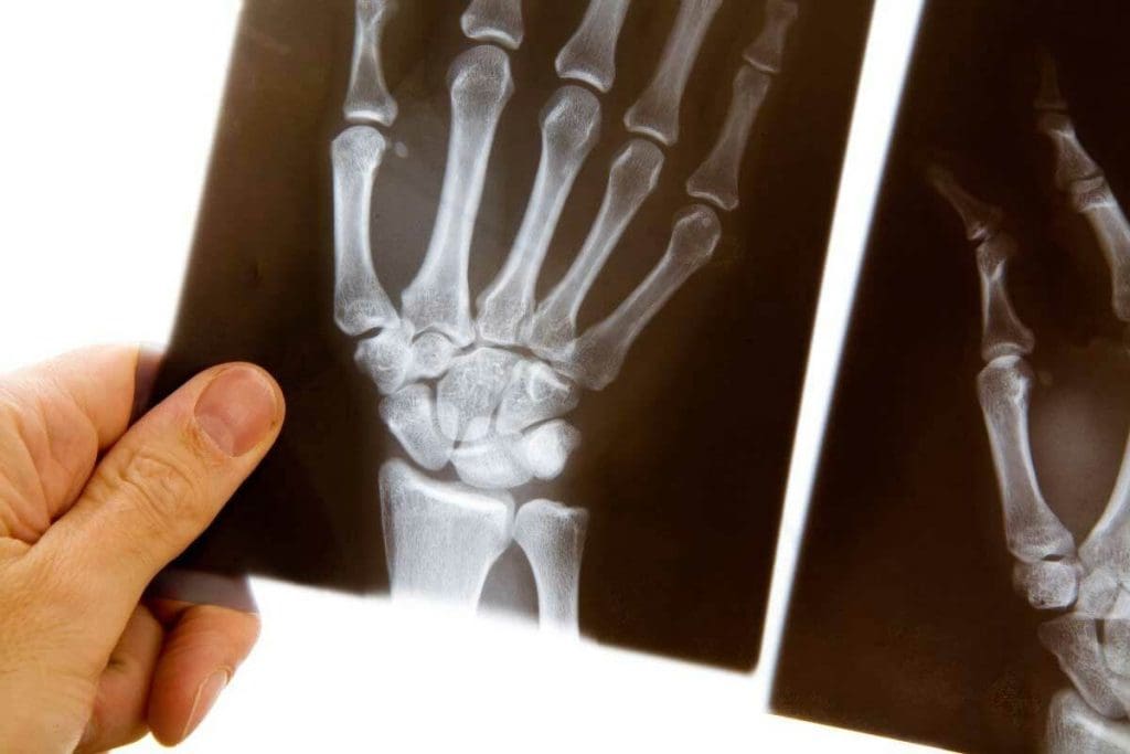
X-rays are a common tool for finding bone fractures. They are quick and effective. This makes them a go-to choice for doctors.
Types of Fractures Easily Detected by X-rays
X-rays work best for fractures where bones are clearly out of place. For example, transverse fractures are easy to spot. These happen at a right angle to the bone’s axis.
Comminuted fractures are also clear on X-rays. These are when bones break into many pieces. This shows up because the bone structure is broken.
Advantages of X-rays for Acute Bone Imaging
X-rays give quick results. They are often used first in emergency rooms and clinics. They also use low levels of radiation, which is good for patients.
Limitations and Blind Spots in X-ray Technology
But, X-rays can miss some fractures. Hairline fractures and stress fractures might not show up right away. This is true for early stages or in tricky spots like the wrist or spine.
X-rays also don’t show soft tissues well. This can make it hard to see the full picture of an injury.
Can MRI Show Broken Bones? Understanding Its Capabilities
MRI technology has changed how we diagnose bone injuries. It gives detailed images of bones and soft tissues. This helps doctors find and check bone fractures that other tests can’t see.
We use MRI to understand bone injuries fully. It shows both bones and soft tissues. This is key for finding complex fractures and related injuries.
How MRI Visualizes Bone Structures and Fractures
MRI uses a strong magnetic field and radio waves to create detailed images. It can spot different bone injuries, even those X-rays or CT scans miss.
MRI is great at showing bone marrow edema and soft tissue injuries around fractures. It helps doctors see how bad the injury is. This information helps plan the best treatment.
Types of Fractures Best Detected by MRI
MRI is best at finding stress fractures, hidden fractures, and other bone injuries. It’s also good at spotting ligament and tendon injuries with fractures.
Some fractures MRI is good at finding include:
- Stress fractures
- Occult fractures
- Growth plate fractures
- Tibial plateau fractures
For more info on MRI vs. X-ray for joint problems, visit this page.
Sensitivity and Specificity Rates for Fracture Detection
Studies show MRI is very good at finding fractures. It’s better than X-rays in many cases. Here are some key findings:
| Imaging Modality | Sensitivity | Specificity |
| MRI | 95% | 90% |
| X-ray | 70% | 85% |
As the table shows, MRI is better at finding fractures than X-rays. This makes MRI a valuable tool in orthopedics.
Detecting Subtle Bone Injuries: Where MRI Excels
Subtle bone injuries are hard to spot with regular imaging. But MRI can find them easily. At Liv Hospital, we use MRI to catch these injuries early. This helps prevent them from getting worse.
Visualizing Microfractures and Hairline Fractures
Microfractures and hairline fractures are tiny and hard to see on X-rays. But MRI can show them clearly. It’s great for spotting these small injuries early.
Key benefits of MRI in detecting microfractures include:
- High-resolution imaging of bone structures
- Ability to detect changes in bone marrow signal
- Visualization of surrounding soft tissue injuries
Identifying Bone Bruises and Bone Marrow Edema
Bone bruises and bone marrow edema are not always seen on X-rays. MRI is good at finding these because it shows changes in the bone marrow. This helps doctors understand the injury better.
Early Detection of Stress Fractures Before They Progress
Stress fractures are small cracks in the bone from too much stress. MRI can spot them early. This means patients can get help fast, which helps them heal quicker.
| Feature | X-ray | MRI |
| Detection of Microfractures | Limited | Highly Sensitive |
| Visualization of Soft Tissues | No | Yes |
| Early Detection of Stress Fractures | Difficult | Effective |
At Liv Hospital, we use MRI to give the best care for patients with bone injuries. This way, we make sure they get the right diagnosis and treatment.
Soft Tissue Visualization Around Broken Bones
Understanding the damage to soft tissues is key when treating fractures. When a bone breaks, the soft tissues around it can get hurt too. MRI is great at showing these soft tissues, helping doctors see the full injury.
Ligament and Tendon Injuries Associated with Fractures
Ligament and tendon injuries often happen with fractures. MRI helps doctors see these injuries clearly. This is important for treatment planning, as ignoring these injuries can cause long-term pain and slow healing.
- Ligament tears: MRI can spot tears in ligaments, which is important for joints.
- Tendon injuries: It can show tendonitis, tendinosis, or ruptures, helping with recovery plans.
- Associated injuries: It also checks for muscle strains or bruises.
Cartilage Damage Assessment in Joint Fractures
Cartilage damage is common with joint fractures. MRI gives a detailed look at cartilage health. This helps doctors understand how the joint might work in the future and plan the best treatment.
- It can spot cartilage thinning or defects.
- Osteochondral lesions, where cartilage and bone are damaged, can be seen.
- It finds loose cartilage pieces or joint bodies.
Evaluating Nerve and Blood Vessel Involvement
Nerve and blood vessel injuries can happen with fractures. These can lead to serious problems if not caught early. MRI can see these injuries, helping doctors act fast.
Nerve injuries: It can find nerve damage, helping avoid long-term nerve problems.
Vascular injuries: It spots blood vessel damage, like pseudoaneurysms or fistulas, to prevent serious bleeding or lack of blood flow.
Clinical Cases: When X-rays Miss What MRI Finds
MRI often outshines X-rays in diagnosing complex fractures and soft tissue injuries. We see many cases where X-rays can’t provide clear results. In these situations, MRI uncovers important details that X-rays miss.
Scaphoid and Other Wrist Fractures
Diagnosing wrist fractures, like those of the scaphoid bone, is tough with X-rays alone. MRI, on the other hand, excels in spotting these fractures, even when they’re new.
A study showed MRI found scaphoid fractures in 97% of cases, while X-rays only found 64%. This shows MRI’s key role in diagnosing scaphoid fractures, even when X-rays are negative.
Tibial Plateau and Growth Plate Fractures
MRI shines in diagnosing tibial plateau fractures and growth plate injuries in kids. X-rays often can’t fully show the extent of these injuries, missing soft tissue and growth plate details.
MRI gives a detailed look at bone and soft tissue injuries. For tibial plateau fractures, it shows how deep the articular surface is depressed and any ligament injuries. This info is vital for planning surgery.
Vertebral Compression Fractures and Spinal Injuries
MRI beats X-ray in diagnosing vertebral compression fractures and spinal injuries. It spots subtle fractures, checks the posterior ligamentous complex, and looks for spinal cord or nerve root compression.
A study found MRI revealed extra fractures not seen on X-ray in over 30% of cases. It also gave vital info on spinal stability and nerve involvement, helping guide treatment.
| Fracture Type | X-ray Detection Rate | MRI Detection Rate |
| Scaphoid Fractures | 64% | 97% |
| Tibial Plateau Fractures | 70% | 95% |
| Vertebral Compression Fractures | 60% | 90% |
These cases highlight MRI’s importance in diagnosing fractures and soft tissue injuries missed by X-rays. MRI’s detailed assessment helps doctors create better treatment plans, leading to better patient outcomes.
Recent Advancements in MRI Technology for Fracture Detection
Advances in MRI technology have changed how we find and diagnose fractures. At Liv Hospital, we lead in medical tech. This ensures our patients get the most accurate and quick diagnoses.
New MRI tech has made finding fractures more precise. We use high-field strength MRI, special bone imaging sequences, and AI for fracture detection.
High-Field Strength MRI Capabilities
High-field strength MRI is a big step forward in finding fractures. It uses stronger magnetic fields to create clearer images. This lets us spot even the smallest fractures that other methods might miss.
This tech gives us better views of bones and changes in bone marrow. It helps us diagnose fractures more accurately and plan better treatments.
Specialized Sequences for Bone Imaging
There are special MRI sequences for better bone imaging. These highlight bone and fracture details. They give radiologists the info they need for accurate diagnoses.
These sequences help us see how big fractures are and check soft tissues. They also spot any complications. This info is key for making the right treatment plans for each patient.
Artificial Intelligence Applications in Fracture Detection
AI in MRI fracture detection is a big step. AI algorithms learn to spot fracture patterns. This helps radiologists find issues faster and more accurately.
AI helps speed up image analysis. This means quicker diagnoses and treatments. It also cuts down on human mistakes, making diagnoses more reliable.
At Liv Hospital, we use the latest MRI tech for top-notch care. With high-field MRI, special sequences, and AI, we find fractures precisely and quickly. This leads to better care for our patients.
Fracture Classification and Treatment Planning with MRI
MRI helps us classify fractures better and plan treatments. It shows both bone and soft tissue details. This lets us fully understand the injury.
Improving Fracture Classification Accuracy
MRI gives us clear images of fractures. It shows the full extent and complexity of the break. This is key for fractures that X-rays can’t see well.
With MRI, we can see the fracture line, how much it’s moved, and soft tissue damage. Knowing this helps us classify the fracture right. This guides our treatment choices.
| Fracture Type | X-ray Visibility | MRI Visibility |
| Simple Fracture | High | High |
| Comminuted Fracture | Moderate | High |
| Stress Fracture | Low | High |
Determining Fracture Age and Healing Status
MRI helps us figure out when a fracture happened and how it’s healing. It looks at bone marrow and soft tissue signals. This tells us the fracture’s age and healing progress.
This info is key for choosing the right treatment. It could be surgery or just rest. MRI shows us how the bone is healing. This helps us decide if we need to change the treatment plan.
Impact on Surgical vs. Conservative Treatment Decisions
MRI’s detailed images change how we decide between surgery and rest. It helps us understand the fracture’s severity. This lets us pick the best treatment.
For example, MRI can show if a fracture needs surgery because it’s not stable. Or if it can heal with rest. This makes treatment more precise. It helps patients heal better and avoids complications.
We use MRI to make sure patients get the right care. This approach focuses on each patient’s needs. It shows our dedication to quality, patient-centered care.
Time, Cost, and Accessibility Considerations
Choosing between MRI and X-ray for bone fractures is not just about accuracy. It also involves cost and how easy it is to get the test. Healthcare providers must think about how these choices affect patient care and how resources are used.
Availability and Wait Times for Different Imaging
X-ray machines are everywhere, from emergency rooms to clinics. MRI machines, though, are rarer and need their own space. This means MRI scans often take longer to get, making wait times longer in some places.
In some areas, MRI appointments can take weeks. X-rays, though, can be done quickly, even right away in emergencies. This wait time difference can affect how quickly patients get the care they need.
Cost Comparison Between X-ray and MRI
X-rays are cheap and many insurances cover them. MRI scans, being more complex, cost more. They need special facilities and staff.
Because of the cost, doctors might start with X-rays for fractures. If more detail is needed, they might choose MRI. But the higher cost and longer wait times make this choice careful.
Insurance Coverage and Reimbursement Factors
Insurance and how much it covers also matters. Most plans cover both X-rays and MRIs, but how much they cover can vary. Some plans might need you to get approval for MRI scans first, but X-rays are often okay without it.
How much doctors get paid for these tests can also affect who offers them. Knowing this helps both doctors and patients understand the options better.
Patient Experience and Safety Factors
When we compare MRI and X-ray for finding fractures, patient needs are key. It’s important to look at how well they work and how safe and comfortable they are for patients.
Radiation Exposure Comparison
MRI doesn’t use harmful radiation like X-rays do. X-rays are a worry for people who need many tests or are sensitive to radiation. MRI, on the other hand, uses magnetic fields and radio waves to make images without radiation.
Radiation Exposure: X-ray vs. MRI
| Imaging Modality | Radiation Exposure | Typical Use |
| X-ray | Yes | Initial fracture assessment |
| MRI | No | Detailed soft tissue and bone marrow evaluation |
Contraindications and Special Considerations for MRI
MRI is usually safe, but there are some things to watch out for. People with metal implants or pacemakers can’t have MRI. Also, those who are scared of tight spaces might need special help, like open MRI machines or sedation.
Duration and Comfort During Imaging Procedures
MRI scans usually take longer than X-rays, from 15 to 90 minutes. Making sure patients are comfortable is very important. Things like earplugs, headphones, and sedation can help a lot.
In summary, MRI is safer because it doesn’t use radiation. But, we must think about who can’t have MRI and make sure patients are comfortable during the test.
Liv Hospital’s Integrated Approach to Fracture Diagnostics
At Liv Hospital, we’re proud of our way of diagnosing fractures. We use the latest technology and focus on the patient. Our team works together to give each patient a diagnosis and treatment plan that fits their needs.
Multidisciplinary Assessment of Bone Injuries
Our team includes orthopedic specialists, radiologists, and more. They work together to look at bone injuries from all sides. This teamwork helps us find the best treatment for each patient.
A study on PubMed Central shows our approach improves patient care.
Our team’s work has many benefits:
- Comprehensive diagnosis: We get a clearer picture of a patient’s condition.
- Personalized treatment plans: We tailor treatments to each patient’s needs.
- Improved patient outcomes: Our teamwork leads to better results and faster recovery.
Combining Imaging Modalities for Complete Diagnosis
We use different imaging methods at Liv Hospital. This includes X-ray, MRI, and CT scans. It helps us understand a patient’s condition fully.
- Enhanced diagnostic accuracy: Using various imaging methods helps avoid mistakes.
- Comprehensive assessment: We can see the full extent of a patient’s condition.
Patient-Centered Care in Diagnostic Decision-Making
At Liv Hospital, we put patients first. We make sure our care is open, collaborative, and listens to patients. This way, we create treatment plans that meet each patient’s needs and goals.
Our patient-centered care includes:
- Clear communication: We make sure patients understand their diagnosis and options.
- Personalized care: We tailor treatments to each patient’s unique situation.
Conclusion: The Complementary Role of MRI and X-ray in Bone Injury Diagnosis
At Liv Hospital, we understand the importance of MRI and X-ray in diagnosing bone injuries. X-rays give a fast and effective first look at bone fractures. MRI, on the other hand, shows more detail of both bone and soft tissue injuries.
Using MRI and X-ray together helps doctors fully understand the injury. This is key for creating a good treatment plan. By combining these methods, we can make diagnoses more accurate and help patients get better faster.
We are dedicated to top-notch healthcare, shown in our use of MRI and X-ray for fracture diagnosis. This approach helps us give complete care, from diagnosis to recovery. Our goal is to ensure our patients get the best care possible.
In summary, MRI and X-ray together are a strong tool for diagnosing bone injuries. We keep using these technologies to improve patient care and outcomes.
FAQ
What does an MRI show that an X-ray can’t when detecting broken bones?
MRI shows more than X-rays. It can see both bone and soft tissue. This means it can find small bone injuries and soft tissue damage. X-rays might miss these.
Can MRI detect broken bones that X-rays miss?
Yes, MRI can find fractures and injuries that X-rays can’t. It’s very good at spotting subtle or complex bone problems.
Will an MRI show a broken bone if it’s present?
Usually, yes. MRI can show broken bones, even if they’re not clear on X-rays. It’s great at showing bone and soft tissue details.
Does MRI show bone fractures that are not visible on X-ray?
Yes, MRI can show fractures and injuries not seen on X-rays. This is true for hairline, non-displaced fractures and soft tissue issues.
What are the advantages of using MRI over X-ray for fracture detection?
MRI has big advantages. It can spot more injuries, show complex anatomy, and help plan treatment better.
Are there any limitations to using MRI for detecting broken bones?
While MRI is great, it’s not always needed. Cost, access, and patient conditions like metal implants or claustrophobia can limit its use.
How does MRI compare to X-ray in terms of radiation exposure?
MRI is safer because it doesn’t use ionizing radiation. This makes it better for patients needing repeated scans or who are sensitive to radiation.
Can MRI be used to assess the healing status of a fracture?
Yes, MRI can tell how old a fracture is and if it’s healing. It shows bone and tissue details, helping with treatment choices.
What role does MRI play in treatment planning for fractures?
MRI’s detailed images help classify fractures and check soft tissue injuries. This guides treatment choices, making it key in planning care.
How does Liv Hospital approach fracture diagnostics?
Liv Hospital uses a team approach. They combine MRI and X-ray for a full diagnosis. They focus on patient care.
Reference
- Liu, X. D., et al. (2020). Comparison between computed tomography and magnetic resonance imaging in the clinical diagnosis of tibial plateau fractures. Journal of Healthcare Engineering. https://pmc.ncbi.nlm.nih.gov/articles/PMC7520768/


