Last Updated on November 27, 2025 by Bilal Hasdemir
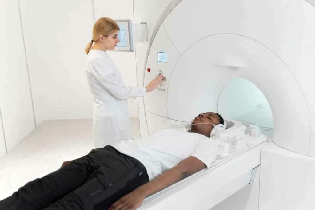
At Liv Hospital, we use cutting-edge imaging to find and treat complex diseases. The FDG PET scan combines positron emission tomography (PET) with fluorodeoxyglucose (FDG) to see how tissues and organs function, providing detailed insights for accurate diagnosis and treatment planning.
An FDG PET scan is a top-notch test. It shows how the body’s tissues and organs work. This helps find cancer, inflammation, and other issues.
We use this key technology to give our patients the best care. Our team of experts uses FDG PET imaging to spot different medical problems with great accuracy.
Key Takeaways
- FDG PET scans help diagnose and manage complex diseases like cancer and inflammation.
- The radiotracer FDG is used to examine the metabolic activity of tissues and organs.
- LivHospital utilizes advanced FDG PET imaging techniques for precise diagnosis.
- FDG PET scans aid in detecting abnormal tissue behavior and identifying metastasis.
- Our team of experts uses FDG PET imaging to deliver world-class healthcare services.
The Science and Purpose of FDG PET Scanning
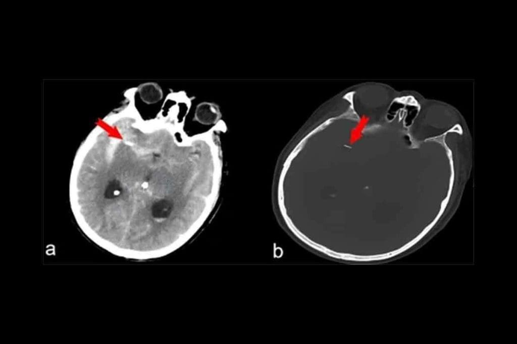
FDG PET scanning has changed medical imaging a lot. It gives doctors important insights into how our bodies work and what might be wrong. This helps them diagnose and treat many health issues better.
Definition and Core Principles
FDG PET scanning uses a special sugar called FDG to see how active our cells are. It works because cancer cells use more sugar than healthy cells do. This helps doctors find cancer.
First, a tiny bit of radioactive FDG is given to the patient. It goes to all cells in the body. Then, a PET scanner picks up the signals from the FDG. This makes detailed pictures of where the sugar is being used.
Historical Development of Medical Imaging Technology
The start of FDG PET scanning was a big step forward in medical imaging. The idea of PET scanning began in the 1950s. But it wasn’t until the 1970s that the first scanners were made. The 1980s brought FDG into use, making PET scans common in hospitals.
As technology got better, so did PET scans. Now, FDG PET/CT scans are key in finding and treating diseases like cancer, brain problems, and heart issues. They help doctors a lot in planning treatments and checking how well they work.
| Year | Milestone |
| 1950s | Concept of PET scanning emerges |
| 1970s | First PET scanners developed |
| 1980s | Introduction of FDG as a tracer |
| 2000s | Integration of PET with CT scanning |
What Does FDG Mean in Medical Terms?
Fluorodeoxyglucose, or FDG, is a key tracer in PET imaging. It helps us see how cells use glucose. This is key for diagnosing and tracking many health issues.
The Chemistry of Fluorodeoxyglucose
FDG is a special glucose-like molecule. It has a hydrogen atom where glucose has a hydroxyl group. This makes it perfect for PET scans.
Its structure is close to glucose, so cells absorb it. But, unlike glucose, FDG gets stuck in cells. This lets us see where cells are most active.
FDG as a Medical Abbreviation Explained
In medicine, FDG means fluorodeoxyglucose. It’s used to see how the body’s cells work. It’s a key tool in diagnosing and treating diseases, like cancer.
FDG shows us where cells are working hard. This is often a sign of disease. Knowing what FDG means helps doctors understand PET scans better.
| Term | Abbreviation | Medical Significance |
| Fluorodeoxyglucose | FDG | Used in PET scans to assess cellular metabolism |
| Positron Emission Tomography | PET | Imaging technique using tracers like FDG |
| Glucose Analog | – | Mimics glucose uptake in cells, used for diagnostic purposes |
Understanding FDG’s chemistry and medical use shows its importance. It helps improve how we diagnose and treat patients.
How FDG PET Imaging Works in Clinical Practice
In clinical practice, FDG PET imaging is key for checking how cells and tissues work. It uses the idea that cancer cells use more glucose than regular cells.
The Metabolic Basis of Glucose Uptake
Fluorodeoxyglucose (FDG) is a glucose-like substance that cells take up based on their glucose use. Cancer cells, with their high metabolism, take up more FDG than normal cells. This helps doctors see which tissues are most active.
Doctors give FDG through a vein in the hand or arm. It spreads through the body, where cells absorb it. The more FDG a tissue or lesion takes up, the more active it is.
The Technical Process Behind the Scan
The FDG PET scan process starts with giving FDG and letting it spread. Then, the patient goes into the PET scanner. The scanner picks up gamma rays from the FDG, making detailed images of metabolic activity. Advanced algorithms turn these signals into images that show where FDG is most active.
Integration with CT Scanning: The Combined Approach
Combining FDG PET with CT scanning is a big step forward in imaging. It mixes the metabolic info from PET with the body’s structure from CT. This gives doctors a clearer picture of what’s going on inside the body.
A leading nuclear medicine expert says, “Merging PET and CT images helps pinpoint active lesions. This is key for cancer staging and checking how treatments work.”
“The mix of PET and CT has changed oncology, leading to better diagnoses and treatments.”
Clinical Applications of FDG PET in Oncology
In cancer care, FDG PET imaging is key for detecting, staging, and managing cancers. FDG PET scans are vital in oncology, showing how tumors work and grow.
Cancer Detection and Initial Diagnosis
FDG PET scans help diagnose cancers like Hodgkin lymphoma, non-Hodgkin lymphoma, and lung cancer. They spot cancer cells early, even when they’re small. For more on PET scans, check healthdirect.gov.au.
Tumor Staging and Treatment Planning
Knowing the cancer stage is key for the right treatment. FDG PET imaging shows how far cancer has spread. This helps doctors plan the best treatment.
Monitoring Treatment Response and Recurrence
FDG PET scans track how well treatments work. They check if tumors are shrinking. They also find cancer coming back early, so doctors can act fast.
FDG PET has made cancer care better by finding cancers early and guiding treatments. As cancer care grows, FDG PET will help even more, making treatments more precise and personal.
Differentiating Between Benign and Malignant Lesions with FDG PET
FDG PET scans are key in telling benign from malignant lesions. They show how active the cells are. Cancer cells are more active, making them easier to spot.
We use FDG PET scans to check if a lesion is benign or malignant. Knowing this helps us make the right diagnosis and treatment plan.
Metabolic Signatures of Malignancy
Malignant cells take up more glucose because they’re very active. This is why FDG PET scans are used in cancer care. FDG is like glucose and shows how active tumors are.
“The use of FDG PET in oncology has revolutionized the way we diagnose and manage cancer, providing valuable insights into the metabolic characteristics of tumors.”
The way a lesion takes up FDG tells us a lot. High FDG uptake usually means it’s cancerous. Low uptake might mean it’s not.
Standardized Uptake Value (SUV) Interpretation
The Standardized Uptake Value (SUV) measures how much FDG a lesion takes up. It helps us tell if a lesion is benign or malignant. An SUV above a certain level often means it’s cancer, but this can change based on the tumor.
| SUV Value | Interpretation |
| Low (<2.5) | Likely benign |
| Moderate (2.5-4.0) | Indeterminate; requires further evaluation |
| High (>4.0) | Likely malignant |
Understanding SUV values needs careful thought. False positives and negatives can happen. For example, inflammation can make a lesion look like cancer on a scan.
By using FDG PET scans with other tests, we can make more accurate diagnoses. This helps us plan better treatments.
Understanding “FDG-Avid” Findings and Their Significance
It’s key to know what FDG-avid lesions mean for diagnosis and treatment. When looking at FDG PET scans, “FDG-avid” spots show high fluorodeoxyglucose (FDG) uptake. This can mean different things, like tumors or inflammation.
What Makes a Lesion FDG-Avid?
A spot is called FDG-avid if it uses more glucose than nearby tissues. This is often seen in fast-growing cells, like cancer. But, not all spots are cancer; inflammation or infection can also show up as FDG-avid.
FDG is taken in by cells like glucose. Inside the cell, it’s changed into FDG-6-phosphate. This can’t be broken down further, so it stays in the cell. This makes it visible on PET scans.
Non-Cancerous Causes of High FDG Uptake
High FDG uptake isn’t always cancer. It can also be from:
- Inflammation
- Infections
- Granulomatous diseases
- Benign tumors
- Normal tissue activity
To correctly read FDG PET scans, knowing these causes is important. Doctors need to look at the whole picture, including the patient’s history and other scans, to make the right call.
| Condition | Typical FDG Uptake Pattern | Clinical Context |
| Malignant Tumor | High, often uneven uptake | Known or suspected cancer |
| Inflammation | Variable, often widespread uptake | Recent surgery, infection, or inflammatory disease |
| Infection | High uptake, often in one area | Signs of infection, fever, high WBC count |
| Granulomatous Disease | High uptake, often in a specific pattern | Known granulomatous disease, like sarcoidosis |
Knowing why FDG spots are active helps doctors make better diagnoses and plans. This detailed knowledge is key to giving patients the best care.
FDG PET Applications in Neurology
FDG PET scanning has changed neurology by giving us deep insights into brain metabolism. We use this advanced imaging to diagnose and manage many neurological disorders. This improves patient care and treatment results.
Alzheimer’s Disease and Dementia Evaluation
In neurology, FDG PET is key for checking Alzheimer’s disease and other dementias. It looks at glucose metabolism in the brain. This helps us spot patterns that show different types of dementia. This helps us make more accurate diagnoses and treatment plans.
“FDG PET imaging is vital for diagnosing and managing Alzheimer’s disease,” says a leading neurology expert. “It helps doctors tell it apart from other causes of memory loss.”
Epilepsy and Other Neurological Disorders
FDG PET is also great for checking epilepsy and other brain conditions. It finds abnormal brain activity, which is key for planning treatments. We’ve seen big improvements in managing epilepsy with FDG PET, leading to better patient results.
FDG PET’s role in neurology shows its wide use and importance in today’s medicine. As we learn more about brain disorders with FDG PET, we can give better care to our patients.
Using FDG PET in neurological checks helps us diagnose and treat complex conditions better. This not only helps patients but also helps us improve neurological care.
The Role of FDG PET in Cardiology
Cardiologists use FDG PET scans to check if heart muscle is alive and to find heart inflammation. This tool is key in cardiology, helping doctors make treatment plans.
Myocardial Viability Assessment
FDG PET helps see if heart muscle is alive, mainly in those with heart disease or after a heart attack. It looks at how much glucose the heart muscle takes in. This shows if some heart muscle might get better with new blood flow.
To do this, a tiny bit of radioactive glucose (FDG) is given to the patient. The FDG goes to heart muscle cells. A PET scanner then finds this glucose, showing how active the heart is.
Key Benefits of FDG PET in Myocardial Viability Assessment:
- It finds heart muscle that might get better with new blood flow.
- It helps doctors decide the best treatment by checking heart muscle health.
- It’s very helpful for people with heart disease or after a heart attack.
| Condition | FDG PET Findings | Clinical Implication |
| Hibernating Myocardium | Preserved or increased FDG uptake | Potential benefit from revascularization |
| Scar Tissue | Reduced or absent FDG uptake | Limited benefit from revascularization |
Cardiac Inflammation and Infection Detection
FDG PET is also great for finding heart inflammation and infection. This includes conditions like myocarditis or endocarditis. It spots these because the heart muscle takes up more glucose when it’s inflamed or infected.
By using FDG PET with CT scans, doctors get a full picture of the heart. This helps them diagnose and treat complex heart problems better.
- Assessment of myocardial viability
- Detection of cardiac inflammation and infection
- Guiding treatment decisions in complex cardiac cases
The Patient Experience: Before, During, and After an FDG PET Scan
We know that getting an FDG PET scan can be stressful. So, we’re here to help you through every step.
Pre-Scan Preparations and Requirements
To make sure your FDG PET scan goes well, you need to prepare. Don’t do hard exercise for a couple of days before because it can change how FDG spreads. Also, you should not eat for at least four hours before the scan, but you can drink water.
Tell your doctor about any medicines you take and any health issues like diabetes. For more info on getting ready for a PET scan, check out RadiologyInfo.org.
| Pre-Scan Requirement | Description |
| Avoid strenuous exercise | For a couple of days before the scan to prevent altered FDG distribution. |
| Fasting | For at least four hours before the scan. |
| Medication disclosure | Inform your healthcare provider about any medications you’re taking. |
What to Expect During and After the Procedure
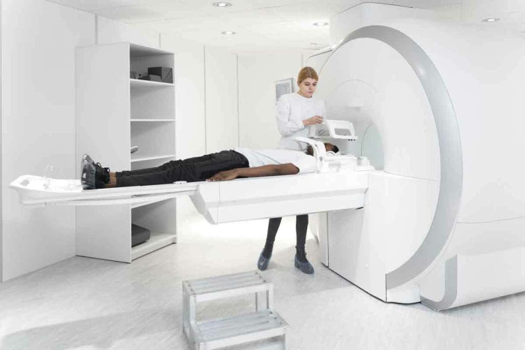
During the FDG PET scan, you’ll lie on a table that slides into a big PET scanner. The scan is painless and lasts about 30 minutes to an hour. After it’s done, you can usually go back to your normal day unless your doctor says not to.
You might feel tired or hungry after fasting. Eating something after the scan can help. Our team wants to make sure you’re comfortable and stress-free during the FDG PET scan.
Limitations and Safety Considerations of FDG PET
FDG PET scans are a powerful tool for diagnosis. Yet, they have their limits and safety concerns. It’s key to know these to use them safely and effectively.
False Positives and Negatives: Clinical Challenges
FDG PET scans can sometimes show false positives and negatives. False positives cause unnecessary worry and extra tests. False negatives can lead to late diagnosis and treatment.
Several things can cause these false results. For example, inflammation or infection can show up as increased FDG uptake, not just cancer.
| Causes of False Positives | Causes of False Negatives |
| Inflammatory processes | Small tumor size |
| Infection | Low metabolic activity |
| Physiological uptake (e.g., brown fat) | Technical issues (e.g., motion artifacts) |
Radiation Exposure and Risk Assessment
FDG PET scans involve radiation exposure. The dose is low, but we must weigh the risks against the benefits. We consider the patient’s age, the scan’s necessity, and other imaging options.
PET-CT scans, which add CT to PET, have extra risks. These include allergic reactions and kidney problems from contrast. Choosing the right patients and preparing them well helps reduce these risks.
Knowing the limits and safety of FDG PET scans helps us use them better. This way, we ensure patients get the most from this technology while keeping risks low.
Advanced Techniques and Future Directions in FDG PET
The field of FDG PET imaging is growing fast. New technologies and uses are leading the way. We’re seeing big steps forward in FDG PET technology.
Technological Innovations in Scanner Design
PET scanners are getting better. They now have higher resolution, are more sensitive, and scan faster. Digital PET scanners, for example, improve image quality and cut down scan times. This makes the process easier for patients.
Another big change is combining PET with MRI and CT. This hybrid imaging gives us more detailed views. It mixes PET’s metabolic info with CT or MRI’s body details.
| Technological Innovation | Description | Clinical Impact |
| Digital PET Scanners | Enhanced detector technology for improved resolution and sensitivity | Better image quality, reduced scan times |
| PET/CT Integration | Combination of metabolic and anatomical imaging | More accurate diagnosis and staging of diseases |
| PET/MRI Integration | Simultaneous assessment of metabolic and soft tissue information | Enhanced evaluation of complex conditions, such as cancer and neurological disorders |
Emerging Clinical Applications and Protocols
New uses and protocols are making FDG PET even more useful. Tracers like quinoline-based compounds and 68Ga-labeled FAP inhibitors are being tested. They might help us see diseases in new ways.
For instance, FAPI PET could help doctors understand tumor behavior and how well treatments work. These new tracers could lead to more precise imaging. This means better diagnosis and treatment plans for patients.
Looking ahead, FDG PET will keep being a key tool in medical imaging. Research and development will likely bring more breakthroughs. These will help improve patient care and our understanding of diseases.
Conclusion: The Evolving Role of FDG PET in Modern Medicine
Medical technology keeps getting better, and FDG PET imaging is at the heart of it. It’s key in fields like oncology, neurology, and cardiology. It gives doctors the info they need to diagnose and treat patients.
FDG PET scanning is a must-have for spotting and managing diseases. It shows how active cells are, helping doctors tell good cells from bad. This helps them see how well treatments are working and if the disease is coming back.
FDG PET is set to stay a top choice in medical imaging. As we learn more about how cells use glucose, FDG PET will keep giving doctors important clues. These clues help make treatment plans better.
The future of FDG PET imaging is bright. New tech and ways of using it will make it even better at finding diseases. So, FDG PET will keep being a big part of modern medicine. It will help make patients’ care even better.
FAQ
What is an FDG PET scan, and what does FDG stand for in medical imaging?
An FDG PET scan is a test that uses Fluorodeoxyglucose (FDG) to see how active cells are in the body. It helps doctors find and manage diseases like cancer, brain disorders, and heart problems.
How does FDG PET imaging work, and what is its significance in clinical practice?
FDG PET imaging finds active cells by using Fluorodeoxyglucose, a sugar-like substance. It’s important because it shows how tissues work. This helps doctors diagnose, plan treatments, and check how well treatments are working.
What is the role of FDG PET in cancer detection and management?
FDG PET is key in finding and managing cancer. It spots areas where cells are very active, which often means cancer. It also helps see how well treatments are working and if cancer comes back.
How is FDG PET used in neurology, particularily in evaluating Alzheimer’s disease and epilepsy?
In neurology, FDG PET looks at brain activity. It helps diagnose and manage Alzheimer’s and epilepsy by finding where glucose metabolism is different.
What are the applications of FDG PET in cardiology?
In cardiology, FDG PET checks if heart muscle is alive and finds heart infections. It gives important info for patient care.
What preparations are required before undergoing an FDG PET scan?
Before an FDG PET scan, you need to fast, avoid hard exercise, and manage diabetes. Doctors give specific instructions for the best results.
What are the potencial risks and limitations associated with FDG PET scans?
Risks include radiation exposure. Limitations include false positives and negatives. Knowing these helps use FDG PET safely and effectively.
How is the Standardized Uptake Value (SUV) interpreted in FDG PET scans?
SUV measures how much FDG is taken up by tissues. Higher SUV values often mean cancer. It helps doctors tell the difference between good and bad lesions.
What makes a lesion FDG-avid, and what are the implications?
A lesion is FDG-avid if it takes up a lot of FDG. This can mean cancer, but also other issues like inflammation. Getting it right is key for treatment decisions.
What are the future directions and emerging applications of FDG PET?
New tech and uses are coming for FDG PET. It will help doctors diagnose and plan treatments even better in the future.
Reference
- Lmuhaideb A, et al. 18F-FDG PET/CT imaging in oncology. Ann Saudi Med. 2011 https://www.ncbi.nlm.nih.gov/pmc/articles/PMC3159211/


