Last Updated on November 27, 2025 by Bilal Hasdemir

At Liv Hospital, we use advanced imaging to help diagnose and treat diseases. The fluorodeoxyglucose (FDG) PET scan is key in finding cancer and understanding how it develops. The link between PET scan sugar and cancer is important because cancer cells use more sugar than normal cells, allowing the scan to detect abnormal activity in the body.
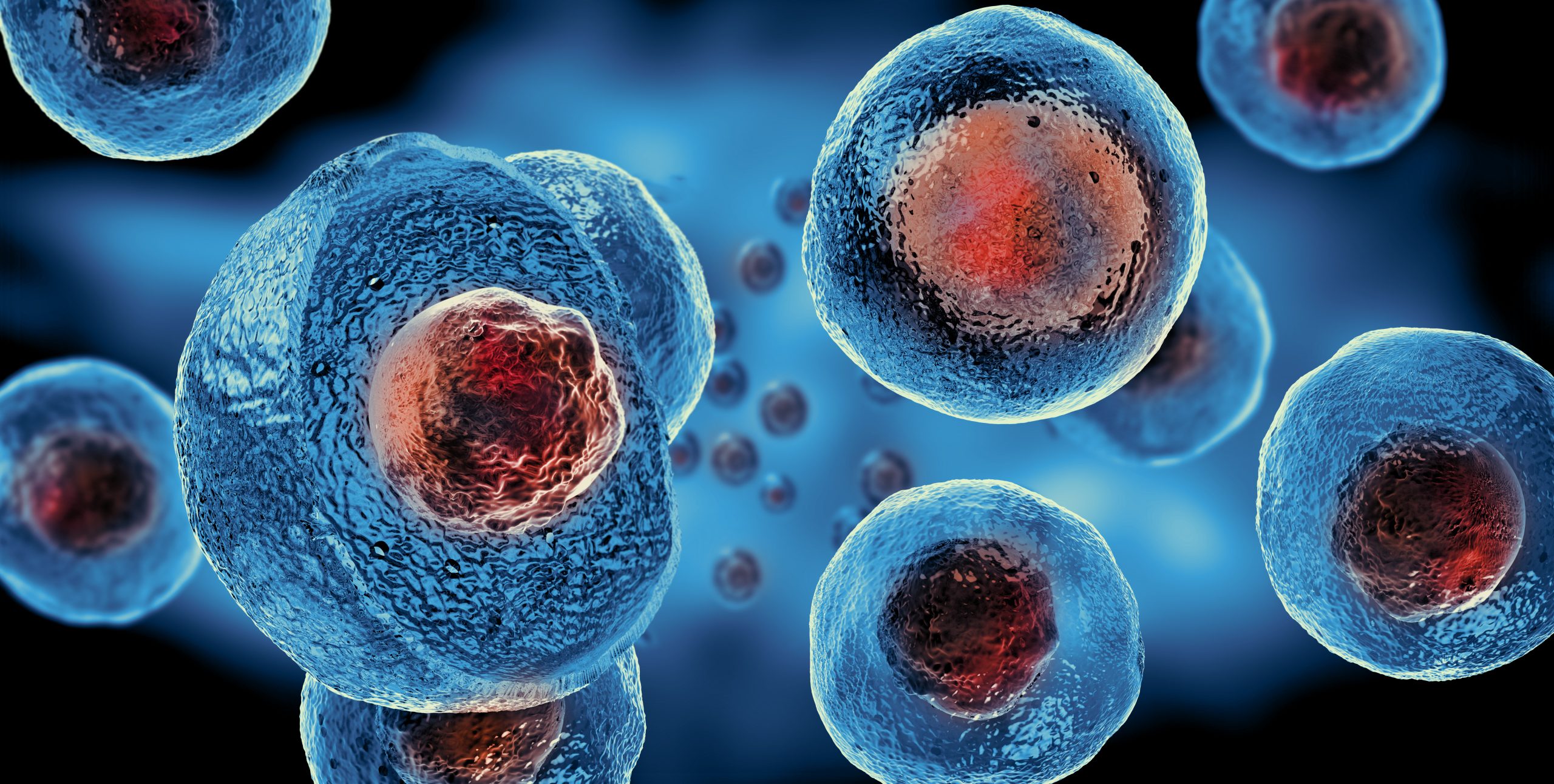
Cancer cells use more sugar than normal cells. When we add FDG, it goes to these cells. This makes them show up bright on the scan, helping us find and watch cancer.
Key Takeaways
- FDG PET scans detect cancer by highlighting abnormal metabolic activity.
- Cancer cells have higher glucose metabolism rates than normal cells.
- FDG accumulates in cancerous areas, appearing as bright spots on the scan.
- Understanding FDG activity is key for accurate cancer diagnosis and treatment.
- Liv Hospital uses advanced PET scan technology for effective cancer care.
What is FDG in Medical Terms? The Special Sugar Used in PET Scans

Fluorodeoxyglucose, or FDG, is a special sugar that has changed nuclear medicine. It’s a glucose-like molecule with radioactive fluorine-18 (18F) for PET scans. This sugar is used by cells instead of regular glucose, helping find cancer.
The Chemical Structure of Fluorodeoxyglucose
FDG is a glucose molecule with a fluorine-18 change. This change lets it be used by cells but not broken down. It builds up in cells that use a lot of glucose.
Chemical Formula: C6H11FO5 for FDG, with fluorine-18 incorporated.
| Chemical Property | Glucose | FDG |
| Chemical Formula | C6H12O6 | C6H11FO5 |
| Molecular Weight | 180.16 g/mol | 181.15 g/mol (without F-18) |
| Metabolic Pathway | Undergoes glycolysis | Trapped in cells after phosphorylation |
How FDG Mimics Glucose in the Body
FDG acts like glucose by entering cells through glucose transporters. Inside, it’s changed into FDG-6-phosphate by hexokinase. Unlike glucose, FDG-6-phosphate can’t move forward in the glycolytic pathway. It stays in the cell, showing up in areas that are very active.
For more detailed information on glucose metabolism and its relation to FDG, we recommend exploring resources such as NCBI’s book on PET Imaging.
The Role of Radioactive Fluorine-18 in Imaging
Fluorine-18 in FDG is key for PET imaging. It’s a positron-emitting radionuclide with a half-life of about 110 minutes. When it decays, it emits positrons that create gamma rays. These rays are picked up by the PET scanner, making detailed images of FDG in the body.
Knowing about FDG’s structure, how it works like glucose, and the role of fluorine-18 helps us see its value. It’s used in PET scans to diagnose and manage conditions, like cancer.
The FDG-PET Scanning Process: From Injection to Image

The FDG-PET scan process starts long before the scan itself. It involves detailed preparation and precise steps. FDG-PET imaging is a complex tool that needs teamwork between the patient, radiology team, and nuclear medicine experts.
Patient Preparation for an FDG-PET Scan
Getting ready for an FDG-PET scan is key for clear images. Patients usually fast for hours before the scan. This ensures their body is ready for the best FDG uptake.
They’re also told to avoid hard exercise and stay calm before and after the FDG radiotracer injection.
The Injection and Uptake Period
The FDG radiotracer is given through a vein in the arm. Then, there’s a wait time for the FDG to spread in the body’s tissues. This wait is about 60 minutes.
During this time, patients must stay very calm and not move much. This helps avoid muscle activity that could mess up the scan.
How the PET Scanner Detects FDG Activity
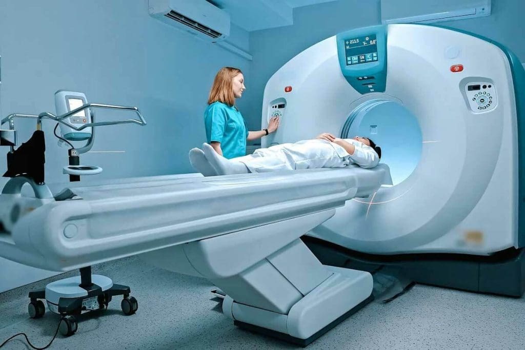
The PET scanner finds the FDG activity in the body during the scan. It moves around the patient, taking detailed pictures of where the FDG is. These pictures show where the FDG has built up.
Image Processing and Interpretation
After the scan, the data is processed to make the final images. These images are then looked at by nuclear medicine experts. They check the FDG uptake patterns to spot issues, like cancer.
The experts look at how much and where the FDG is to understand the patient’s health better.
Interpreting FDG Activity on PET Scans
Understanding FDG uptake on PET scans is key. It shows how cancer cells use glucose. This helps spot tumors and see how aggressive they are.
What Do “Hot Spots” Indicate in Cancer Detection?
“Hot spots” on PET scans mean high glucose use. But, they can also show inflammation, not just cancer. We must check these spots carefully.
Key factors in interpreting hot spots include:
- The intensity of FDG uptake
- The location and distribution of hot spots
- Correlation with other imaging modalities like CT or MRI
Standardized Uptake Values (SUV) Explained
Standardized Uptake Values (SUV) measure FDG uptake. They help compare PET scans from different places. A high SUV often means a tumor is more aggressive.
But, SUV values can change due to:
- Patient preparation and glucose levels
- Time between FDG injection and scanning
- Scanner characteristics and image reconstruction methods
Common Patterns of FDG Uptake in Different Cancer Types
Each cancer type shows FDG uptake differently. For example, aggressive lymphomas have high uptake. But, some prostate cancers might show less uptake.
Knowing these patterns helps us tell cancer types apart. We also look at other signs and symptoms to make accurate diagnoses.
Clinical Applications of FDG-PET Imaging in Oncology
FDG-PET imaging has changed oncology by giving key info for cancer diagnosis and treatment planning. We use FDG-PET scans to see how active tumors are. This is key for understanding cancer’s extent and behavior.
Initial Cancer Diagnosis and Staging
FDG-PET imaging is key in cancer diagnosis and staging. It finds the main tumor site, checks how far the disease has spread, and spots metastases. This info is vital for picking the right treatment.
- Accurate Staging: FDG-PET scans give accurate staging info, which is key for the right treatment approach.
- Tumor Characterization: By looking at tumor metabolism, FDG-PET helps understand cancer aggressiveness.
Treatment Response Monitoring
Watching how a patient responds to treatment is another big use of FDG-PET imaging. By comparing scans before and after treatment, doctors can see if the treatment is working. They can then adjust the treatment as needed.
- Early Response Assessment: FDG-PET scans can spot early changes in tumor metabolism, helping adjust treatment plans quickly.
- Personalized Medicine: By looking at treatment response for each patient, FDG-PET supports personalized medicine.
Recurrence Detection
Finding cancer recurrence early is key for effective management. FDG-PET scans are very good at spotting recurrence, even before other imaging can see it.
- Early Detection: FDG-PET imaging helps find recurrence early, improving treatment chances.
- Surveillance: Regular FDG-PET scans can watch for recurrence in high-risk patients, catching it early.
Radiation Therapy Planning Using FDG-PET
FDG-PET imaging is also used in planning radiation therapy. It helps define tumor boundaries more accurately and targets therapy better.
- Precise Targeting: FDG-PET gives detailed info on tumor metabolism, helping target tumors during radiation therapy.
- Reduced Toxicity: Accurate tumor volume info from FDG-PET can reduce radiation to healthy tissues, lowering toxicity.
Distinguishing Between PET Scan Inflammation or Cancer
Detecting inflammation versus cancer on FDG-PET scans is a big challenge. FDG-PET scans are great at finding cancer. But, they can also pick up inflammation because inflammatory cells take up FDG too.
Why Inflammatory Cells Also Consume FDG
Inflammatory cells like macrophages and lymphocytes take up FDG. This is because they are active and use glucose, just like FDG. So, inflammation can look like cancer on PET scans, which can lead to wrong diagnoses.
“The overlap between inflammatory and malignant processes on FDG-PET scans complicates the diagnostic process,” as noted by medical professionals. Understanding this overlap is key for correct diagnosis.
Characteristic Patterns of Inflammatory FDG Uptake
Inflammation shows different patterns on PET scans than cancer. Inflammation tends to spread out, while cancer is more focused. Spotting these patterns is vital for making the right diagnosis.
- Diffuse uptake patterns are more commonly associated with inflammation.
- Focal uptake is more typical of malignant lesions.
- The intensity of FDG uptake can also provide clues, with higher uptake often seen in cancer.
False Positives and False Negatives in FDG-PET
False positives happen when inflammation is mistaken for cancer. False negatives occur when cancer is missed because it’s not very active. Knowing why these mistakes happen is important for better diagnosis.
False positives can be caused by infections, granulomatous diseases, and inflammation after surgery. False negatives can be due to small tumors, low-grade cancers, or tumors that don’t use much glucose.
Normal Physiologic FDG Uptake Patterns
It’s also important to know the normal patterns of FDG uptake. For example, the brain uses a lot of glucose, so it shows high uptake. The heart, liver, and muscles also show some uptake.
Knowing these normal patterns helps doctors tell the difference between harmless and harmful activity. This makes FDG-PET scans more accurate for diagnosing.
Advances in FDG-PET Technology and Techniques
Recent changes in FDG-PET technology have changed how we diagnose and monitor cancer. These updates have made PET scans more accurate and effective in fighting cancer.
FDG PET-CT and PET-MRI Hybrid Imaging
PET scans now combine with CT and MRI for better images. FDG PET-CT is key in cancer staging and checking how treatments work. It shows both how tumors work and their location.
PET-MRI gives even clearer images of soft tissues than CT. This is great for some cancers. It lets doctors see how active tumors are and their exact location at the same time.
Time-of-Flight and Digital PET Technology
New PET scanner tech, like Time-of-Flight (TOF) and digital PET, has made images clearer and more accurate. TOF measures when photons arrive, improving contrast and finding tumors better.
Digital PET scanners are also more sensitive and detailed. These updates help doctors diagnose and stage cancer more accurately.
Artificial Intelligence in FDG-PET Image Interpretation
Artificial Intelligence (AI) is changing how we read FDG-PET scans. AI helps measure PET images, spot tumors, and guess treatment results. It looks for patterns in big data that humans might miss.
AI with FDG-PET could make diagnoses more precise, save time, and help tailor treatments to each patient.
Beyond FDG: The Future of PET Imaging in Cancer Care
PET imaging is set to become even more important in cancer care. At Liv Hospital, we aim to lead in Turkey and worldwide. We focus on top-notch patient care and constant improvement.
FDG-PET scans have changed how we diagnose and treat cancer. Now, scientists are working on specialized tracers. These tracers aim to find specific cancers more accurately, leading to better treatments.
Specialized Tracers for Specific Cancer Types
New tracers are being made to find certain cancers, like prostate cancer. These tracers have shown great promise in tests. They could lead to more precise diagnoses and treatments.
PSMA-targeting tracers are good at finding prostate cancer that has come back. Tracers for neuroendocrine tumors also work well.
Emerging Applications in Personalized Cancer Medicine
PET imaging is key in personalized cancer care. It uses special tracers to see how tumors work. This helps doctors understand how aggressive the cancer is and how it might react to treatment.
This info helps doctors make treatment plans that fit each patient. For example, PET imaging can show who will get the most from targeted therapies. This can avoid side effects for others.
PET imaging is getting better, and we’ll see new uses in cancer care soon. The future looks bright for better patient results and new discoveries in oncology.
Conclusion: The Vital Role of FDG-PET Scans in Modern Cancer Management
FDG-PET scans are key in fighting cancer today. They help doctors diagnose, plan treatments, and check how well treatments work. This pet scan sugar and cancer tech has changed how we fight cancer, making treatments more precise.
We use fdg pet scan tech to see how cells use sugar, which cancer cells do differently. This helps doctors find and treat cancer better. It’s a big help in making treatment plans that work.
As we keep improving in fighting cancer, FDG-PET scans will play an even bigger role. They help doctors understand cancer better, leading to better care for patients. This tech is a big step forward in cancer treatment.
Adding pet scan sugar and cancer tech to how we treat cancer has made a big difference. As tech gets better, we’ll see even more accurate and personal ways to fight cancer.
FAQ
What is FDG in PET scans?
FDG, or fluorodeoxyglucose, is a special sugar used in PET scans to find cancer. It acts like glucose in the body. This helps doctors spot areas with high activity, like cancer.
How does FDG detect cancer?
Cancer cells use more energy than normal cells. They take up more FDG. This makes FDG a key tool for finding cancer with PET scans.
What is the role of radioactive fluorine-18 in FDG-PET imaging?
Radioactive fluorine-18 is added to FDG. This makes it visible to PET scanners. It helps in imaging where FDG is taken up in the body.
How should I prepare for an FDG-PET scan?
You’ll need to fast before the scan for the best results. Your doctor will give you more specific instructions.
What do “hot spots” on an FDG-PET scan indicate?
“Hot spots” show where FDG is taken up a lot. This could mean cancer or other active conditions.
How is FDG activity measured on PET scans?
PET scans use Standardized Uptake Values (SUV) to measure FDG activity. This gives a number for how much FDG is taken up in tissues.
Can FDG-PET scans distinguish between inflammation and cancer?
Both inflammation and cancer can show up on scans. But, patterns and details can help tell them apart. Doctors and radiologists are key in making these calls.
What are the clinical applications of FDG-PET in oncology?
FDG-PET helps in diagnosing cancer, figuring out how far it has spread, and checking how treatments are working. It’s also used to find cancer that comes back and to plan radiation therapy.
How have advancements in technology improved FDG-PET imaging?
New tech like PET-CT and PET-MRI, better PET scanners, and AI have made FDG-PET scans more accurate. These advancements help doctors diagnose better.
What is the future of PET imaging in cancer care beyond FDG?
New tracers for different cancers and AI in medicine are on the horizon. These could lead to treatments that fit each patient’s needs better.
What is the significance of FDG-PET scans in modern cancer management?
FDG-PET scans are essential for finding, staging, planning, and tracking cancer. They help improve how well patients do.
References
- Zadeh, M. Z., et al. (2023). Clinical application of 18F-FDG-PET quantification in hematological malignancies. European Journal of Haematology, 111(1), 25-37. https://www.sciencedirect.com/science/article/abs/pii/S2152265023002276


