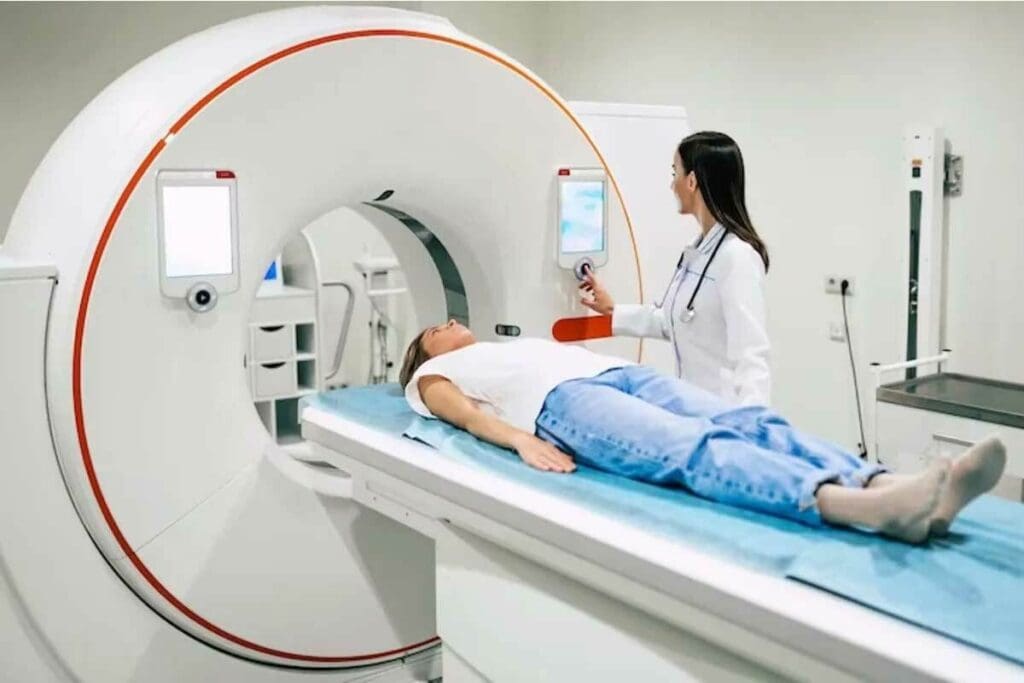
Medical diagnostics have seen big leaps forward with the creation and development of positron emission tomography (PET). The history of positron emission tomography dates back to the 1950s, with early innovations in detecting positron emissions to visualize metabolic activity. This method uses special substances to see how our bodies work, helping doctors understand changes in our metabolism.
In the early 1950s, scientists first showed how positron-emitting radiotracers could help in medical imaging. This breakthrough changed how we diagnose diseases. We’ll look at important moments and the people who made PET scans better.
At Liv Hospital, we aim for the best in healthcare. We make sure PET scan technology keeps improving. This way, we help patients get the best care and trust in our services.
Key Takeaways
- Understanding the historical context of PET scan development
- Recognizing the significance of PET scans in modern diagnostics
- Identifying key milestones in PET scan technology advancement
- Acknowledging the role of innovators in shaping PET scan history
- Liv Hospital’s commitment to excellence in PET scan technology
The Origins of Positron Emission Tomography

To understand PET, we must go back to the basics of science. We explore how positrons and radiation detection work. The story of PET starts with physics discoveries from the early 20th century.
Understanding the Positron: Scientific Foundations
In 1928, Paul Dirac suggested the existence of positrons, the electron’s opposite. Carl Anderson proved them real in 1932. This was key for PET imaging.
Early Radiation Detection Methods
The early days of detecting radiation were vital for PET. The Geiger counter was invented early in the 20th century. Later, scintillation detectors and gamma cameras helped detect gamma rays from positron annihilation.
Conceptual Beginnings of Emission Tomography
In the 1960s, combining images from different sources was explored. This was before CT machines existed. David Kuhl and Michel Ter-Pogossian were key in developing this idea. They helped create modern PET scanners.
| Year | Event | Contributor |
| 1928 | Proposal of positron existence | Paul Dirac |
| 1932 | Experimental confirmation of positrons | Carl Anderson |
| 1960s | Conceptual beginnings of emission tomography | David Kuhl, Michel Ter-Pogossian |
Milestone 1: Pioneering Work in the 1950s
In the 1950s, scientists started working with positron-emitting radiotracers. This was the start of PET scan development. It was a key time for the technology that would later be vital in medical imaging.
First Experiments with Positron-Emitting Radiotracers
The first tests with positron-emitting radiotracers were important. They showed how these tracers could help see metabolic processes in the body. Researchers found that PET technology had great promise for the future.
A pioneer in the field said, “The development of PET technology was a gradual process that relied on the convergence of several scientific disciplines.”
“The ability to image metabolic processes in vivo was a significant breakthrough, giving new insights into the human body.”
Early Clinical Imaging Potentials
PET technology had huge early clinical imaging possibilities. Researchers saw it as a tool for diagnosing and treating diseases. It could help find conditions like cancer early.
Technical Limitations of First-Generation Equipment
The first PET equipment had big technical issues. Its resolution and sensitivity were not as good as today’s. Improving these areas was a key focus for future research.
| Technical Aspect | First-Generation PET | Modern PET |
| Resolution | Low | High |
| Sensitivity | Limited | Advanced |
The 1950s were a critical time for PET technology. As we kept innovating and solving problems, PET scans became essential in medical diagnostics.
Milestone 2: David Kuhl and the Birth of Emission Reconstruction Tomography
David Kuhl’s research was a big step forward. David Kuhl played a central role in advancing PET technology through his work.
Kuhl’s Groundbreaking Research
David Kuhl worked on making images from emission data. His research started the path for today’s nuclear medicine. He created algorithms to make accurate images from emission tomography scanners.
Development of SPECT Principles
Kuhl also helped with Single Photon Emission Computed Tomography (SPECT). SPECT shows how well a patient’s body is working. The SPECT/CT has made nuclear medicine scans more accurate.
Laying the Groundwork for PET Technology
Kuhl’s work wasn’t directly on PET, but it helped a lot. His ideas were used by others to improve PET scanners.
David Kuhl’s work was key in medical imaging, including PET. His work helps doctors diagnose better and care for patients.
Milestone 3: The Ter-Pogossian Era and Technological Breakthroughs
Michel Ter-Pogossian’s time in PET scan history is marked by major tech leaps. He made big improvements in the PET scanner’s abilities and speed.
Michel Ter-Pogossian’s Contributions
Michel Ter-Pogossian was key in boosting PET tech. He worked on making PET scanners better and more useful. Ter-Pogossian’s work led to better detectors and algorithms, making images clearer.
Washington University Research Team
The Washington University Team, led by Ter-Pogossian, was vital in PET tech growth. They built the PETT scanner, a big step in using PET in hospitals. Their work improved PET tech and showed its medical uses.
Development of the PETT Scanner
The PETT scanner was a major tech leap. It was one of the first real uses of PET imaging. The PETT scanner’s design had new features like multiple detector rings, making it better. It paved the way for today’s PET/CT scanners.
Some key features of the PETT scanner included:
- Multiple detector rings for improved sensitivity
- Advanced reconstruction algorithms for enhanced image quality
- Innovative design allowing for transaxial tomography
Milestone 4: Phelps and Hoffman – The Fathers of Modern PET
In the history of Positron Emission Tomography, Michael Phelps and Edward Hoffman are key figures. Their work has greatly improved diagnostic imaging.
Revolutionary Approach to PET Imaging
Michael Phelps made big strides in PET imaging. He improved scanner technology and algorithms for better images. His work led to modern PET scanners that show more detail and are more accurate.
Technical Innovations in PET
Edward Hoffman’s work was vital in creating the ECAT scanner. This was a big step forward in PET technology. It allowed for more precise and detailed scans.
The ECAT Scanner Development
The ECAT scanner was a breakthrough in PET history. It combined advanced computers with new detector systems. This led to better image quality and more accurate diagnoses.
| Feature | ECAT Scanner | Previous PET Scanners |
| Image Resolution | High | Low-Moderate |
| Diagnostic Accuracy | Improved | Limited |
| Scanning Time | Faster | Slower |
Michael Phelps and Edward Hoffman’s work in PET technology has made a big difference. Their innovations keep improving PET scanning today. This helps doctors diagnose better all over the world.
Milestone 5: Commercial PET Scanners of the 1970s
The 1970s saw the first commercial PET scanners hit the market. This was a big leap forward in medical imaging. It moved from research tools to devices ready for clinical use.
First Market-Ready PET Systems
Several major players in medical imaging introduced the first commercial PET scanners. These systems were the result of years of hard work. Companies like EG&G ORTEC (now part of PerkinElmer) and EMI Limited (later part of Thorn EMI) led the way.
Technical Specifications and Capabilities
The first commercial PET scanners had better resolution and sensitivity than earlier models. They featured:
- Multiple detector rings to boost sensitivity
- Advanced algorithms for clearer images
- Wider fields of view for whole-body scans
Early Adoption Challenges in Clinical Settings
Introducing these scanners to clinics was tough. High costs, specialized training needs, and access to radiopharmaceuticals were big hurdles. But as technology improved and prices fell, PET scanners became more common in clinics.
The arrival of commercial PET scanners in the 1970s was a major milestone. It set the stage for the advanced diagnostic tools we use today.
The Complete History of Positron Emission Tomography Development
The journey of Positron Emission Tomography (PET) has been incredible. It started with simple beginnings and grew into a key tool in medicine today.
Timeline of Critical Discoveries
PET’s history is filled with important discoveries. These milestones have shaped the technology and its use in medicine.
- The discovery of positrons and the understanding of their role in radiation.
- Early experiments with positron-emitting radiotracers in the 1950s.
- The development of emission reconstruction tomography by David Kuhl.
- Technological breakthroughs achieved by Michel Ter-Pogossian and his team.
These discoveries have been key in making PET technology what it is today.
Evolution of Scanner Designs
PET scanner designs have changed a lot over time. Early scanners were simple but have evolved into advanced tools.
- The first-generation PETT (Positron Emission Transaxial Tomography) scanners.
- The introduction of the ECAT (Emission Computed Axial Tomography) scanner.
- Modern PET/CT and PET/MRI hybrid systems that combine functional and anatomical imaging.
These updates have made PET scans more accurate and useful.
Key Patents and Intellectual Property Milestones
PET technology has led to many patents and important discoveries. These patents cover key areas like:
- The development of Bismuth Germanate (BGO) crystals for detectors.
- Advances in Time-of-Flight (TOF) PET technology.
- Innovations in PET/CT and PET/MRI integration.
These patents have helped protect innovations and encouraged more research in PET technology.
Milestone 6: PET-CT Integration in the 1990s
In the 1990s, PET-CT integration changed the game. It combined PET and CT scans into one. This new tech gave doctors detailed images and info on how the body works, making diagnoses better.
Combining Anatomical and Functional Imaging
PET and CT scans were merged to get both body structure and function info at once. This made diagnoses more accurate. It was a big win for cancer care, helping doctors stage tumors and track treatments.
Technical Challenges Overcome
Creating PET-CT scanners was tough. It needed to sync PET and CT data, handle image quality differences, and fuse images well. Shimadzu was key in solving these problems, making PET-CT a reality.
Clinical Impact of Hybrid Imaging
PET-CT has changed patient care a lot. It gives doctors both body structure and function information in one scan. This has helped detect and stage cancers better, guide treatments, and track how well treatments work. It’s a big step towards better diagnostic tools.
The 1990s PET-CT integration was a breakthrough. It improved diagnosis and opened doors for more hybrid imaging innovations.
Milestone 7: Detector Material Revolution – BGO and Beyond
Advances in detector materials, like BGO, have greatly improved PET scans. New materials have made PET scans more sensitive and accurate. This has changed how we diagnose diseases.
Bismuth Germanate (BGO) Implementation
Bismuth germanate (BGO) is key in PET detector tech because it’s very good at detecting gamma rays. BGO’s dense crystal structure helps absorb gamma rays well. This makes images clearer. BGO has made PET scans much more accurate.
Advances in Crystal Technology
New crystal technologies have made PET scans even better. Innovations in crystal manufacturing have improved light yield and speed. This leads to sharper images and better health outcomes.
Impact on Scan Quality and Patient Experience
Using BGO and other advanced materials has greatly improved scans and patient care. Patients now have shorter scans and more accurate diagnoses. Doctors also get clearer images. We’re seeing even more improvements in detector tech for the future.
Modern PET Technology and Applications
Today, PET scans can show images in amazing detail, thanks to new tech. Modern positron emission tomography equipment has changed how we do diagnostic imaging. It gives us more precise and confident diagnoses.
Whole-Body PET Scanning Capabilities
One big step forward in PET tech is whole-body PET scanning. It lets us see the whole body in one go. This is super helpful for finding cancer and checking how treatments are working.
Whole-body PET scanning is now a key tool in medicine. It gives doctors a clear view of how diseases spread and grow.
Time-of-Flight PET Advancements
Time-of-flight PET is another big leap in PET tech. It measures when photons arrive, making images clearer and more accurate. This is really helpful for people with bigger bodies.
Current Market Size and Growth
The market for positron emission tomography equipment is getting bigger. This is because more people want better diagnostic tools. As health systems update, the market size of PET tech will keep growing.
Things like new tech, more chronic diseases, and the need for PET scans in making decisions are driving this growth. In short, modern PET tech has made huge progress. It offers whole-body PET scanning and time-of-flight PET for better care. As the market grows, we’ll see even more improvements in healthcare.
Conclusion: The Enduring Legacy of PET Innovation
The history of positron emission tomography shows our endless drive for medical progress. It has grown from simple beginnings to a key part of today’s medical world.
PET scans have changed how we see inside the body. They help doctors find and treat diseases better. Looking back, it’s clear that PET scans are essential for patient care.
The PET scan market keeps growing. This is thanks to better scanners, materials, and ways to see inside the body. PET tech will keep leading in medical breakthroughs, helping patients everywhere.
Looking ahead, PET’s impact will only grow. It will keep shaping how we diagnose and treat diseases for many years.
FAQ
When was the PET scan developed?
The PET scan was developed in the 1970s. The first commercial PET scanners were introduced during this time.
Who invented the PET scanner?
Several researchers contributed to the development of the PET scanner. Michel Ter-Pogossian, Michael Phelps, and Edward Hoffman are key pioneers in this field.
What is the history of positron emission tomography?
The history of PET started in the 1950s with early experiments. The technology improved in the 1970s and 1990s. This included the first commercial PET scanners and the combination of PET and CT scans.
What is the significance of PET scans in medical diagnostics?
PET scans are vital in medical diagnostics. They help doctors see how the body works, like glucose use and blood flow. This is key for diagnosing and treating many diseases, including cancer and heart disease.
What are the technical specifications of modern PET scanners?
Modern PET scanners have advanced features. They have high-resolution detectors and new algorithms. These improvements allow for detailed whole-body scans and hybrid imaging like PET-CT.
What is the current market size and growth of the PET scan industry?
The PET scan industry has grown a lot in recent years. This growth is due to more demand for imaging services, better technology, and wider medical uses.
What is the role of bismuth germanate (BGO) in PET technology?
Bismuth germanate (BGO) is used in PET detectors. It’s known for its high density and sensitivity. Using BGO has made PET scans better, with clearer images and faster scans.
How has PET technology evolved?
PET technology has changed a lot. It started with early experiments and grew to include commercial scanners and hybrid imaging. These changes have made scans better and more useful in medicine.
What are the benefits of combining PET and CT scans?
Combining PET and CT scans gives a full view of medical conditions. It helps doctors make more accurate diagnoses and care for patients better. This also makes clinical work more efficient.
References:
- Jones, T. (2017). History and future technical innovation in positron emission tomography. European Journal of Nuclear Medicine and Molecular Imaging, Retrieved from https://pmc.ncbi.nlm.nih.gov/articles/PMC5374360/


