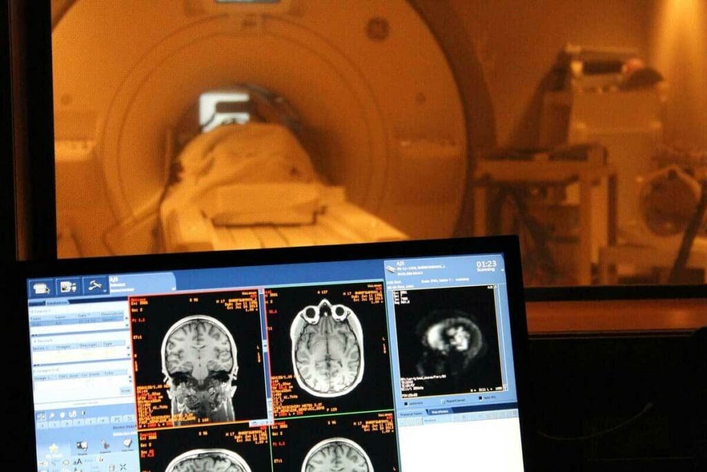
Understanding the Difference Between fMRI and PET Scans
Choosing between fMRI and PET is key for good brain checks. At Liv Hospital, we know how important it is to make smart choices in brain scans.
Functional MRI (fMRI) shows brain activity by looking at blood oxygen changes. Positron Emission Tomography (PET) uses radioactive tracers to see how cells work. Knowing how these tools differ helps doctors give better care.
We’ll look at the main differences between fMRI and PET. We’ll talk about their special uses and benefits in brain scans. This guide will help you choose the right tool for your needs when comparing fmri vs pet.
Neuroimaging has changed how we see the human brain. We’ve seen big steps forward in brain imaging, from CT and MRI to fMRI, PET, and EEG. These tools help us see brain areas, check their health, and watch how they work.
The history of brain imaging is filled with new ideas. Early methods laid the groundwork for later ones. Functional MRI (fMRI) and Positron Emission Tomography (PET) are key, letting us see brain activity live. A study on the National Center for Biotechnology Information shows how these tools have improved our brain understanding.
Brain imaging has grown a lot, changing neuroscience. Now, we can map brain connections and see how it reacts to things.
Functional imaging is key in today’s medicine, mainly for brain disorders. fMRI and PET help find brain problems and track disease changes. They’re vital for doctors and researchers.
fMRI detects changes in blood oxygen levels to show how our brains work. It’s a key tool in neuroscience. This method is non-invasive and has changed brain research and diagnosis a lot.
The BOLD effect is the core of fMRI. It’s based on oxygenated and deoxygenated hemoglobin’s different magnetic properties. When brain areas are active, they use more oxygen. This leads to more blood flow and a change in oxygen levels.
This change is what fMRI detects. It lets us see which parts of the brain are active.
fMRI tracks neural activity by looking at blood oxygen changes. Active neurons need more oxygen, which brings more blood. The BOLD signal shows this activity.
This method is great for studying the brain and finding out which areas do what.
To do fMRI scans, you need a few things. A strong magnetic field, usually 1.5 Tesla or more, is key. You also need special radiofrequency coils to pick up brain signals.
The scanner must have advanced software to handle the scan data. This is important for getting accurate results.
| Technical Requirement | Description | Importance |
| Magnetic Field Strength | 1.5 Tesla or higher | High |
| Radiofrequency Coils | Sophisticated coils for signal detection | High |
| Software Capabilities | Advanced data processing | Critical |
In summary, fMRI is a powerful tool for brain studies and diagnosing diseases. It’s non-invasive and shows detailed brain activity. This makes it very useful in research and medical settings.
PET scanning is a powerful tool that uses radioactive tracers to see and measure brain functions. It has changed neuroimaging, giving us new insights into brain work and metabolism.
At the core of PET scanning are radioactive tracers, or radiopharmaceuticals. These tracers join in the body’s metabolic processes, showing us different physiological activities. The most used tracer in brain PET scans is Fluorodeoxyglucose (FDG), a glucose molecule with a radioactive fluorine atom.
As the brain uses glucose for energy, the FDG tracer builds up in active areas. This buildup sends signals to the PET scanner.
The heart of PET scans is positron emission. When the tracer decays, it releases a positron, the opposite of an electron. This positron meets an electron, causing them to annihilate and release gamma photons.
The PET scanner catches these gamma photons. It uses them to make detailed images of the tracer in the brain.
PET scanning needs special equipment and places. The PET scanner is a big, doughnut-shaped machine with many detector rings. These detectors catch the gamma photons from positron annihilation.
Also, PET scanning needs a cyclotron or access to radiopharmaceuticals. A team of experts, like radiologists and nuclear medicine technologists, is required.
The tech needed for PET scanning includes advanced image software and a clean environment. This shows how complex and advanced PET scanning is.
Knowing the differences between fMRI and PET is key for picking the right neuroimaging method. Both have changed neuroscience, giving us new views on the brain.
fMRI and PET are both top tools in neuroimaging, but they shine in different ways. fMRI gives us detailed brain maps without using radiation. This makes it great for studying how the brain works and its connections. PET, on the other hand, shows us how cells are working, which helps in finding and tracking diseases like cancer and brain disorders.
Choosing between fMRI and PET depends on what you’re studying or treating. fMRI is best for mapping the brain and studying thinking because it shows detailed brain activity. PET is better for looking at how cells are working, which is key for spotting diseases early, like Alzheimer’s or cancer.
fMRI and PET have greatly changed how we do research and treat patients in neuroscience. By picking the right tool, we can learn more about the brain and diseases. This helps us make better diagnoses and treatments.
By using the best of both fMRI and PET, we can keep pushing neuroscience forward and help patients more.
It’s important to know how fMRI and PET differ in spatial and temporal resolution. This knowledge helps pick the right neuroimaging method for research or medical use. These differences affect the data collected and insights from studies.
Functional MRI (fMRI) beats PET in spatial detail, pinpointing brain activity more accurately. This precision helps researchers pinpoint brain areas for various tasks and conditions. For example, fMRI has mapped brain areas for language and motor control.
fMRI shines in spatial detail but lags in temporal resolution, taking seconds to show changes. PET, though, can track some metabolic processes quickly, though not as fast as EEG. “Choosing between fMRI and PET depends on the study’s focus,” says a neuroscientist.
It’s vital to think about these aspects when planning studies.
The resolution differences between fMRI and PET affect how accurate diagnoses are. For instance, fMRI’s sharp detail is great for planning brain tumor surgeries. It helps avoid damaging nearby brain areas.
PET, on the other hand, shows metabolic changes early, helping spot diseases like Alzheimer’s. Knowing each method’s strengths helps doctors choose the best tool for each case.
In summary, picking between fMRI and PET depends on the study’s needs. Using each technique’s strengths can deepen our brain function understanding and boost diagnostic accuracy.
Choosing between fMRI and PET for brain imaging depends on radiation exposure. These technologies handle radiation differently, affecting safety.
PET scans use radioactive tracers to see how the body works. These tracers are mostly safe but expose patients to radiation. This raises concerns about long-term health risks, like cancer.
To lower these risks, PET centers follow strict rules. They make sure patients get the least amount of radiation needed for diagnosis.
Precautions for PET scans include careful screening to avoid too much radiation. This is important for pregnant women, breastfeeding moms, and kids. Centers also follow rules to keep doses low while keeping images clear.
fMRI is different because it doesn’t use radiation. It uses magnetic fields and radio waves to see brain activity. This makes it safer for those who need many scans or are worried about radiation.
fMRI’s non-invasive nature boosts safety and allows for more flexible scans. This is great for studies that need many scans without worrying about radiation buildup.
Both fMRI and PET have safety steps. For fMRI, checking for metal implants and watching for claustrophobia are key. For PET, it’s about keeping radiation doses low.
Common safety steps include checking patients before scans, watching them during, and having emergency plans ready. These steps help make imaging safe and effective for patients.
In summary, fMRI is a safer choice than PET for many patients, thanks to its non-invasive nature. Knowing these differences helps in making better choices for brain imaging.
Beyond their technical capabilities, the practical considerations of fMRI and PET are key. These include cost and accessibility. We will look at these factors to see how they affect research and clinical practice.
fMRI needs special MRI equipment and places that can handle strong magnetic fields. PET scans require a cyclotron for making radioactive tracers and facilities for preparing them. The setup needed for both is big, affecting how easy they are to use.
fMRI Facility Requirements:
PET Facility Requirements:
The costs of running fMRI and PET are different. fMRI costs less to run because it doesn’t need radioactive tracers. PET, on the other hand, has ongoing costs for the cyclotron, making tracers, and disposing of waste.
| Cost Factor | fMRI | PET |
| Initial Investment | High (MRI scanner) | Very High (Cyclotron, PET scanner) |
| Operational Costs | Lower (Maintenance, staffing) | Higher (Tracers, cyclotron operation) |
| Patient Costs | Generally lower | Generally higher due to tracer costs |
For more detailed information on PET scanning, including its costs and operational requirements, visit RadiologyInfo.org.
Availability and Wait Times for Procedures
The availability of fMRI and PET services is limited by the need for special equipment and staff. fMRI is more common because MRI tech is widespread. PET, needing a cyclotron and radiopharmacy, is less common and has longer wait times.
“The choice between fMRI and PET often depends on the specific clinical or research question, as well as practical considerations such as availability and cost.”
In conclusion, the practical aspects of fMRI and PET, like facility needs, costs, and availability, are key. Understanding these aspects is vital for using them well in both research and clinical settings.
In the world of neuroimaging, fMRI and PET are leaders. They have changed medicine a lot. Each has its own special uses in different medical situations.
PET scans are key in fighting cancer. They help find tumors, see how big they are, and check if treatments are working. This is because they show how active cells are, which is a sign of many cancers.
Cancer Detection: PET scans are a big help in finding and understanding many cancers. This includes lymphoma, melanoma, and colorectal cancer.
fMRI is great at showing how the brain works. It helps check brain functions and activity. This is very important before surgery and for understanding brain problems.
Functional Mapping: fMRI helps find out which brain parts do what. This is useful for planning surgeries and studying the brain.
Both fMRI and PET are important for brain and nervous system problems. PET shows how cells are working, while fMRI looks at brain activity and connections.
Knowing what each can do helps doctors choose the right tool for each patient.
It’s key to know how fMRI and PET differ in data analysis. Both have changed neuroscience a lot. But they measure and analyze data in different ways.
PET scans can directly measure things like glucose use or blood flow. This is thanks to radioactive tracers. PET’s ability to quantify makes it great for tracking diseases like cancer.
In PET scans, how much tracer is taken up shows how active a tissue is. This helps doctors understand and plan treatments.
fMRI uses stats to figure out brain activity from blood oxygen changes. It analyzes data with complex models to find active brain areas. But, fMRI’s stats need careful handling to avoid mistakes.
New methods like machine learning are being used to improve fMRI’s accuracy. They help spot patterns that old methods miss.
PET and fMRI both struggle with understanding and standardizing data. PET’s tracer uptake and scanner setup can impact its accuracy. fMRI’s results can vary due to different scanners and analysis methods.
To fix these issues, there’s a push for standardizing data collection and analysis. This includes creating guidelines and using standards to ensure scanner consistency.
Knowing the strengths and weaknesses of each method helps us better understand neuroimaging results. This knowledge is vital for making informed decisions in both medical and research fields.
Choosing between fMRI and PET for clinical use requires a deep understanding of each. We must weigh their strengths and weaknesses to pick the best imaging method for each case.
When creating diagnostic tools, we focus on the clinical question at hand. fMRI is great for mapping brain functions and assessing cognitive tasks because it offers detailed brain activity insights. PET, on the other hand, is best for checking metabolic activity, which is key for spotting cancer and tracking treatment effects.
Choosing between fMRI and PET should follow evidence-based guidelines and clinical standards. We also need to think about what imaging options are available and the skills of the healthcare team.
| Imaging Modality | Primary Use | Key Advantages |
| fMRI | Functional mapping, cognitive assessment | High spatial resolution, non-invasive |
| PET | Cancer detection, metabolic disorders | High sensitivity to metabolic changes, quantitative data |
When deciding on imaging, we must think about the patient’s unique needs. Patient comfort and safety are top priorities, and the imaging choice should match the patient’s situation.
We also look at the patient’s medical history and what we’re trying to diagnose. For example, PET might be better for tracking cancer treatment in patients with a cancer history.
In some cases, using both fMRI and PET together can be beneficial. For example, fMRI can show active brain areas, and PET can look at metabolic changes in those spots.
This combined method can give a fuller picture of complex conditions. It helps make better treatment choices and improves patient care.
As we learn more about the brain, fMRI and PET will keep being key in research and treatment. The differences between these technologies are big, from how they work to how they’re used in medicine.
Functional neuroimaging has changed how we diagnose and plan treatments. fMRI shows brain function without using radiation, making it great for brain checks. PET scans, though, help see how cells work, helping find and check cancer.
The future of brain imaging looks bright, with these tools getting even better. This will help doctors get better at diagnosing and treating brain problems. As we learn more, these tools will become even more important in brain research.
Knowing the good and bad of fMRI vs PET helps doctors make better choices. This leads to better care for patients. The growth in brain imaging will keep changing how we study and treat brain diseases.
fMRI and PET scans work differently. fMRI looks at brain activity by watching blood flow changes. PET scans use radioactive tracers to see how cells work.
fMRI has better detail than PET. It shows brain structures and activity more clearly.
PET is better for finding cancer and metabolic issues. It spots areas with high activity.
PET scans use radioactive tracers, which can be risky. fMRI is safer because it doesn’t use radiation.
Costs and access vary. fMRI is more common in hospitals. PET needs a special machine to make tracers.
Yes, using both fMRI and PET together can give a fuller picture. This is helpful in complex cases.
fMRI is great for mapping the brain. It shows blood flow changes in real-time.
PET uses direct measurements of tracer uptake. fMRI uses stats to figure out brain activity from BOLD signal changes.
The choice depends on the study’s goals. It’s about what part of the brain you want to study and the detail needed.
Yes, some conditions are better studied with one over the other. For example, fMRI is good for connectivity. PET is better for metabolic activity in diseases.
Subscribe to our e-newsletter to stay informed about the latest innovations in the world of health and exclusive offers!