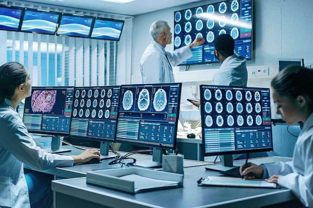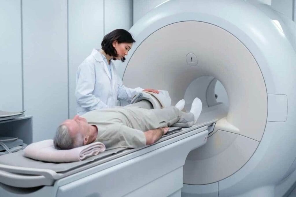
We are on the cusp of a medical breakthrough. It’s rooted in the pioneering work on positron emission tomography (PET). The PET scan technology has changed diagnostics. It lets healthcare professionals see inside the human body.
The first PET camera for humans was made in 1973. Edward Hoffman, Michael M. Ter-Pogossian, and Michael E. Phelps did it at Washington University. This was a big step in PET imaging history. It opened doors for better healthcare.
As we look into the history of PET scans, we’ll see important milestones. We’ll learn about the people who made this technology. And how it has helped patients.
Key Takeaways
- The first PET camera was developed in 1973 by Edward Hoffman, Michael M. Ter-Pogossian, and Michael E. Phelps.
- PET scan technology has revolutionized diagnostics and multidisciplinary healthcare.
- The development of PET imaging has enabled healthcare professionals to gain valuable insights into the human body.
- PET scans have become a vital tool in modern medical imaging.
- The history of PET scans is marked by significant milestones and innovations.
The Evolution of Medical Imaging Before PET

Medical imaging has seen a lot of changes over the years. It started with X-rays and has grown to include more advanced techniques. These tools have greatly helped us understand the human body.
Early Diagnostic Imaging Technologies
Wilhelm Conrad Roentgen discovered X-rays in 1895. This was a big step in medical imaging. It led to the creation of fluoroscopy, which lets doctors see inside the body in real time.
Later, ultrasound and CT scans were developed. Each of these technologies brought new ways to diagnose diseases.
Fluoroscopy helped doctors see how organs move and helped with some procedures. Ultrasound uses sound waves to show what’s inside the body. It’s very useful for checking on babies and heart health.
The Limitations of Static Imaging Methods
Even with these new tools, there were big problems. Early X-rays and CT scans could only show what the body looked like at one moment. They couldn’t show how the body works or what’s happening inside.
Doctors had to guess how things were working based on what they saw. This wasn’t always accurate and made it hard to plan treatments.
The Need for Functional Metabolic Imaging
Doctors needed a better way to see how the body works. They wanted a tool that could show not just what the body looks like but also how it’s functioning.
This need led to the creation of Positron Emission Tomography (PET). PET scans would change how doctors diagnose diseases by showing how the body’s cells are working.
The Conceptualization of Positron Tomography Emission in the 1950s

The 1950s were a key time for PET technology. It was a time that changed how we diagnose diseases. This period was important for the start of modern medical imaging.
Theoretical Foundations of Positron Physics
Positron physics was the basis for PET. Positrons, the opposite of electrons, were first thought of by Paul Dirac in 1928. Scientists soon realized that positrons could be used for medical imaging.
When a positron meets an electron, they destroy each other, releasing energy. This energy can be detected.
Understanding positrons was not just about them. It was also about creating math models for their behavior in materials, like human tissue.
Early Experiments with Positron Detection
In the 1950s, scientists started to detect positrons. A famous medical expert was a key figure in this area. He looked into using positron detection for medical imaging.
“The detection of positrons and their application in medical imaging represented a significant advancement in our ability to understand and diagnose diseases at a molecular level.”
These early experiments showed that positrons could be detected well enough for medical imaging.
First Attempts at Translating Theory to Practice
Turning positron theory into real imaging tech was hard. The first steps were making detectors that could catch the photons from positron and electron interactions.
| Year | Milestone | Researcher(s) |
| 1950 | Proposal of positron detection for medical imaging | Dr. Gordon Brownell |
| 1953 | First experiments with positron detection | Dr. William Sweet |
The early 1950s were full of important steps in PET tech development. Many researchers helped it grow.
The path from idea to use was long and hard. But the 1950s were key to the PET scans we use today.
Pioneering Work: David E. Kuhl’s Foundational Contributions
David E. Kuhl was a true pioneer in the field of medical imaging. His work in the 1950s and 1960s was key to creating the PET scan technology we know today. His research laid the foundation for Positron Emission Tomography (PET).
Kuhl’s Innovations in Tomographic Imaging
David E. Kuhl made big strides in tomographic imaging. He worked on improving how we can see inside the body. His focus on better image reconstruction was essential for PET technology.
Thanks to his work, doctors could diagnose diseases more accurately. This brought us closer to the PET scan technology that changed diagnostic medicine.
The Collaborative Efforts of Luke Chapman and Roy Edwards
Kuhl didn’t work alone. He teamed up with Luke Chapman and Roy Edwards. Together, they sped up the creation of PET scan technology. They combined their knowledge of nuclear medicine and imaging.
Their teamwork was key to solving the technical hurdles of making a working PET scanner.
Early Prototype Development and Testing
Building the first PET scan prototypes was a major step. Kuhl and his team faced many challenges but kept pushing forward. They designed and tested these early versions.
| Year | Milestone | Contributors |
| 1950s | The theoretical foundations of PET were laid | David E. Kuhl, others |
| 1960s | Innovations in tomographic imaging | David E. Kuhl, Luke Chapman, Roy Edwards |
| Early 1970s | Development of early PET prototypes | David E. Kuhl, collaborators |
The table shows the important milestones in PET technology. It highlights David E. Kuhl and his team’s contributions.
Critical Advancements in the 1960s
The 1960s were key for PET imaging. This decade saw big research efforts. These efforts helped create the advanced PET technology we use now.
Gordon Brownell and William Sweet’s Research at Massachusetts General Hospital
Gordon Brownell and William Sweet worked at Massachusetts General Hospital. They improved early PET systems. Brownell was key in making detectors and data processing better.
Brownell said, “PET technology needed better detectors and data analysis.” This shows the big challenges early PET researchers faced.
Technical Challenges in Early Positron Imaging
Early PET imaging had big technical challenges. Detectors were not very sensitive. Also, making images from data was hard.
| Challenge | Description | Impact on PET Development |
| Limited Detector Sensitivity | Early detectors had a limited ability to detect positron annihilation events. | Reduced image quality and accuracy. |
| Complex Image Reconstruction | The process of reconstructing images from detected events was complex. | Required significant computational resources and sophisticated algorithms. |
The Precursors to Modern PET Technology
The 1960s were important for PET technology. Innovations in design, data, and image making helped a lot. Gordon Brownell and William Sweet were key in solving these problems.
Looking back, the 1960s were vital for PET scans. Their work has shaped today’s technology. It also keeps influencing new PET tech and uses.
Who Invented the PET Scan: The Breakthrough of 1973
In 1973, a breakthrough in medical tech was made by Michael E. Phelps, Michel Ter-Pogossian, and Edward Hoffman. They created the first working PET scanner. This changed how doctors could see inside the body.
The Collaborative Genius
Phelps, Ter-Pogossian, and Hoffman worked together to solve PET tech’s big problems. Their skills in physics, engineering, and medicine helped make a system that could see body processes well.
The hexagonal detector array was a big part of their success. It made the PET scanner better at showing detailed images.
Development of the Revolutionary Hexagonal Detector Array
The hexagonal detector array was a key part of the PET scanner’s improvement. It lets the team get more data, making images clearer and more accurate.
PETT III: The First Clinically Viable PET Scanner
The PETT III scanner was the first to be used in hospitals. It was a big step forward in medical imaging. It helped doctors diagnose and track many conditions better.
| Feature | PETT III | Previous PET Scanners |
| Detector Array | Hexagonal | Linear or Circular |
| Sensitivity | Significantly Improved | Limited |
| Clinical Viability | First Clinically Viable | Experimental |
The creation of PETT III was a major achievement. It led to PET tech becoming a key part of medical imaging. Now, PET scans help diagnose and treat many diseases, like cancer and brain disorders.
The Evolution of Positron Emission Tomography PET Through the Decades
Over the years, PET has grown a lot. This is thanks to better detectors, computers, and ways to make images. Now, PET is a key tool in medicine, helping doctors diagnose and treat diseases.
Key Technological Improvements: 1970s-1990s
In the early days of PET, detector technology improved a lot. New materials and designs made PET scanners more sensitive and clear. For example, switching to BGO scintillators made detectors work better.
| Decade | Technological Advancement | Impact on PET Technology |
| 1970s | Introduction of BGO scintillators | Improved detector efficiency and image quality |
| 1980s | Advancements in detector array design | Enhanced sensitivity and resolution |
| 1990s | Development of new reconstruction algorithms | Better image quality and reduced noise |
Computational Advancements and Image Quality Enhancements
Computers have also played a big role in improving PET images. Faster computers and better algorithms have led to clearer, less noisy images. This is thanks to new ways of making images, like iterative reconstruction.
Dr. Michael E. Phelps, a PET pioneer, said, “Computers and algorithms have made PET useful in clinics.” This shows how important new tech is for PET’s growth.
Commercial Development and Industry Adoption
As PET got better, it became more available. At first, scanners were made for research, but soon, companies started making them. This made PET easier for hospitals to use.
By the late 1990s and early 2000s, PET was widely used. It was paired with other imaging, like CT, making it even more useful. This led to the creation of hybrid imaging systems.
Today, PET keeps getting better. Scientists are working on new tracers, detectors, and image-making methods. As it evolves, PET will likely become even more important in fighting diseases.
Understanding How PET Imaging Works
PET imaging detects positrons from radiotracers in the body. It shows how the body’s tissues and organs work. This technology has changed how doctors diagnose diseases.
The Science Behind Positron Electron Tomography
PET imaging finds positrons, the opposite of electrons. When a positron meets an electron, they destroy each other. This creates two gamma photons that the PET scanner catches.
Key Components of PET Imaging:
- Radiotracers: Substances that emit positrons, used to trace metabolic processes.
- PET Scanner: The device that detects the gamma photons emitted during positron-electron annihilation.
- Image Reconstruction: The process of creating detailed images from the detected annihilation events.
Radiotracers and the Critical Role of 2-Fluorodeoxyglucose (FDG)
Radiotracers are key in PET imaging. 2-Fluorodeoxyglucose (FDG) is a common one, used a lot in cancer research. It acts like glucose and shows where cells are most active.
| Radiotracer | Application | Metabolic Process |
| 2-Fluorodeoxyglucose (FDG) | Oncology, Neurology | Glucose Metabolism |
| Oxygen-15 Water | Cardiology | Blood Flow |
| Fluorodopa | Neurology | Dopamine Metabolism |
Image Reconstruction Techniques and Interpretation
The PET scanner’s data is turned into images with special algorithms. These images show how active different parts of the body are. Doctors need to know a lot to understand these images.
PET imaging is very important in healthcare. It helps find and manage many diseases. It’s used for cancer and heart problems, among others.
Global Adoption and Standardization of PET Technology
PET scans are used all over the world. This shows how far medical imaging has come. The standardization of PET technology has helped it spread globally.
Regulatory Approvals and Clinical Implementation
The journey to global use started with strict regulatory approvals. In the United States, the FDA was key in approving PET scans for different uses. These approvals made PET scans legit and helped get insurance coverage.
As PET technology became more accepted, setting up standard protocols was vital. These included rules for patient prep, image taking, and how to read the data.
Development of International Protocols and Standards
Working together internationally helped create PET standards. The Society of Nuclear Medicine and Molecular Imaging (SNMMI) led in making guidelines. These ensure PET imaging quality is the same everywhere.
These standards cover making and using radiotracers, scanner quality checks, and how to interpret images.
Training and Education in PET Emission Tomography
Training and education are key to PET technology’s global use. These programs help many professionals, from doctors to researchers.
There’s been a big push for more education. This includes workshops, online courses, and certifications. They aim to improve skills in PET imaging.
Clinical Applications of Modern Pet Imaging
PET scans are now used in many medical areas. They help doctors understand the body’s metabolic processes. This is key to diagnosing and treating diseases.
Oncology: Revolutionary Impact on Cancer Detection and Staging
PET imaging has changed how we find and understand cancer. 2-Fluorodeoxyglucose (FDG)-PET spots active cancer cells early. This helps in diagnosing cancers like lymphoma and melanoma.
It also lets doctors see how well treatments are working. This is important for catching cancer early.
Neurology: Brain Function Assessment and Dementia Diagnosis
PET scans are key in studying the brain. FDG-PET shows how the brain uses glucose. This helps in diagnosing dementia and Alzheimer’s.
They also help with brain tumors and epilepsy. This information is vital for planning surgeries and treatments.
Cardiology: Heart Function and Myocardial Viability
In cardiology, PET scans check the heart’s health. Rubidium-82 PET looks at blood flow to the heart. This helps find heart disease and see if the heart muscle is working.
This info is key to choosing the right treatment for heart patients.
Emerging Applications in Other Medical Fields
PET imaging is also used in other areas. It’s being studied for looking at inflammation and infections. This could lead to new ways to diagnose and treat diseases.
As technology gets better, we’ll see more uses of PET imaging. This will help doctors better diagnose and treat complex diseases.
The Development of Hybrid Technologies and Future Innovations
PET has changed how we diagnose diseases. It has led to big steps forward in medical imaging, thanks to hybrid technologies. Positron Emission Tomography (PET) is a key player in this area.
PET-CT systems are a big deal in medicine. They mix PET’s ability to see how tissues work with CT’s view of body structures. This mix makes diagnosis more accurate.
The Integration of PET-CT Systems: A Diagnostic Revolution
PET-CT systems are vital in fighting cancer, studying the brain, and heart health. They combine PET’s metabolic insights with CT’s detailed images. Key benefits include:
- Enhanced diagnostic accuracy
- Improved cancer staging and monitoring
- Better assessment of neurological disorders
- More accurate evaluation of cardiac viability
PET-MRI and Other Advanced Combinations
PET-MRI is another hybrid technology. It merges PET’s metabolic views with MRI’s soft tissue details. This gives us a deep look into tissue health and structure.
Recent studies show that ET-MRI is great for brain and cancer studies. It helps doctors make better treatment plans.
Recent Advancements in PET Technology
PET tech has improved a lot in recent years. Better materials, algorithms, and designs have boosted its sensitivity and image quality.
Some notable advancements are:
- Time-of-Flight (TOF) PET, which improves image quality and cuts down on noise
- Advanced algorithms that make images clearer and more accurate
- New materials that make PET more sensitive and cheaper
The Future Frontier of Positron Tomography
The future of PET looks bright. We expect more integration with other imaging and better radiotracers. These changes will help PET reach more patients and improve care.
PET’s future is exciting. We might see:
- Personalized medicine with custom-made radiotracers
- Artificial intelligence to analyze images better
- Even better scanners for clearer and more detailed images
Conclusion: The Enduring Legacy of PET Scan Innovation
We’ve looked into the amazing history of Positron Emission Tomography (PET) scans. This technology has changed how we diagnose and treat diseases. It has greatly helped doctors and patients.
Researchers like David E. Kuhl, Michael E. Phelps, and Michel Ter-Pogossian made big steps in PET technology. Their work has helped us understand the body better. This means we can find and treat serious diseases sooner.
PET scans are now a key part of healthcare and keep getting better. Looking ahead, PET technology will keep being a big help in fighting diseases. It will improve how well patients do and their quality of life. We’re building on what these pioneers started, making medical imaging and diagnostics even better.
FAQ
What is Positron Emission Tomography (PET)?
PET is a medical imaging method. It shows how active tissues and organs are. This helps doctors diagnose and treat diseases like cancer, brain disorders, and heart issues.
Who are the pioneers behind the development of PET scan technology?
Key figures like David E. Kuhl and Michael E. Phelps helped create PET scans. They worked on making PET scans useful for medical use.
What was the significance of the 1973 breakthrough in PET technology?
In 1973, Michael E. Phelps and others made the first working PET scanner. This was a big step forward for PET technology.
How does PET imaging work?
PET imaging uses a radioactive tracer to find active areas. It shows detailed images of how tissues and organs work.
What are the clinical applications of modern PET imaging?
Today, PET scans help in many areas, like cancer, brain, and heart diseases. They help doctors diagnose and track diseases better.
What are hybrid PET technologies, and how have they advanced medical imaging?
Hybrid PET technologies, like PET-CT and PET-MRI, mix PET’s function with other imaging’s detail. This makes diagnosis more accurate and complete.
When was the PET scan invented?
The idea of PET scans started in the 1950s. The first working PET scanner was made in 1973.
What is the role of radiotracers in PET imaging?
Radiotracers, like 2-Fluorodeoxyglucose (FDG), are key in PET scans. They help find active areas, making detailed images possible.
How has PET technology evolved over the decades?
PET technology has grown a lot. It’s gotten better detectors and algorithms. Now, there are hybrid PET systems, making it even more useful.
References
- Hoffman, E. J., Ter-Pogossian, M. M., & Phelps, M. E. (1973). Development of the first human PET scanner. This major milestone is documented in historical reviews and memorial articles. Retrieved from https://pmc.ncbi.nlm.nih.gov/articles/PMC3653214/


