Last Updated on November 27, 2025 by Bilal Hasdemir
Medical imaging has changed how we diagnose and treat injuries over the last 30 years.Tools like X-rays, CT scans, and MRIs each have their own uses and strengths.

ct scan vs mri vs xray
X-rays give quick, 2D images that are great for finding bone fractures. But, they can’t see tendon or ligament damage. Other imaging tools can show more about soft tissue injuries.
Key Takeaways
- Knowing the differences between X-Ray, CT scan, and MRI is key for accurate diagnosis.
- X-rays are best for finding bone fractures.
- Other imaging tools can spot soft tissue injuries.
- Choosing the right imaging tool helps in quick recovery.
- Liv Hospital focuses on patient-centered care.
Understanding Medical Imaging Technologies
Medical imaging has changed how we diagnose diseases. It lets doctors see inside the body clearly. We use different imaging methods to find and treat health problems.
The Evolution of Diagnostic Imaging
Diagnostic imaging has grown a lot. Wilhelm Conrad Röntgen discovered X-rays in 1895. Later, CT scans and MRI came along. These tools help us find and treat injuries and diseases better.
“The development of new imaging technologies has transformed the field of medicine, enabling healthcare providers to offer more accurate diagnoses and targeted treatments.”
Radiologists highlight
The Role of Imaging in Injury Detection
Medical imaging is key in finding injuries. It helps doctors see problems in bones, soft tissues, and organs. With different methods, doctors can:
- Check how bad an injury is
- Decide the best treatment
- Watch how an injury heals
X-rays are often used for bone fractures. MRI is better for soft tissue injuries. CT scans give detailed images of complex injuries.
| Imaging Modality | Primary Use | Key Benefits |
| X-Ray | Bone fractures, foreign objects | Quick, widely available, low cost |
| CT Scan | Complex injuries, internal injuries | Detailed cross-sectional images, fast scanning |
| MRI | Soft tissue injuries, certain bone conditions | High-resolution images of soft tissues, no radiation |

X-Ray Technology: Capabilities and Limitations
It’s important to know what X-Ray technology can and can’t do. X-Rays are key in medical imaging, helping to spot bone injuries.
How X-Ray Imaging Works
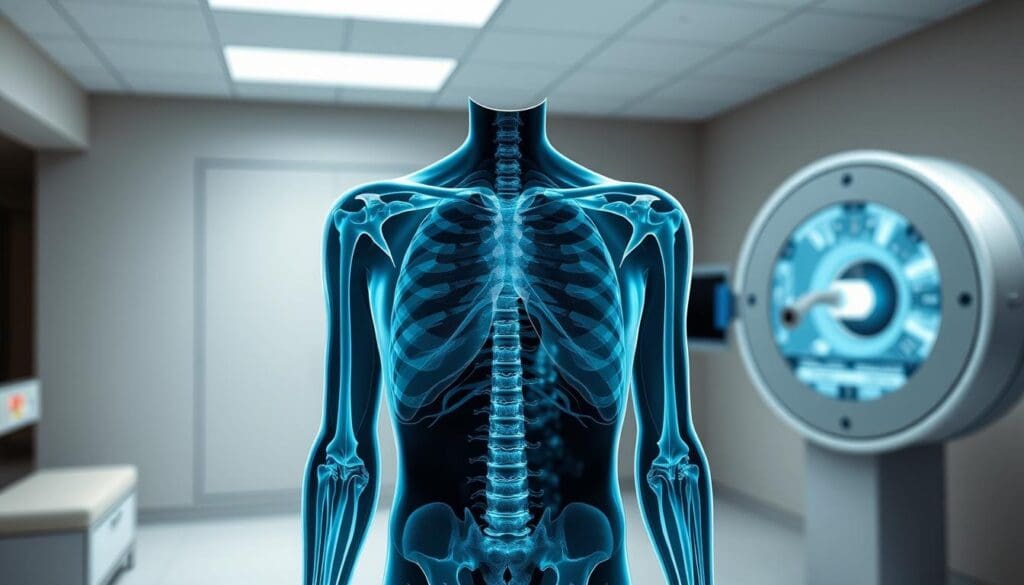
X-Ray imaging uses beams that go through the body. Tissues absorb these beams differently, making it possible to see inside. X-Ray technology is great for bones because they block more X-Rays than soft tissues.
We use X-Rays to check for fractures, dislocations, and bone problems. It’s fast, taking just a few minutes, and is found in many hospitals.

What X-Rays Can and Cannot Detect
X-Rays are good at finding bone fractures and foreign objects. They can also spot some soft tissue issues. But, they can’t see ligaments and tendons well because bones block more X-Rays.
- X-Rays can spot bone fractures and dislocations.
- They can find some soft tissue problems, like calcifications.
- But, they’re not good for checking soft tissues closely.
Do X-Rays Show Ligament Damage?
Ligament damage is hard to see on X-Rays. But signs like joint instability might hint at a problem. To really know, you might need an MRI.
It’s best to talk to your doctor about what imaging test you need. If you think you have ligament damage, more tests might be needed.
“The choice of imaging modality depends on the clinical context and the specific injury being evaluated.” – A specialist notes.
Can X-Rays Show Tendon Damage?
It’s important to know what X-Rays can and can’t do when it comes to tendon damage. Tendon injuries are common, and X-Rays are often the first choice for imaging. But, they’re not the best at showing soft tissues like tendons.
Doctors use different imaging tools to find and treat injuries. But, X-Rays are mainly good for bones, not soft tissues. Tendon damage needs more detailed imaging.
Why Tendons Are Not Visible on X-Rays
Tendons are soft tissues that X-Rays can’t show well. This is because X-Rays mainly capture bone images. The technology works by passing through the body, and soft tissues like tendons don’t block X-Rays as much as bones do.
“X-Rays are not effective in visualizing soft tissues like tendons and ligaments,” says a key fact about X-Ray technology’s limits in diagnosing injuries.
When Additional Imaging Is Necessary
When tendon damage is thought of, we need more imaging. MRI (Magnetic Resonance Imaging) and ultrasound are good for this. MRI is great at showing soft tissues, like tendons and ligaments, giving detailed images for diagnosis.
For suspected tendon injuries, we often suggest more imaging. As said,
“The diagnostic pathway for suspected injuries often involves a combination of clinical assessment and imaging studies.”
This way, patients get the right care for their injury.
Knowing X-rays’ limits in showing tendon damage helps us choose the best imaging for patients.
CT Scan Technology: Advanced X-Ray Imaging
CT scan technology is a big step forward in medical imaging. It gives detailed views of the body’s inside. This tech is key for spotting complex injuries and conditions.
The Science Behind CT Scanning
CT scanning combines many X-ray images from different angles to show the body’s inside. This process involves rotating an X-ray source and detectors around the patient, capturing data that is then turned into images.
The science of CT scanning is based on X-ray absorption. Different tissues absorb X-rays at different levels. This helps doctors see different parts of the body.
3D Reconstruction and Cross-Sectional Views
CT scans are great because they show 3D reconstructions and cross-sectional views of the body’s inside. This is super helpful for checking complex fractures, vascular diseases, and soft tissue injuries.
By looking at the body in slices, doctors can understand injuries or diseases better. This helps them plan the right treatment.
Soft Tissue Visualization Capabilities
CT scans are not just for bones; they’re also good at showing soft tissues. By changing the imaging settings, CT scans can tell different soft tissues apart.
- CT scans can see organs and soft tissue structures.
- They help find soft tissue injuries and diseases.
- Contrast agents make some structures more visible.
In summary, CT scan technology has changed how we diagnose with detailed, cross-sectional views of the body. Its 3D and soft tissue imaging skills make it a top tool in medicine today.
MRI Technology: Magnetic Resonance Imaging Explained
MRI technology is a key part of medical imaging. It helps us see complex injuries clearly. MRI gives us detailed views of the body’s soft tissues, which are vital for making accurate diagnoses.
How MRI Uses Magnets and Radio Waves
MRI machines use strong magnets and radio waves to show us what’s inside the body. First, they align hydrogen atoms with a magnetic field. Then, they use radio waves to disturb these atoms and catch signals. These signals help create detailed images.
“The MRI’s ability to differentiate between various soft tissues is unparalleled,”a leading radiologist notes. “It’s a game-changer for diagnosing injuries that are not visible on X-rays or CT scans.”
Superior Soft Tissue Imaging
MRI is great at showing soft tissues like tendons, ligaments, and organs. This is key for spotting issues like tendonitis, ligament sprains, and joint disorders.
Detecting Tendon and Ligament Injuries
MRI is super helpful for finding tendon and ligament injuries. These are hard to spot with other methods. MRI lets doctors see these soft tissues clearly, helping them plan better treatments.
In sports medicine, MRI is often used to check for ACL tears or tendon ruptures. This allows for quick and right treatment.
We see MRI as essential in today’s diagnostics. It greatly improves our ability to give accurate and effective care for soft tissue injuries.
CT Scan vs MRI vs X-Ray: The 7 Key Differences
Medical imaging technologies like CT scans, MRI, and X-Rays have changed how we diagnose diseases. Each technology works differently. Knowing these differences helps healthcare providers and patients make better choices.
Difference #1: Imaging Technology
X-Rays, CT scans, and MRI use different ways to create images. X-Rays use ionizing radiation to see bones and dense tissues. CT scans combine X-Rays and computer tech for detailed cross-sections of the body.
MRI uses powerful magnets and radio waves to show both bones and soft tissues in great detail.
Difference #2: Radiation Exposure
Another big difference is radiation levels. X-Rays and CT scans use ionizing radiation. CT scans give more radiation because they use many X-Ray beams. MRI, being radiation-free, is safer for those needing many scans or are sensitive to radiation.
Difference #3: Image Detail and Resolution
Each technology shows different levels of detail. CT scans show more than X-Rays, great for complex bones. MRI, with its high resolution, is best for soft tissues like tendons and organs.
Difference #4: Soft Tissue Visualization
MRI is the best for seeing soft tissues. It clearly shows tendons, ligaments, and other soft tissues. X-Rays and CT scans can’t show these as well. MRI is key for diagnosing soft tissue injuries.
Understanding these differences helps healthcare providers pick the right imaging for each patient. This ensures accurate diagnoses and effective treatments.
Bone Injury Detection: Which Imaging Method Is Best?
Choosing the right imaging method is key for accurate diagnosis and treatment of bone injuries. The injury’s type and severity determine the best imaging modality.
Simple Fractures: When X-Rays Are Sufficient
For simple fractures, X-rays are usually the first choice. They are quick, easy to get, and show bone integrity well. X-rays work best for fractures in arms and legs but don’t show soft tissue damage well.
Key benefits of X-rays for simple fractures include:
- Rapid results
- Low cost compared to other imaging modalities
- Wide availability in emergency settings
Complex Fractures: The Value of CT Scans
For complex fractures, CT scans are essential. They give detailed views and 3D reconstructions. This helps surgeons plan the best treatment. CT scans are great for fractures in hard-to-reach areas like the spine, pelvis, or joints.
“CT scans provide a level of detail that is not available with conventional X-rays, making them indispensable for complex fracture assessment.”
As noted by medical imaging experts.
When MRI Might Be Needed for Bone Injuries
While X-rays and CT scans are good for bones, an MRI is needed for soft tissues. MRI shows soft tissues like ligaments, tendons, and muscles. It can spot bone bruises, hidden fractures, and soft tissue injuries.
Reasons to choose MRI for bone injuries include:
- Detailed visualization of soft tissues
- Ability to detect bone marrow edema and occult fractures
- Assessment of ligament and tendon injuries
In conclusion, picking the right imaging method depends on the injury’s details. Knowing each method’s strengths helps healthcare providers make the best choices for patients.
The Difference Between X-Ray and CT Scan
It’s important to know the difference between X-Ray and CT scan for accurate injury diagnosis. Both are used in medical imaging but serve different purposes. They offer unique benefits.
From 2D to 3D: The Fundamental Distinction
X-Rays give 2D images, good for initial checks of bone fractures and lung issues. CT scans, on the other hand, create 3D images by combining X-rays from different angles. This 3D view helps see complex injuries and internal structures better.
For complex fractures or internal injuries, CT scans are better. They provide a detailed view.
Radiation Dose Considerations
CT scans use more radiation than X-Rays. This is because they take many X-Ray images to make a 3D image. But, new CT technology has made them safer by reducing radiation doses.
When to Choose CT Over X-Ray
The choice between X-Ray and CT scan depends on the situation. CT scans are better for:
- Detailed views of complex structures
- Checking internal injuries or bleeding
- Guiding treatments or interventions
| Imaging Modality | Dimensionality | Radiation Dose | Clinical Use |
| X-Ray | 2D | Low | Initial assessment of fractures, lung conditions |
| CT Scan | 3D | Higher | Complex injuries, internal bleeding, detailed structural assessment |
In conclusion, X-Ray and CT scan are both important for diagnosis but differ in many ways. Knowing these differences helps healthcare providers choose the best imaging for each patient.
Alternatives to MRI for Patients with Contraindications
Some patients can’t have an MRI because of claustrophobia or medical implants. We know not everyone can get an MRI. It’s important to have other ways to diagnose.
CT Arthrography: An Alternative for Joint Imaging
CT arthrography is a good choice when MRI isn’t possible. It’s great for looking at joints. You get a contrast material in the joint, then a CT scan.
This method shows the joint’s details well. It’s perfect for checking joint injuries or problems like ligament tears. It’s a top pick when MRI isn’t an option.
Ultrasound for Soft Tissue Evaluation
Ultrasound imaging is another good choice. It uses sound waves to see inside the body. It’s great for soft tissue injuries like tendonitis or muscle tears.
Ultrasound is safe, easy, and not too expensive. It’s also good for guiding injections or other procedures.
Options When Claustrophobia or Implants Prevent MRI
For those who can’t do MRI because of claustrophobia or implants, there are other ways. We look at each patient’s situation to find the best option.
- CT scans are good for many things, like checking bone fractures or soft tissue issues.
- X-rays are often used first for bone injuries or conditions.
- Ultrasound is flexible and works for many soft tissue checks.
We help patients find the best imaging method for them. This ensures they get the best care possible.
Clinical Decision Making: Choosing the Right Imaging Test
Choosing the right imaging test is key for good patient care. We take a careful and informed approach to pick the best imaging method.
The Diagnostic Pathway for Suspected Injuries
The path to diagnosing injuries starts with the patient’s symptoms and medical history. Effective clinical decision making combines clinical wisdom and guidelines to find the right imaging test.
For example, X-rays are often first for suspected bone fractures because they’re cheap and easy to get. But for soft tissue injuries or complex fractures, advanced imaging techniques like MRI or CT scans are needed for a detailed look.
| Imaging Modality | Typical Use Cases | Advantages |
| X-Ray | Bone fractures, foreign objects | Quick, low cost, widely available |
| CT Scan | Complex fractures, internal injuries | High detail, 3D reconstruction |
| MRI | Soft tissue injuries, tendon and ligament damage | Superior soft tissue visualization |
Cost-Effectiveness Considerations
Cost is a big factor in choosing an imaging test. We aim to find the best balance between getting accurate results and keeping costs down. Often, the first test should give enough info to decide on treatment.
Patient-Specific Factors in Imaging Selection
Each patient’s unique situation affects the imaging choice. For instance, some patients with metal implants can’t have MRI scans. This means we need to find other ways to image them.
By thinking about these factors and using our knowledge, we can choose the best imaging tests for our patients. This improves care quality and efficiency.
Advanced Imaging at Liv Hospital: Commitment to Quality Care
At our hospital,we prioritize exceptional care through our cutting-edge imaging technology. We never stop improving to give you the best diagnosis and treatment plans.
State-of-the-Art Imaging Technologies
We use the latest MRI and CT scans to see inside your body clearly. Our top-notch gear lets us get super sharp images. This means we can spot problems fast and often avoid more tests.
Our imaging tech has some cool features:
- High-resolution imaging: Our gear gives us clear pictures for accurate diagnoses.
- Advanced software: We use smart software to make images even clearer and help with tough diagnoses.
- Fast scanning times: Our tech scans quickly, making it less uncomfortable for you and helping us move faster.
Patient-Centered Approach to Diagnostic Imaging
Our well-trained staff at Liv Hospital puts your comfort and safety first when you get scanned. We make sure you get care that fits your needs and situation.
We do this by:
- Compassionate staff: Our team is trained to be kind and caring, making you feel more at ease.
- Clear communication: We make sure you know what’s happening and what you need to do before your scan.
- Comfortable facilities: Our scanning rooms are cozy and calm, helping to reduce your stress and anxiety.
Innovation in Medical Imaging Practices
We’re always looking to improve in medical imaging, keeping up with the latest tech and methods. This means we can offer you the best care possible, using the most advanced tools.
Our commitment to innovation shows in:
- Participation in clinical trials: We join research to check out new imaging ways and tools.
- Collaboration with industry leaders: We team up with top makers to learn about new trends and tech.
- Continuous education and training: Our team keeps learning to stay up-to-date with imaging advancements.
Conclusion: Making Informed Decisions About Medical Imaging
Knowing the differences between X-Ray, CT Scan, and MRI is key. We’ve looked at what each can do and their limits. This helps us understand their roles in finding injuries in bones and soft tissues.
Choosing the right imaging test is vital for accurate diagnosis and treatment. Healthcare experts consider radiation, image detail, and soft tissue visibility. This helps them pick the best test for each patient.
Liv Hospital aims to provide top-notch healthcare. We offer international patient support and guidance. Our advanced imaging and patient-focused care ensure the best results. By making smart choices about medical imaging, we help patients get the best care possible.
FAQ
Can X-Rays detect tendon tears?
No, X-rays can’t find tendon tears. They mainly show bones, not soft tissues like tendons.
What’s the difference between X-Ray, CT scan, and MRI?
X-Rays use radiation for 2D bone images. CT scans create 3D images of bones and some soft tissues. MRI shows detailed soft tissue images, like tendons and ligaments, using magnetic fields and radio waves.
Can an X-Ray show ligament damage?
X-Rays aren’t great for seeing ligament damage directly. They’re better for bones. But, they might show signs like joint instability or bone fractures that suggest ligament issues.
Do X-Rays show tendons?
No, X-Rays can’t see tendons. They’re soft tissues that X-Rays can’t show.
Are there alternatives to MRI for diagnosing soft tissue injuries?
Yes, you can use CT arthrography or ultrasound instead of MRI. They’re good for soft tissue checks, even when MRI isn’t an option.
What’s the difference between a CT scan and an X-Ray?
X-Rays give 2D bone images. CT scans offer 3D views of bones and some soft tissues. This makes CT scans more detailed.
Can X-Rays show torn ligaments?
X-Rays aren’t the go-to for finding torn ligaments. You usually need MRI or other advanced tests for a clear diagnosis.
Is there an alternative to MRI for patients with claustrophobia or implants?
Yes, for those with claustrophobia or implants, CT arthrography or ultrasound can be good options. They help check soft tissue injuries without MRI.
How do I know which imaging test is right for me?
Choosing the right test depends on your injury, medical history, and personal factors. A healthcare professional will help decide based on these.
What are the advantages of MRI over CT scan and X-Ray?
MRI is better for soft tissue views, like tendons and ligaments. It doesn’t use harmful radiation. This makes MRI great for injury diagnosis.
References
- Siddiqui, M., et al. (2022). Magnetic Resonance Imaging Versus Computed Tomography for Diagnostic and Treatment Planning in Orthopedic Care: A Review. PMC. https://pmc.ncbi.nlm.nih.gov/articles/PMC9305220/
- Mineralized tissue visualization with MRI: Practical insights and limitations. (2024). Journal / Review Article. https://www.sciencedirect.com/science/article/pii/S2211568424002560


