Last Updated on November 27, 2025 by Bilal Hasdemir
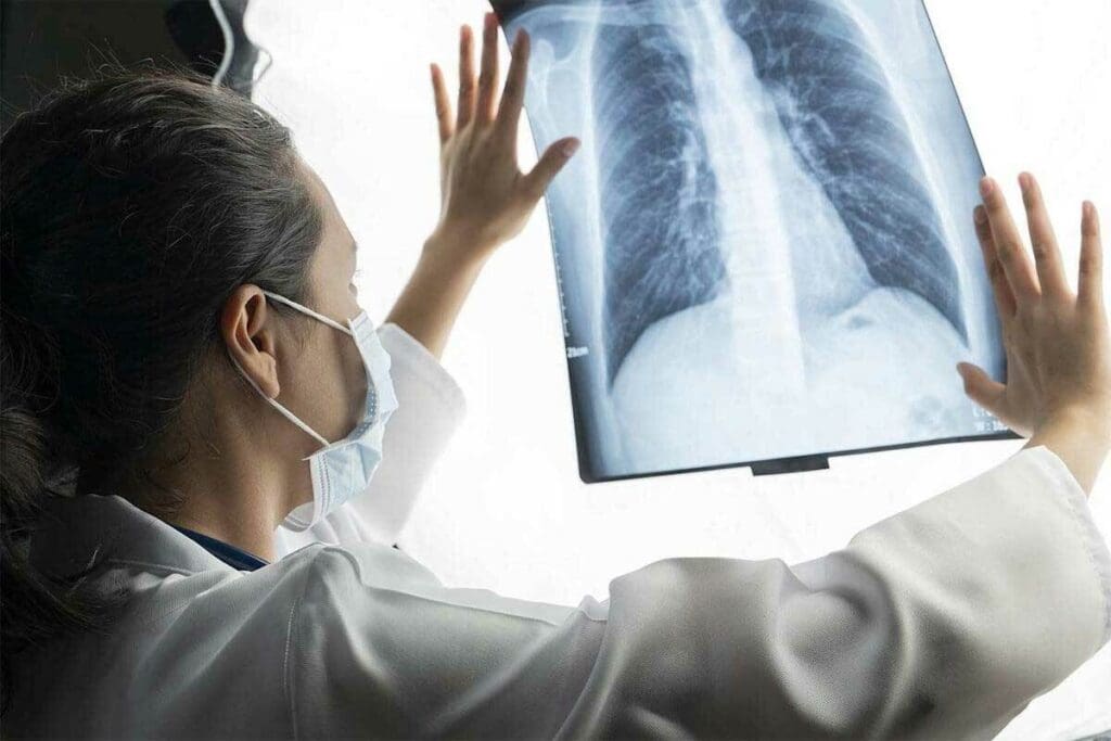
Medical imaging, like X-rays, is key for diagnosing and treating health issues. Yet, there’s worry about the risks, like cancer.
X-rays and gamma rays are ionizing radiation. Many people ask x rays cause cancer, and it is true that exposure to high doses of these radiations can increase cancer risk. It’s important to understand this risk for both patients and doctors so that medical imaging is used judiciously, balancing benefits and potential harms.
At Liv Hospital, we use safe protocols to lower radiation while getting accurate diagnoses. This way, we balance the benefits of imaging with the risks.
Key Takeaways
- X-rays are a form of ionizing radiation that can potentially increase cancer risk.
- The risk associated with X-rays depends on the dose and frequency of exposure.
- Medical imaging protocols are designed to minimize radiation exposure.
- Understanding the balance between the benefits and risks of X-rays is key.
- Liv Hospital follows international standards for safe and effective imaging.
The Science Behind X-Rays and Radiation
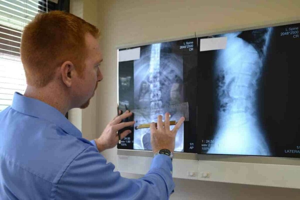
X-rays and gamma rays are types of electromagnetic radiation used in medicine. Knowing their basics helps us understand their risks and benefits.
What Are X-Rays and How Do They Work?
X-rays are a kind of ionizing radiation. They can go through soft tissues but get stopped by denser things like bone. This makes them great for medical imaging, letting us see inside the body.
When X-rays go through the body, a detector catches them. This creates an image of what’s inside.
Types of Medical Radiation: X-Rays vs. Gamma Rays
Both X-rays and gamma rays are used in medical imaging, but they come from different sources. X-rays are made by an X-ray machine. Gamma rays, on the other hand, come from radioactive isotopes.
Gamma rays have more energy and are used in cancer treatments. The main differences are:
- Energy Level: Gamma rays have higher energy than X-rays.
- Source: X-rays are produced by an X-ray machine; gamma rays come from radioactive decay.
- Application: X-rays are commonly used for diagnostic imaging; gamma rays are often used in cancer treatment.
Knowing these differences helps us see the unique risks and benefits of each type of radiation.
Radiation as a Carcinogen: The Classification Evidence
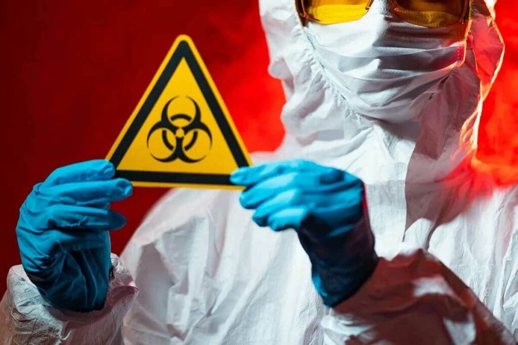
Health organizations have classified X-rays and gamma rays as carcinogens. This is based on a lot of research into how radiation affects our bodies. It shows their link to cancer.
How X-Rays and Gamma Rays Are Classified by Health Organizations
The International Agency for Research on Cancer (IARC) says X-rays and gamma rays are human carcinogens. They have a lot of evidence from studies and experiments. This shows that these rays can cause cancer in people.
The Biological Mechanism of Radiation Damage
Radiation damage happens when X-rays or gamma rays hit DNA in cells. This can lead to genetic mutations. These mutations can mess up how cells work and lead to cancer.
The way radiation damages cells is key. It can break DNA strands. If these breaks aren’t fixed, they can cause genetic changes that might lead to cancer.
This evidence shows why we need to be careful with radiation. It’s very important in medical settings where these rays are used a lot.
Can X-Rays Cause Cancer? Examining the Evidence
Looking into whether X-rays cause cancer means checking out old studies on high-dose radiation and new ones on low-dose radiation.
Historical Studies on Radiation Exposure
Old studies, like those on atomic bomb survivors, have given us a lot of information. They show a strong link between high radiation doses and more cancer.
Table: Cancer Risk Associated with Radiation Exposure
| Radiation Dose (mSv) | Cancer Risk Increase |
| 0-100 | Minimal |
| 100-500 | Low |
| 500-1000 | Moderate |
| >1000 | High |
Modern Research Findings on Low-Dose Radiation
New studies have looked at low-dose radiation, like what you get from most X-rays. They found that while the risk is small, it’s not zero. This is true for kids and people who get scanned a lot.
The research shows that high doses of radiation definitely raise cancer risk. But, the risk from low doses in X-rays is much lower. It’s something to think about, though, for people who are more at risk.
High-Dose Radiation Exposure: Clear Cancer Links
High-dose radiation exposure is linked to a higher risk of cancer. This is shown by historical events and medical studies. Studies on survivors of big radiation incidents and patients who got high doses of radiation support this link.
Lessons from Atomic Bomb Survivors
The atomic bombings of Hiroshima and Nagasaki in 1945 gave us real data on radiation effects. The survivors, called hibakusha, showed a big rise in cancer cases. This was due to the high radiation they got.
Chernobyl and Other Radiation Disasters
The Chernobyl nuclear disaster in 1986 is another key example. Those who got high doses of radiation suffered a lot. Children exposed to radioactive iodine saw a big jump in thyroid cancer. Other disasters have also shown the dangers of high radiation.
High-Dose Medical Radiation Treatments
In medicine, high-dose radiation is used to fight cancer. But, it can also raise the risk of getting another cancer. Studies on these treatments have taught us a lot about radiation’s impact on health.
| Event | Year | Primary Health Impact |
| Atomic Bombings | 1945 | Increased cancer incidence |
| Chernobyl Disaster | 1986 | Thyroid cancer increase |
| High-Dose Medical Radiation | Ongoing | Risk of secondary cancers |
The evidence from these sources clearly shows a link between high-dose radiation and cancer risk. Knowing this is key for doctors and the public.
Typical Medical X-Ray Procedures and Cancer Risk
X-rays are a common tool in medicine, but it’s important to know how they affect cancer risk.
They are used in many tests, like dental X-rays, chest X-rays, mammograms, and bone X-rays. Most studies say that typical X-rays don’t raise cancer risk much.
Dental X-Rays: Risk Assessment
Dental X-rays help find problems not seen during a regular check-up. The risk from dental X-rays is low. This is because they use little radiation and technology has improved to reduce doses.
Chest X-Rays and Mammograms: What the Data Shows
Chest X-rays and mammograms are key for spotting issues in the chest and breast cancer. Even though they use radiation, the benefits are often greater. This is true for catching problems early and treating them.
Bone X-Rays and Other Common Procedures
Bone X-rays help find fractures and other bone problems. The radiation from these tests is usually small. The benefits of diagnosing these issues are big.
To understand the risks better, look at this table comparing X-ray doses:
| Procedure | Typical Radiation Dose (mSv) |
| Dental X-ray | 0.005 |
| Chest X-ray | 0.1 |
| Mammogram | 0.4 |
| Bone X-ray | 0.001-0.01 |
In summary, X-rays do carry some risk, but their benefits are huge. The risks are usually low.
Does Gamma Radiation Cause Cancer?
Gamma radiation is a type of ionizing radiation linked to health risks, including cancer. It’s used in medicine for treatments and in industry for various tasks.
Gamma Radiation Sources in Medicine and Industry
In medicine, gamma radiation is used for cancer treatment, like gamma knife radiosurgery, and for imaging. In industry, it’s used to sterilize medical tools and food.
Gamma Radiation and Cancer: Research Findings
Studies show gamma radiation can raise cancer risk. Research on nuclear accident survivors and those exposed for medical reasons shows its dangers.
| Exposure Source | Cancer Risk | Study Findings |
| Nuclear Accidents | Increased | Studies on nuclear accident survivors show a higher incidence of cancer. |
| Medical Exposure | Variable | Risk depends on dose and duration of exposure; some studies indicate an increased cancer risk. |
| Industrial Exposure | Controlled | Strict regulations minimize exposure risks in industrial settings. |
The evidence points to gamma radiation causing cancer, mainly at high doses. But, the risk at low doses, like from medical imaging, is debated and under research.
CT Scans and Cancer Risk: Understanding the Numbers
CT scans are used more often in healthcare, and knowing their impact on cancer risk is key. They help doctors see inside the body, which is good for diagnosing many conditions. But, the radiation from these scans worries people about long-term risks.
How CT Scans Differ from Regular X-Rays
CT scans and regular X-rays are not the same when it comes to radiation. X-rays send out a focused beam, but CT scans use many beams that move around the body. This makes CT scans give out more radiation than X-rays.
Research on CT Scan Radiation Exposure
Many studies have looked into how CT scan radiation affects cancer risk. They found that CT scans can raise cancer risk, mainly in kids and young adults. One big study said about 103,000 cancers could happen because of CT scans, based on how often they’re used.
The 103,000 Future Cancers Projection: Context and Meaning
The idea that 103,000 cancers might come from CT scans is a big deal. It shows a possible rise in cancer cases from these scans. But, we must see this number in the bigger picture of cancer risk and how CT scans help doctors diagnose.
Understanding the risks and benefits of CT scans is important. While there’s a chance of more cancer, CT scans are very useful. Doctors are working to use less radiation while keeping images clear.
How Many X-Rays Cause Cancer? Dose-Dependent Risk
X-rays, like other ionizing radiation, have a cancer risk that depends on the dose. Knowing how radiation dose and cancer risk are linked is key to understanding the safety of medical imaging.
Radiation Dose Measurement and Comparison
The dose from X-rays is measured in millisieverts (mSv). A chest X-ray is about 0.1 mSv. CT scans can be 2 to 10 mSv or more, depending on the scan and body part.
This helps us see the risks of different scans. For example, the average yearly background radiation in the U.S. is 3 mSv. This helps patients and doctors understand the doses better.
Cumulative Effects of Multiple X-Ray Exposures
Getting many X-rays can increase cancer risk. The total dose over time is what matters. Important points include:
- The risk of cancer from radiation exposure is generally considered to be cumulative.
- Patients who have many scans may get more radiation.
- Doctors should keep track of a patient’s radiation history to avoid too much exposure.
Risk Thresholds and Safety Margins
High doses of radiation clearly raise cancer risk. But low doses are less understood. Key points include:
- The linear no-threshold (LNT) model is often used to estimate cancer risk from low doses of radiation.
- There is ongoing debate about the validity of the LNT model for very low doses.
- Safety margins are built into medical imaging protocols to minimize radiation exposure while maintaining diagnostic quality.
Understanding X-ray risks and using safety measures helps doctors. This way, they can use imaging while keeping risks low.
Putting X-Ray Cancer Risk in Perspective
The risk of cancer from X-rays is a concern that needs to be put into perspective. To understand this risk, it’s essential to compare it to other known risks and benefits associated with medical imaging.
Comparing to Background Radiation Exposure
One way to contextualize the cancer risk from X-rays is by comparing it to background radiation exposure. Background radiation is the level of ionizing radiation that is typically present in the environment. It’s usually measured in areas remote from any known radiation sources. The average annual background radiation exposure in the United States is about 3.1 millisieverts (mSv).
A typical chest X-ray has a dose of about 0.1 mSv. This means that a chest X-ray is equivalent to about 10 days of background radiation exposure.
Contextualizing Against the 37-44% Lifetime Cancer Risk
The lifetime risk of developing cancer for the general population is about 37-44%. When evaluating the risk from X-rays, it’s important to understand that this risk is cumulative. It should be considered in the context of the overall lifetime cancer risk.
For instance, having a few X-rays over a lifetime contributes a relatively small amount to the overall cancer risk compared to other factors.
Risk vs. Benefit Analysis in Medical Imaging
When considering medical imaging, the benefits often outweigh the risks. X-rays and other imaging modalities provide critical diagnostic information. This information can lead to timely and effective treatments.
A risk vs. benefit analysis involves weighing the cancer risk against the diagnostic benefits. In many cases, the information gained from an X-ray can be lifesaving or significantly improve treatment outcomes.
Some key points to consider in this analysis include:
- The diagnostic value of the X-ray or imaging procedure
- The presence of alternative imaging methods with lower radiation doses
- The patient’s overall health condition and risk factors
By understanding these factors and comparing the risks to background radiation and the general lifetime cancer risk, patients and healthcare providers can make informed decisions about the use of X-rays and other medical imaging.
Special Risk Considerations: Who Should Be Extra Cautious?
X-rays are usually safe, but some people need to be more careful. This is because they are more sensitive to radiation. Some groups are more at risk from X-ray exposure.
Children and Radiation Sensitivity
Children are more sensitive to radiation because their bodies are growing. This makes them more likely to be harmed by radiation. Doctors and radiologists use the lowest dose needed for X-rays on kids.
Pregnant Women and Fetal Exposure
Pregnant women need to be careful about X-ray exposure. While the risk is low, it’s best to avoid it if possible. Doctors will think carefully before doing X-rays on pregnant women.
People with Multiple Scans or Genetic Predispositions
Those who have had many X-rays or are genetically sensitive should be cautious. Getting too much radiation can harm them more. Doctors might suggest other imaging methods or careful dose control for these individuals.
| Group | Special Considerations |
| Children | Use lowest possible dose, careful dose management |
| Pregnant Women | Avoid unnecessary exposure, weigh benefits vs. risks |
| Multiple Scans or Genetic Predispositions | Consider alternative imaging, strict dose management |
Radiation Safety Measures in Modern Medical Imaging
Keeping patients safe during medical imaging is a big deal. Today’s hospitals use many ways to cut down on radiation while keeping images clear.
Minimizing Radiation Exposure in Medical Facilities
Hospitals take steps to lower radiation doses. They use the least amount of radiation needed for tests, called ALARA. This means picking the right tests and adjusting equipment to get good images with less radiation.
The American Cancer Society says new tech has helped a lot. For example, digital X-rays need less radiation than old film-based ones to get great pictures.
Technological Advances Reducing Radiation Doses
New tech has made it safer to use radiation. For CT scans, new algorithms let doctors use less radiation without losing image quality. Better detectors also mean less radiation is needed for clear images.
A study showed CT scanners with new tech cut doses by 30% in two years. This shows a big push to use less radiation in medical imaging.
Regulatory Oversight and Safety Standards
Groups like the U.S. Nuclear Regulatory Commission (NRC) make rules for safe radiation use. They cover things like keeping equipment in good shape, training staff, and watching doses.
| Regulatory Aspect | Description | Impact on Safety |
| Equipment Maintenance | Regular checks to ensure imaging devices are functioning correctly | Reduces risk of radiation exposure due to faulty equipment |
| Staff Training | Education on proper use of imaging technology and radiation safety protocols | Enhances adherence to safety protocols, minimizing exposure |
| Dose Monitoring | Tracking radiation doses to ensure they remain within safe limits | Helps in maintaining doses as low as reasonably achievable |
A top expert in radiological protection says, “The key to safety is a safety-first culture in medicine. This is backed by strong rules and new tech.”
“The cornerstone of radiation safety is a culture of safety within the medical community, supported by robust regulatory frameworks and continuous technological innovation.”
In short, combining new tech, strict safety rules, and watchful regulators has made medical imaging safer. This not only keeps patients safe but also helps doctors give better care with less risk of radiation.
Conclusion: Balancing Medical Benefits and Radiation Risks
Medical imaging like X-rays has changed healthcare a lot. They give doctors important info to help patients. But, we must think about the risks of radiation too.
X-rays and other imaging help doctors find and treat many health problems. Even though there’s a risk of radiation, studies say the good they do is often more important. This is true when used right.
Medical places need to keep using safety steps and new tech. Doctors should make smart choices about when to use these tools. This way, patients get the benefits of X-rays but with less radiation.
Using medical imaging wisely means knowing both its good and bad sides. By weighing these, doctors can give patients great care. This care is safe and effective.
FAQ
Do X-rays cause cancer?
X-rays can increase cancer risk, but the danger is low for most medical X-rays.
How many X-rays can cause cancer?
The risk from X-rays depends on the dose. It’s hard to say exactly how many X-rays are needed to cause cancer. But, getting many X-rays can raise the risk.
Are X-rays and gamma rays the same thing?
X-rays and gamma rays are both ionizing radiation. But, they come from different sources. X-rays come from machines, while gamma rays come from radioactive materials.
Can gamma radiation cause cancer?
Yes, gamma radiation can cause cancer. It’s used in medicine and industry, but it’s a known carcinogen.
How do medical facilities minimize radiation exposure?
Medical places use the least dose needed for tests. They follow safety rules and use new tech to lower doses.
Are children more susceptible to radiation-induced cancer?
Yes, kids are more at risk because their bodies are growing and they have more years to develop cancer.
Can pregnant women safely undergo X-ray procedures?
Pregnant women can get X-rays when it’s really needed. But, doctors try to keep the baby safe. They weigh the X-ray’s benefits against the risks.
How do CT scans differ from regular X-rays in terms of cancer risk?
CT scans use more radiation than X-rays, which raises cancer risk. But, they give clearer images and are used for important tests.
What is the lifetime risk of developing cancer from background radiation?
The risk of cancer from background radiation is low. Most people get about 2.4 millisieverts of it each year.
How do I know if I’ve had too many X-rays?
Doctors keep track of X-ray doses. You can ask your doctor about your radiation history.
Are there any genetic predispositions that increase the risk of radiation-induced cancer?
Yes, some genetic conditions make you more likely to get cancer from radiation. People with a family history or certain genes should be careful.
Can the benefits of medical imaging outweigh the risks?
Yes, the good from medical imaging often outweighs the bad. This is true when imaging is used wisely and safely.
Reference
- Brenner, D. J., & Hall, E. J. (2007). Computed tomography — An increasing source of radiation exposure. New England Journal of Medicine, 357(22), 2277–2284. https://pubmed.ncbi.nlm.nih.gov/18046031/


