Last Updated on October 31, 2025 by

Getting a correct prostate cancer diagnosis is key to effective treatment. For years, prostate biopsy was the main method used to detect cancer. However, new research has revealed the truth about prostate biopsy—while it’s useful, it can sometimes lead to unnecessary procedures.
That’s why MRI for prostate cancer is becoming more popular. It offers a safer and more precise approach, helping doctors decide who truly needs a biopsy. This reduces the number of unnecessary tests and potential complications.
Recent studies show that MRI can accurately detect prostate cancer, making biopsies less frequent and more targeted. This shift marks an important step forward in prostate cancer care, improving both accuracy and patient outcomes.
Knowing how prostate cancer is detected is key to treating it well. Prostate cancer is a big health issue. Finding it early is very important.
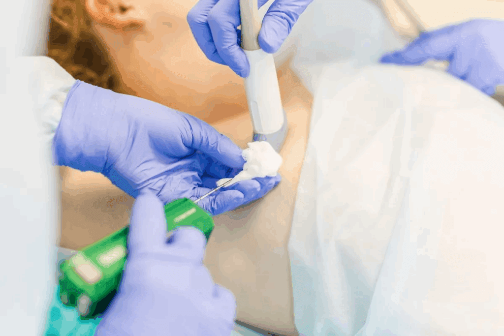
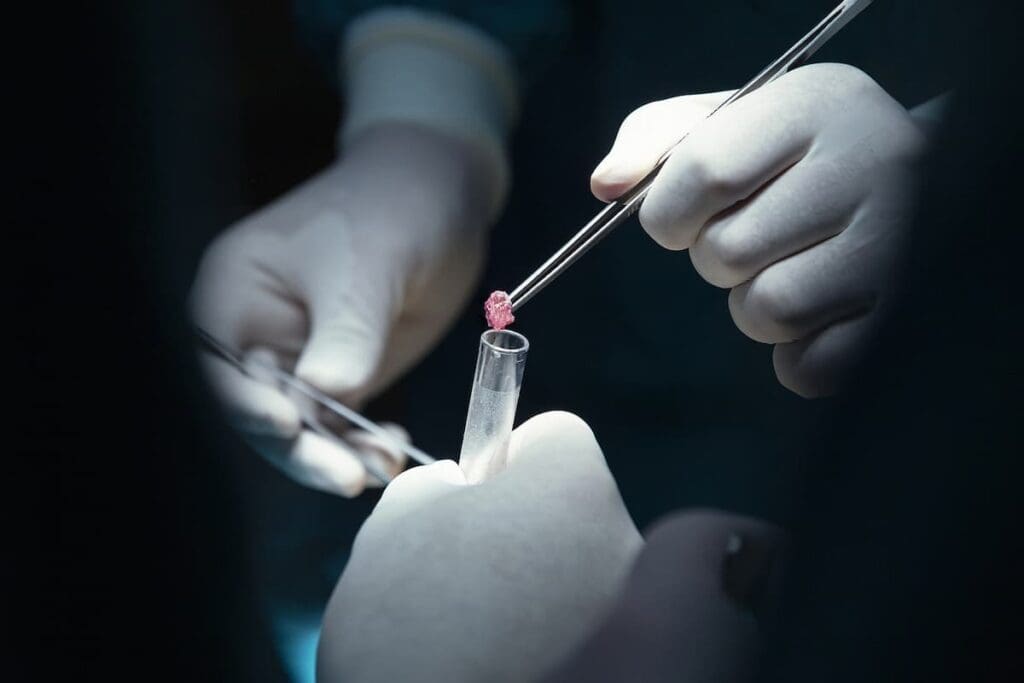
A traditional prostate biopsy takes tissue from the prostate gland with a needle. This tissue is then checked for cancer cells. The whole process is done under local anesthesia and takes samples from different parts of the prostate. The skill of the doctor and the method used affect how accurate the results are.
Before the biopsy, you might need to stop certain medicines. You might also have to prepare your bowel and take antibiotics. It’s very important to follow your doctor’s advice to make sure the biopsy goes well.
An MRI of the prostate uses strong magnets and radio waves to show detailed images of the prostate and nearby areas. This test is non-invasive and helps find signs of cancer. An MRI can give doctors important information for making treatment plans.
For an MRI, you lie on a table that moves into the MRI machine. The whole test takes about 30 to 60 minutes. Some tests might use a contrast agent to make the images clearer.

Both prostate biopsy and MRI are important for finding prostate cancer. Knowing about these tests can help you feel more ready for your diagnosis and treatment.
Knowing the limits of prostate biopsy is key to wise choices about prostate health. Biopsy is vital for diagnosis, but it has its downsides.
A standard systematic biopsy might miss cancer or not show how aggressive it is. This happens because it only looks at a small part of the prostate.
Key accuracy issues include:
Like any procedure, prostate biopsy has risks and side effects. These can be mild or serious and include:
For those over 70, the risks might be too high. Biopsies are rarely done on people over 70 because of these risks.
Experts suggest getting an MRI before a biopsy. MRI can spot areas that need a closer look during biopsy. This makes the biopsy more accurate.
An MRI-guided biopsy can:
By knowing these limits and using MRI, patients and doctors can make better choices about prostate health.
We look into how well MRI spots prostate cancer. This is key for today’s health checks. MRI is a non-invasive way to see the prostate and find tumors.
Getting the most from prostate MRI results needs a good grasp of the images. MRI scans use the Prostate Imaging-Reporting and Data System (PI-RADS). This system helps doctors understand the findings better. The PI-RADS score goes from 1 to 5, with higher numbers meaning a greater chance of cancer.
Key aspects of prostate MRI results include:
One big plus of MRI for prostate cancer is its 96% negative predictive value (NPV). This means a negative MRI is 96% sure there’s no big cancer. This is very reassuring for patients and doctors.
This high NPV is important for several reasons:
Even with its benefits, MRI has its downsides. Some issues include:
Despite these challenges, MRI is a key tool in fighting prostate cancer. New tech and better ways to read scans will make it even better.
MRI-targeted biopsy has changed how we find prostate cancer. It beats old methods in many ways. This helps patients get better care and avoid too many tests.

MRI-targeted biopsy finds serious cancers better than old biopsies. It looks at specific spots shown by MRI. This means doctors can spot cancers that need quick action.
A study on the National Center for Biotechnology Information shows MRI-targeted biopsy is very precise. It finds more cancers that need treatment.
“The integration of MRI into the diagnostic pathway has the potential to significantly improve the detection of clinically significant prostate cancer.” Doctors agree MRI-targeted biopsy is a big step forward.
MRI-targeted biopsy also finds fewer low-risk cancers. These cancers often don’t need treatment. By focusing on MRI spots, we avoid treating cancers that won’t harm us.
This approach makes care better and saves money. It’s a win for patients and the healthcare system.
MRI-TRUS fusion-guided biopsy is very successful. It uses MRI’s clear images and TRUS’s live view. This lets doctors target cancer spots very accurately.
Studies show this method works well. It’s a big step up in finding prostate cancer. MRI and TRUS together make the diagnosis more accurate and reliable.
In short, MRI-targeted biopsy is better than old methods in many ways. It finds serious cancers better and finds fewer low-risk ones. As doctors keep using this tech, patients will get even better care.
Using MRI-first pathways in prostate cancer diagnosis brings big benefits. It cuts down on unnecessary biopsies, lowers complications, and saves money. This makes healthcare better and more cost-effective.
Studies show a big drop in unnecessary biopsies by up to 41% with MRI-first. This means fewer risks, less worry for patients, and better results overall.
Looking at the costs, MRI-first pathways save a lot. They save about £75,000 a year on prostate cancer diagnosis and £11,000 on fewer complications. This smart use of resources means better care and results for patients.
A prostate MRI is a non-invasive test that uses magnetic fields to show detailed images of the prostate gland. A biopsy, on the other hand, involves removing tissue samples from the prostate for microscopic examination.
During a prostate MRI, you lie on a table that slides into a large machine. The whole process takes 30-60 minutes. Sometimes, contrast dye is used to make the images clearer.
Experts suggest getting an MRI before a biopsy. It helps spot areas of concern and might avoid unnecessary biopsies. This can lower risks and improve accuracy.
MRI is very good at ruling out significant prostate cancer. A negative MRI result can reliably show there’s no serious cancer.
MRI-targeted biopsy finds more significant cancers and misses fewer low-risk ones. It also makes cancer diagnosis more accurate.
An MRI-TRUS fusion-guided biopsy uses MRI images and ultrasound to target specific prostate areas. This improves the accuracy of tissue sampling.
While MRI is very accurate, it can’t replace biopsy yet. But it can guide biopsy decisions and cut down on unnecessary biopsies.
MRI has its limits, like false-negative results and variability in interpretation. It also needs top-notch equipment and skilled experts.
To prepare for a prostate MRI, you might need to follow dietary restrictions and avoid certain medications. Your healthcare provider will give you specific instructions for the best results.
A radiologist interprets your MRI results. They look for abnormalities and provide a report to your healthcare provider. This report might include a score on the cancer likelihood.
Subscribe to our e-newsletter to stay informed about the latest innovations in the world of health and exclusive offers!