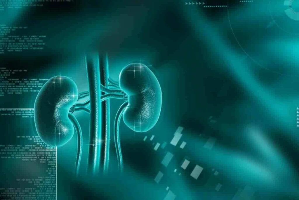
At Liv Hospital, we use advanced renal CT scans. They give us detailed images of the kidneys and urinary tract. This helps us spot and check on structural problems, masses, cysts, and signs of kidney disease. Understand what does a renal ct scan show including kidney abnormalities, stones, and contrast findings.
A CT scan of the kidneys is key for finding many kidney issues. This includes tumors, lesions, blockages from kidney stones, abscesses, polycystic kidney disease, and birth defects. Thanks to this technology, we can give our patients the best care possible.
Key Takeaways
- Detailed cross-sectional images of the kidneys and urinary tract are obtained through renal CT scans.
- Detection and assessment of structural abnormalities, masses, and cysts are possible with CT scan technology.
- Liv Hospital uses advanced renal CT scans for precise diagnosis and treatment planning.
- CT scans help identify various kidney issues, including tumors, lesions, and polycystic kidney disease.
- Accurate diagnosis through CT scans enables healthcare providers to develop effective treatment strategies.
Understanding Renal CT Scan Technology

The technology behind renal CT scans helps doctors find kidney problems accurately. CT scans use X-rays and computers to show detailed kidney images. This makes them a key tool for checking kidney health.
How CT Imaging Works for Kidney Visualization
CT imaging uses X-rays to make pictures of the kidneys. The patient lies on a table that moves into a CT scanner. The scanner spins around the patient, taking pictures from different sides.
Then, a computer puts these images together to show detailed kidney pictures.
Key components of CT imaging for kidney visualization include:
- X-ray technology to capture images from multiple angles
- Advanced computer algorithms to reconstruct detailed images
- Contrast agents (optional) to enhance the visibility of certain structures
Advantages of CT Scans Over Other Kidney Imaging Methods
CT scans have many benefits over other methods. They show more detail than regular X-rays. They can spot many kidney problems, like stones, cysts, and tumors.
| Imaging Method | Detail Level | Ability to Detect Abnormalities |
| Standard X-ray | Low | Limited |
| CT Scan | High | High |
| Ultrasound | Moderate | Moderate |
A medical expert says, “CT scans are vital in nephrology. They give unmatched detail and help diagnose better.”
“The use of CT scans in diagnosing kidney diseases has revolutionized the field, enabling earlier detection and treatment of renal conditions.”
— Senior Nephrologist
We see how important CT scan technology is for kidney health. Knowing how CT scans work and their benefits helps doctors diagnose and treat kidney issues better.
What Does a Renal CT Scan Show: A Detailed Look

A renal CT scan gives a detailed view of the kidneys. It helps doctors spot problems and check how well the kidneys are working. This scan is key for finding kidney issues like tumors, cysts, and blockages.
Detailed Anatomical Structures Visible on Kidney CT
A kidney CT scan shows important parts, including:
- The renal cortex and medulla help to check the kidney tissue
- The renal pelvis and calyces, key to urine collection and flow
- The ureters, which carry urine to the bladder
- Adrenal glands and nearby tissues
These clear images help doctors spot any oddities or changes in the kidneys. This is vital for making the right diagnosis and treatment plan.
Resolution Capabilities for Detecting Small Abnormalities
Today’s CT scanners can spot small issues in the kidneys. They can find:
- Small cysts or tumors not seen by other scans
- Small stones or calcifications causing problems
- Minor changes in kidney size or shape that hint at disease
The high detail of CT scans is great for catching early signs of kidney disease. It also helps find small stones causing pain or blockages.
Normal Kidney Appearance on CT Images
Knowing what normal kidneys look like on CT scans is key. Healthy kidneys usually appear as:
- Symmetrical, bean-shaped with smooth edges
- Even density, with the cortex being less dense than the medulla
- Normal size, about 10-12 cm long
By comparing patient images to these standards, doctors can spot any oddities. This helps them diagnose kidney problems or other issues.
Types of Renal CT Scans and Their Specific Applications
Different types of renal CT scans are used for various kidney issues. The choice of scan depends on the need, patient condition, and what doctors need to know.
Kidney CT Without Contrast: Uses and Limitations
A kidney CT without contrast is great for finding kidney stones and some other issues. It’s good when contrast can’t be used or might be harmful.
Uses: This scan is mainly for spotting kidney stones, calcifications, and some structural problems. It’s also good for patients who can’t have contrast.
Limitations: While it’s good for some things, it might not show soft tissue problems or tumors well. Contrast is needed for better views of these.
CT Scan Kidneys With Contrast: Enhanced Visualization
CT scans with contrast use a special agent to make certain kidney areas clearer.
Enhanced Visualization: The agent makes the kidney’s blood vessels and tissue stand out. This helps find tumors, cysts, and other issues. It’s also great for checking the kidney’s blood vessels.
CT Stonogram for Kidney Stone Detection
A CT stonogram is a special method for finding and figuring out kidney stones.
Diagnostic Benefits: It gives detailed images. These help figure out the stone’s size, where it is, and what it’s made of. This info is key for choosing the right treatment.
Specialized Protocols for Specific Kidney Conditions
There are special CT scan protocols for certain kidney problems, like complex cysts, tumors, or blood vessel issues.
| Condition | CT Scan Protocol | Diagnostic Benefit |
| Kidney Stones | Non-contrast CT or CT Stonogram | Accurate detection and characterization of stones |
| Renal Tumors or Cysts | CT with Contrast | Enhanced visualization of soft tissue abnormalities |
| Vascular Abnormalities | CT Angiography | Detailed imaging of renal vasculature |
Knowing about the different renal CT scans and their uses shows how versatile and useful CT technology is for kidney health.
The Role of Contrast in Kidney Imaging
Contrast agents have changed how we diagnose and treat kidney problems. They help us see kidney anatomy and function clearly. This is important for spotting issues that might not show up on regular scans.
Enhancing Visualization with Kidney Contrast CT Scan
A kidney contrast CT scan makes it easier to see kidney structures. This includes blood vessels, tumors, and cysts. The contrast agent makes these features stand out, helping us find and understand problems.
The contrast agent shows up differences in tissue density. This lets us see kidney details more clearly. It’s great for spotting small issues that might not show up on regular scans.
CT Renal With Contrast: Indications and Benefits
A CT renal with contrast is used for many kidney issues. This includes tumors, cysts, and problems with blood vessels. The main benefits are:
- It helps find small problems.
- It makes it easier to understand kidney masses.
- It shows the kidney’s blood vessels better.
These advantages lead to more accurate diagnoses and better treatment plans. This is good for patient care.
Risks and Considerations for Contrast Use
While contrast agents are mostly safe, there are risks. For example, they can cause kidney failure in some people, like those with kidney disease. It’s important to talk to your doctor about any worries before a scan.
We decide if contrast is needed based on each case. We consider the benefits and risks. People with certain health issues, like kidney disease or diabetes, need extra care.
Detecting Kidney Stones Through Specialized Imaging
CT scans are key in finding kidney stones and understanding their type. We use top-notch CT scan tech to get clear images of the kidneys. This helps us spot and treat kidney stones well.
Renal Stone CT Scan Techniques and Protocols
The renal stone CT scan is a special imaging method for finding kidney stones accurately. We follow certain steps to get the best images, making sure even tiny stones show up. “The use of CT scans for kidney stone detection has become the gold standard due to its high sensitivity and specificity,” as noted by recent medical research.
Identifying Calcifications and Their Significance
Calcifications in the kidneys often mean kidney stones. We spot these calcifications with CT scans, which show up as bright white spots. Knowing where and how big these spots are helps us figure out how serious the problem is and what treatment to use.
Assessing the significance of calcifications involves evaluating their size, number, and distribution within the kidneys. This info is key for deciding the best way to handle kidney stones.
Assessing Stone Size, Location, and Composition
Knowing the size, location, and makeup of kidney stones is important for planning treatment. CT scans help us measure stone size and find out where they are in the urinary tract. They also tell us what kind of stone it is, like calcium oxalate or uric acid, which helps us choose the right treatment.
The composition of kidney stones can significantly influence the treatment approach. For example, some stones might need special treatments or diet changes.
CT on Kidneys for Evaluating Stone-Related Complications
CT scans are not just for finding kidney stones but also for checking for complications. We use them to see if there’s hydronephrosis, where a stone blocks the flow and swells the kidney. “Early detection of such complications is critical for preventing long-term kidney damage,” say doctors.
By using CT scans to find kidney stones and check for complications, we can treat patients quickly and effectively. This improves their health outcomes.
Identifying Structural Abnormalities in the Kidneys
CT scans are key in spotting kidney problems. They help see both birth defects and issues that come later. This info is key to keeping kidneys healthy and finding the right treatment.
Congenital Kidney Variations Visible on CT Scan of Kidneys
Being born with kidney issues is not rare. CT scans can find these problems easily. These include:
- Horseshoe kidney: where kidneys join together
- Renal ectopia: kidneys not in their usual spot
- Renal agenesis: missing one kidney
These issues are often found by chance during scans for other reasons. CT scans give detailed views of the kidneys and any linked problems.
Acquired Structural Changes
Later in life, kidneys can change due to disease. CT scans are good at spotting:
- Polycystic kidney disease (PKD): lots of cysts in the kidneys
- Renal scarring: from long-term infections or reflux
- Changes from chronic blockage or inflammation
These changes can hurt how well the kidneys work. CT scans help track these issues and see how they affect the kidneys.
Hydronephrosis Detection and Evaluation
Hydronephrosis, or swelling of the kidney due to blockage, is easy to spot on CT scans. They allow for:
- Checking how much swelling there is
- Finding where and why the blockage is
- Looking at any other problems, like thinning of the kidney tissue
CT scans give all the info needed to plan the best treatment. This could be surgery or other treatments.
By finding and understanding kidney problems, CT scans are very important. They help doctors diagnose and treat kidney issues, leading to better health for patients.
Renal Mass Evaluation and Characterization
CT scans are key in figuring out what a renal mass is and what treatment it needs. We use advanced imaging to see if a mass is benign or malignant.
Differentiating Benign from Malignant Lesions
Telling apart benign and malignant lesions is very important. We use CT scans to look at the size, shape, and how they react to contrast of renal masses.
Key features that help differentiate benign from malignant lesions include:
- Size and growth pattern
- Presence of calcifications or fat
- Enhancement characteristics with contrast
- Margins and interface with surrounding tissue
By looking at these features, we can tell what a renal mass is and plan the right treatment.
Cyst Classification and Assessment on CT of Kidneys
Renal cysts are common on CT scans. We use the Bosniak classification to sort them out based on their look.
| Bosniak Category | Characteristics | Malignancy Risk |
| I | Simple cysts with thin walls, no septa or calcifications | Very low |
| II | Cysts with a few thin septa, fine calcifications | Low |
| IIF | Cysts with multiple thin septa, minimal enhancement | Moderate |
| III | Cysts with thickened irregular walls, septa with enhancement | High |
| IV | Cysts with soft tissue components that enhance | Very high |
This system helps us know which cysts need more checking or treatment.
Solid Tumor Identification Features
Solid renal tumors are a big worry. We look for signs like contrast enhancement, uneven look, and growth into nearby areas on CT scans.
Characteristics of solid renal tumors on CT scans include:
- Enhancement with contrast
- Heterogeneous appearance
- Invasion into surrounding structures
- Presence of necrosis or calcifications
Spotting these signs helps us diagnose and treat solid renal tumors right.
Vascular Assessment in Kidney CT Imaging
Vascular assessment is key in checking kidney health with CT imaging. We use top-notch CT scanning to see the kidney’s blood vessels. This helps us spot problems and check blood flow, vital for kidney disease diagnosis and treatment.
Renal Artery and Vein Visualization
CT scans with contrast show the renal arteries and veins clearly. This is key for spotting renal artery stenosis or venous thrombosis. Seeing these blood vessels well helps us find and treat kidney issues.
Detecting Vascular Abnormalities with a CT Scan for the Kidneys
CT scans can spot vascular problems like narrowed or blocked arteries. Finding these issues early is key to avoiding more kidney damage. It also helps us choose the right treatment.
Blood Flow Evaluation in Kidney Pathologies
Checking blood flow is vital for kidney health. CT scans give us insights into kidney perfusion. This helps us find areas with low blood flow, which might mean renal infarction or chronic kidney disease.
Can a CT Scan Detect Kidney Disease: Diagnostic Capabilities
We use CT scans to find signs of kidney disease, like acute and chronic conditions. CT scans are key in spotting many kidney problems.
Signs of Acute and Chronic Kidney Disease on Imaging
CT scans show several signs of kidney disease. These include:
- Changes in kidney size and shape
- Alterations in the cortical thickness
- Presence of cysts or tumors
- Calcifications or stones
- Hydronephrosis or other obstructive changes
In chronic kidney disease, CT scans might show smaller kidneys and thinner cortex. Acute kidney injury, on the other hand, can show bigger kidneys and different density.
Detecting Inflammatory and Infectious Conditions
CT scans are also great for spotting inflammatory and infectious kidney issues. These include:
- Pyelonephritis
- Renal abscesses
- Perinephric abscesses
CT imaging can show inflammation, abscesses, and other issues related to these conditions.
Limitations in Functional Assessment
While CT scans give great details about the body, they can’t measure kidney function. They don’t directly check how well the kidneys work.
So, we often use CT scans with other tests to check kidney function. These include serum creatinine levels and glomerular filtration rate (GFR) calculations.
Complementary Diagnostic Tools
Other tools are also important for checking kidney disease. These include:
| Diagnostic Tool | Information Provided |
| Ultrasound | Assesses kidney size, detects cysts, and obstruction |
| Renal Biopsy | Provides a histological diagnosis of kidney disease |
| Serum Creatinine and GFR | Evaluates kidney function |
By using CT scans with these tools, we get a full picture of a patient’s kidney health.
Interpreting Abnormal Kidney CT Scan Results
When a kidney CT scan shows abnormal results, it’s important to understand what it means. These findings can range from minor issues to serious problems that need quick attention.
Common Abnormalities on Kidney CT Scan Findings
Kidney CT scans often find cysts, stones, tumors, and other structural issues. Cysts are fluid-filled sacs that can be simple or complex. Complex cysts might suggest a higher risk of cancer.
Let’s look at some common findings and what they might mean:
| Abnormality Type | Characteristics | Potential Implications |
| Simple Cysts | Fluid-filled, thin-walled | Generally benign, may not require treatment |
| Complex Cysts | Thick-walled, possibly calcified | May indicate increased risk of malignancy, warranting further investigation |
| Kidney Stones | Calcified deposits | Can cause obstruction, pain, or infection |
| Renal Tumors | Solid masses | Potential for malignancy, requiring biopsy or surgical intervention |
Significance of Asymmetric Kidney Appearance
An asymmetric kidney appearance on a CT scan can point to several issues. These include congenital problems, scarring from infections, or blockages. It’s key to match the scan findings with symptoms and medical history to figure out the cause.
When Further Investigation Is Warranted
More tests are needed when CT scan results are unclear or don’t match symptoms. Tests like MRI or biopsy might be required to confirm a diagnosis.
Grasping the meaning of abnormal kidney CT scan results helps both patients and doctors make better choices about care and treatment.
Patient Preparation and Experience for Kidney Imaging
Knowing what to expect before, during, and after a kidney CT scan can help reduce anxiety. We’ll guide you through the steps of preparing for and experiencing a kidney CT scan.
Prep for Kidney CT Scan: Before Your Appointment
To get ready for a kidney CT scan, you’ll need to:
- Take off clothes, jewelry, or objects that might get in the way.
- Wear a hospital gown during the scan.
- Tell your doctor about any allergies or health issues.
- Maybe get contrast dye orally or through an IV to improve image quality.
What to Expect During the CT Scan of Kthe idney
During the CT scan, you’ll lie on a table that slides into a big, doughnut-shaped machine. The scan is quick, lasting just a few minutes. Our team will make sure you’re comfortable and safe.
Key aspects of the scanning process include:
| Aspect | Description |
| Positioning | You’ll lie flat on the table, possibly with your arms up. |
| Contrast Administration | Contrast might be given orally or through an IV. |
| Scanning | The table will slide into the CT scanner, and images will be taken. |
Post-Scan Care and Follow-up
After the scan, you can usually go back to your normal activities unless your doctor says not to. If contrast dye was used, you might be watched for a bit for any bad reactions.
It’s very important to follow up with your healthcare provider to talk about the scan results and what to do next.
Conclusion: The Value of Renal CT in Kidney Health Assessment
We’ve learned how renal CT scans help check kidney health. They can spot kidney problems early. This is because they use CT and MRI scans a lot.
These scans find cysts in up to 27% of people over 50. Also, 85% of solid kidney tumors are cancerous. Renal cell carcinoma, or RCC, is the most common kidney cancer and is growing more common.
A renal CT scan is done in different stages. The Bosniak system helps sort out cystic masses.
The value of renal CT is in spotting and identifying kidney masses. This includes both harmless and harmful growths. Clear-cell RCC is responsible for 90% of kidney cancer spread.
CT urography helps see if a kidney isn’t working right. It also checks for cancer spread in other parts of the body. Knowing how renal CT scans work helps us see their value in keeping kidneys healthy.
Renal CT scans are key for checking kidney health. They help find problems and guide treatment. As medical tech gets better, so will the role of renal CT scans in kidney health checks.
FAQ
What is a renal CT scan, and how does it work?
A renal CT scan is a test that uses X-rays and computer technology to show detailed images of the kidneys and urinary tract. It helps doctors check kidney health and find problems.
What can a renal CT scan show about my kidneys?
A renal CT scan can show detailed kidney structures and find small problems. It can also spot kidney disease signs like stones, cysts, tumors, and blood vessel issues.
What is the difference between a kidney CT scan with and without contrast?
A kidney CT scan without contrast uses only X-rays. A CT scan with contrast uses a special dye to make kidney structures clearer. This is better for finding blood vessel problems and kidney masses.
How does a CT scan help in detecting kidney stones?
A CT stonogram is a special CT scan for finding kidney stones. It shows stone size, location, and type, and checks for stone-related issues.
Can a CT scan detect kidney disease, and what are its limitations?
Yes, a CT scan can find signs of kidney disease, like inflammation and infections. But, it can’t fully check how well the kidneys work. Other tests might be needed for a complete check.
How do I prepare for a kidney CT scan?
To prepare for a kidney CT scan, you might need to follow a special diet or drink lots of water. You’ll also need to remove any metal items. Your doctor or the imaging center will give you specific instructions.
What can I expect during a CT scan of my kidney?
During the scan, you’ll lie on a table that moves into the CT scanner. You might need to hold your breath or stay very quiet. The scan is usually quick and doesn’t hurt.
What happens after a kidney CT scan?
After the scan, you can usually go back to your normal activities. Your doctor will talk to you about the results and what to do next.
How do CT scans evaluate renal masses and vascular abnormalities?
CT scans can tell if kidney growths are harmless or cancerous. They can also check cysts and tumors. Plus, they can see blood vessel problems and how blood flows in the kidneys.
What are the risks and considerations for contrast use in kidney imaging?
Contrast agents are usually safe but can cause allergic reactions or harm the kidneys in some people. Your doctor will weigh the benefits and risks and talk to you about any concerns.
Can an asymmetric kidney appearance on a CT scan be a cause for concern?
An asymmetric kidney appearance might mean kidney disease or other issues. It’s important to get more tests to find out what’s going on.
How does a CT scan of the kidneys help in managing kidney stones?
A CT scan can find kidney stones accurately. It shows how big they are and what they’re made of. This helps doctors decide how to treat them.
References:
- Traub-Weidinger T, Lonsdale MN, Gil-Nagel A, et al. EANM practice guidelines for the appropriate use of PET and SPECT in epilepsy. Eur J Nucl Med Mol Imaging. 2024;51(7):1891-1908. https://pubmed.ncbi.nlm.nih.gov/38393374/
- Ponisio MR, McConathy J. The Role of SPECT and PET in Epilepsy. AJR Am J Roentgenol. 2021;216(1):186-195. https://doi.org/10.2214/AJR.20.23336


