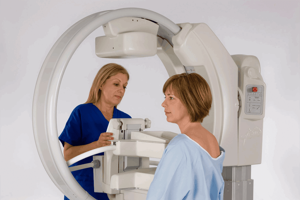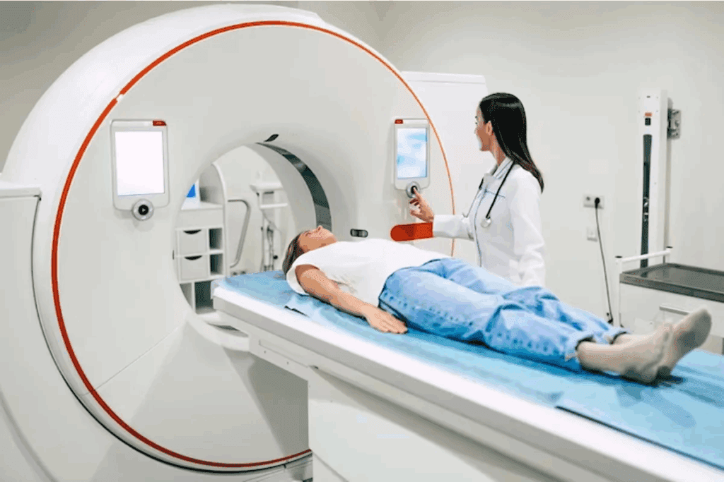
At Liv Hospital, we use the latest in diagnostics. This combines modern medicine with care focused on the patient. A gamma camera scan, or scintigraphy, is a non-invasive test. It uses a gamma camera to find gamma rays from tiny amounts of radioactive tracers given to the patient.
The National Center for Biotechnology Information says gamma cameras are key for radiologists. They let them do “Scintigraphy scans.” These scans give detailed information on how organs work. This tool is important for seeing how serious a disease is and how well treatments are working.
Key Takeaways
- Gamma camera scans are non-invasive diagnostic procedures.
- They use small amounts of radioactive tracers to visualize internal organs.
- Gamma imaging is vital for spotting disease severity.
- It helps in tracking treatment progress.
- Gamma cameras are essential tools in nuclear medicine.
The Fundamentals of Nuclear Medicine Imaging

Nuclear medicine imaging uses radioactive tracers to see how the body works. It helps us understand how organs like the heart and liver function. This is key to spotting diseases early.
This method uses tiny amounts of radioactive material to diagnose and treat diseases. It gives doctors a clear view of organ function. This helps them find problems early.
Definition and Purpose of Nuclear Medicine
Nuclear medicine is a way to use small amounts of radioactive tracers to find and treat diseases. It’s great for looking at how the body’s organs work. This is different from what X-rays and CT scans show.
Radioactive tracers let us see how the body works at a molecular level. This is super helpful for catching diseases early. It also helps check if treatments are working.
Role of Radioactive Tracers in Diagnostic Imaging
Radioactive tracers, or radiopharmaceuticals, are special compounds with a bit of radioactive material. They go to certain parts of the body. A gamma camera then picks up the radiation to make images.
“The development of new radiopharmaceuticals has significantly expanded the diagnostic capabilities of nuclear medicine, enabling the detection of a wide range of diseases and conditions.”
The right tracer depends on what you’re trying to find. Some are good for looking at bones, while others check the heart or find tumors.
| Tracer | Application | Detected Condition |
| Technetium-99m | Bone Scan | Fractures, infections, tumors |
| Iodine-123 | Thyroid Scan | Thyroid disorders |
| Thallium-201 | Cardiac Stress Test | Coronary artery disease |
Nuclear medicine imaging is super important for finding diseases and planning treatments. As technology gets better, nuclear medicine will play an even bigger role in healthcare.
What Is a Gamma Camera Scan?
Gamma camera scans use scintigraphy to look inside the body. They detect gamma rays to show how the body works. This helps doctors understand different body functions.
Definition and Basic Principles of Scintigraphy
Scintigraphy uses tiny amounts of radioactive tracers to find and track health issues. A gamma camera picks up these rays to show where they are in the body. Experts say,
“Scintigraphy has changed nuclear medicine, letting doctors see how organs work and find problems.”
It works because some materials give off gamma rays. The gamma camera catches these rays. This lets us see inside the body.
Historical Development of Gamma Imaging Technology
The history of gamma imaging started in the 1950s. Hal Anger made the first gamma camera. This was a big step for nuclear medicine.
Over time, gamma cameras got better. Now, they can take clearer pictures faster and more accurately. They work with other tools like CT scans, too.
In short, gamma camera scans are key in nuclear medicine. They help us understand the body better. Knowing how they work and their history shows their importance in healthcare.
The Science Behind Gamma Radiation in Medical Imaging
Gamma cameras use gamma radiation from radioactive tracers to make images. This tech is key in nuclear medicine. It helps us see how the body works and find problems.
How Radioactive Tracers Emit Gamma Rays
Substances like Technetium-99m emit gamma radiation. Given to patients, they go to certain body parts. Technetium-99m is popular because it’s good for making images.
Gamma rays come from unstable nuclei in the tracer. As these nuclei get stable, they release energy as gamma radiation. The gamma camera picks up this radiation to make images.
Interaction Between Gamma Rays and Body Tissues
Gamma rays can be absorbed, scattered, or pass through body tissues. The gamma camera catches the rays it’s meant to, ignoring scattered ones. This keeps images clear.
The way gamma rays interact with tissues depends on their energy and the tissue’s density. For example, bone absorbs more rays than soft tissues. This helps in making detailed images of the body.
| Interaction Type | Description | Effect on Imaging |
| Absorption | Gamma rays are absorbed by tissues | Reduce the intensity of detected radiation |
| Scattering | Gamma rays change direction | Can degrade image quality if detected |
| Transmission | Gamma rays pass through tissues without interaction | Contributes to image formation |
Knowing how gamma rays interact with tissues is key. It helps us make better images and understand them better. This improves how we diagnose and treat patients.
Anatomy of a Gamma Camera System
The gamma camera system is a complex tool used in medical imaging. It has several key parts that work together to create detailed images. Knowing about these parts helps us understand how gamma cameras help doctors diagnose diseases.
Key Components and Their Functions
A gamma camera, also known as a scintillation camera, has important parts. These include a detector, usually a sodium iodide crystal, photomultiplier tubes (PMTs), and signal processing software.
The detector is the core of the gamma camera. It turns gamma radiation into visible light. This happens through a sodium iodide crystal that scintillates or emits light when hit by gamma rays.
Sodium Iodide Crystals and Detection Mechanism
The sodium iodide crystal is key to the gamma camera’s detector. When gamma radiation hits the crystal, it creates a light flash. This light is proportional to the gamma ray’s energy.
The crystal’s high density and atomic number make it great at converting gamma rays into visible light. This is how gamma cameras detect radiation.
Photomultiplier Tubes and Signal Processing
The light from the sodium iodide crystal is caught by photomultiplier tubes (PMTs). These tubes are very sensitive and can make weak light signals strong. The PMTs are set up in an array to catch as much light as they can.
The electrical signals from the PMTs are then processed by signal processing software. This software analyzes the data to create images of where the radioactive tracer is in the body. It’s vital for making clear images and giving doctors accurate information.
In summary, a gamma camera system has key parts like sodium iodide crystals, photomultiplier tubes, and advanced software. Together, they help the gamma camera detect gamma radiation and create detailed images for medical use.
Types of Gamma Cameras in Modern Medicine
Modern medicine uses many types of gamma cameras for different needs. These cameras have evolved to meet various medical needs. They help doctors get better images for diagnosis.
Anger Cameras (Traditional Gamma Cameras)
Anger cameras were named after their creator, Hal Anger. They’ve been used in nuclear medicine for years. These cameras use a scintillation crystal to detect gamma rays from radioactive tracers.
Key Features of Anger Cameras:
- Use sodium iodide crystals for gamma ray detection
- Employ photomultiplier tubes for signal amplification
- Give high-quality images for many diagnostic uses
Single-Head vs. Multi-Head Detector Systems
Gamma cameras come in single-head or multi-head types. Single-head systems have one detector. Multi-head systems, like dual or triple-head, are more sensitive and faster.
Benefits of Multi-Head Systems:
- Can detect low-activity tracers better
- Scan faster, helping more patients
- Give better images with more angles
SPECT Gamma Cameras for 3D Imaging
SPECT gamma cameras are a big step forward in nuclear medicine. They rotate around the patient to create 3D images. These cameras are great for looking at complex structures and functions.
Advantages of SPECT Gamma Cameras:
- Offer 3D images for detailed diagnosis
- Help accurately check organ function and structure
- Works well in many areas, like cardiology and oncology
In summary, there are many gamma cameras in modern medicine. Each one is chosen based on the doctor’s needs. From Anger cameras to SPECT systems, they all help improve patient care and accuracy.
How Gamma Camera Scans Are Performed
When a patient gets a gamma camera scan, they’re part of a special test. It uses radioactive tracers and advanced imaging. We make sure every step is done carefully to get accurate results.
Patient Preparation and Safety Protocols
Before the scan, patient preparation is key. Patients should tell their doctor about any medications or allergies. The scan is painless, and the camera and medicine don’t cause discomfort.
Patients get a small dose of a radioactive drug. This makes their body emit a little radiation for a short time. We follow strict safety rules to keep radiation low and protect the patient.
Tracer Administration and Uptake Period
The next step is giving the radioactive tracer. It’s given through injection, ingestion, or inhalation, based on the scan type. After, the tracer builds up in the area of interest.
Patients then wait for a while before the scan. How long it takes depends on the tracer and the condition being checked.
Image Acquisition and Processing Techniques
After waiting, the patient lies under the gamma camera. The image acquisition process starts. The camera picks up gamma rays from the tracer to make detailed body images.
We use top-notch image processing to make the images clear. A radiologist then looks at these images to diagnose and manage health issues.
| Step | Description | Key Considerations |
| Patient Preparation | Informing the healthcare provider about medications and allergies | Safety protocols, minimizing radiation exposure |
| Tracer Administration | Injection, ingestion, or inhalation of radioactive tracer | Type of scan, uptake period |
| Image Acquisition | Positioning under the gamma camera, detecting gamma rays | Image processing techniques, diagnostic quality |
Clinical Applications of Gamma Imaging
Gamma imaging is key in medical diagnostics. It’s also known as scintigraphy. It helps diagnose and manage many health issues.
Cardiac Function Assessment and Perfusion Studies
Gamma imaging is used a lot for heart health checks. It looks at blood flow to the heart muscles. It spots areas of low blood flow and checks the heart’s function.
We use gamma cameras for heart scans. These scans are vital for finding heart disease and checking if treatments work.
| Condition | Gamma Imaging Application | Benefits |
| Coronary Artery Disease | Myocardial Perfusion Imaging | Assesses blood flow, identifies ischemia |
| Cardiac Function | Ejection Fraction Measurement | Evaluates the heart’s pumping efficiency |
Thyroid Disorders Diagnosis and Treatment Planning
Gamma imaging is essential for thyroid issues. It helps find thyroid problems and plan treatments. Thyroid scans show how the thyroid gland works.
We use gamma cameras for thyroid scans. These scans give important information on the thyroid gland’s health.
Bone Scans for Fractures, Infections, and Tumors
Bone scans use gamma imaging to find fractures, infections, and tumors. They’re great for spotting bone cancer in patients.
Gamma cameras do whole-body bone scans. They give a detailed look at the bones.
Cancer Detection, Staging, and Treatment Monitoring
Gamma imaging is important for finding and tracking cancer. PET and SPECT scans check how tumors work and how well treatments are working.
We use gamma imaging to make treatment plans. This ensures patients get the best care.
| Cancer Type | Gamma Imaging Technique | Application |
| Various Cancers | PET/CT | Tumor staging, treatment monitoring |
| Lymphoma | SPECT/CT | Disease assessment, treatment response |
Advantages and Limitations of Gamma Camera Scans
Gamma camera scans help doctors understand how our bodies work. They show how organs function, which is key to diagnosing and treating diseases.
Benefits of Functional Over Structural Imaging
Gamma camera scans are great because they give us functional information about the body. Unlike CT or MRI, which focus on body structure, these scans reveal how organs work. This info is super useful for:
- Checking heart function and blood flow
- Diagnosing thyroid issues
- Finding and tracking cancer
- Seeing how treatments are working
Functional imaging helps catch diseases early, before they show up in body structure scans. This lets doctors start treatments sooner, which can help patients a lot.
Radiation Exposure Considerations and Safety Measures
Gamma camera scans are helpful but do involve radiation. We take many steps to keep radiation doses low for patients. These steps include:
- Using the least amount of radioactive material needed
- Adjusting scan settings to use less radiation
- Keeping gamma camera systems in top shape
We also make sure to weigh the benefits of scans against the risks of radiation. This means choosing who gets scans carefully and looking at other imaging options, too.
Knowing the good and bad of gamma camera scans helps us use them wisely to help patients. Our focus on safety and accuracy means patients get the best results from their scans.
Comparing Gamma Imaging to Other Diagnostic Modalities
Gamma imaging has its own strengths and weaknesses compared to other diagnostic tools. It’s important to know how each technology works. This helps doctors make accurate diagnoses and plan treatments well.
Gamma Cameras vs. CT, MRI, and PET Scanning
Gamma cameras, CT, MRI, and PET scans are all different. Gamma cameras use special tracers to show images of the body’s organs. They work like PET scans, but in a different way.
- CT scans show detailed pictures of the body’s structure using X-rays.
- MRI gives clear images of soft tissues without using radiation. It uses magnetic fields and radio waves.
- PET scans also use tracers but detect coincidence events, not single gamma rays like gamma cameras.
Gamma imaging is great for seeing how the body works, not just its structure. For example, bone scans can find diseases or fractures that CT or MRI might miss.
Complementary Role in Diagnostic Imaging
Each imaging method has its own strengths, but they can work together, too. For example, SPECT/CT combines the body’s function from gamma imaging with CT’s detailed pictures.
- Gamma imaging is best for checking how the body works, like heart function or tumor activity.
- CT and MRI give detailed pictures of the body’s structure, important for surgery planning.
- PET scans show how active the body’s cells are, helping in cancer treatment.
Knowing how these imaging methods work together helps doctors choose the best tests for patients. As gamma imaging gets better, it will play an even bigger role in helping patients.
Technological Advancements in Gamma Camera Technology
Gamma camera technology has seen big changes, making diagnostic nuclear medicine better. We’ve seen better image quality, more accurate diagnoses, and safer patients. New gamma camera machines use the latest tech, like digital detectors and hybrid systems, for top-notch images.
Digital Gamma Cameras and Improved Resolution
Digital gamma cameras are a big step up from old ones. They have higher sensitivity and resolution, helping doctors spot small problems more easily. They also work faster, letting more patients get checked quickly.
These cameras also offer enhanced flexibility in how images are processed. New software can fix issues like scatter and attenuation, making images clearer. This makes doctors more confident in their diagnoses.
Hybrid Imaging Systems (SPECT/CT and SPECT/MRI)
Hybrid systems like SPECT/CT and SPECT/MRI have changed nuclear medicine. They mix functional and anatomical images, giving comprehensive diagnostic information. This helps doctors find problems more accurately and plan treatments better.
SPECT/CT systems are very popular now. They help with improved attenuation correction and enhanced lesion localization. This is key in fields like oncology and cardiology, making diagnoses more precise.
Software Innovations for Enhanced Image Quality
Software has been a big help in making gamma camera images better. New algorithms, like iterative reconstruction, improve image clarity and reduce noise. These algorithms optimize image quality by understanding how gamma rays are detected.
Today’s software also has advanced visualization tools and quantitative analysis capabilities. These tools help doctors get more information from images. They can track disease progression and treatment response, helping tailor care to each patient.
Conclusion: The Future of Gamma Imaging in Medicine
Gamma imaging is key in today’s medicine. It shows how our bodies work in real-time. It’s a big part of making medicine more personal.
As we get better at medical tech, gamma imaging will get even better. We’ll see more accurate diagnoses and better treatments.
Gamma cameras are used in many ways, like checking the heart and finding cancer. New digital cameras and systems like SPECT/CT and SPECT/MRI will make gamma imaging even stronger.
The future of gamma imaging looks bright. We’ll see more precise tests and better treatments. This will help patients a lot and change nuclear medicine for the better.
FAQ
What is a gamma camera scan?
A gamma camera scan, also known as scintigraphy, is a way to see inside the body. It uses a gamma camera to find where a special tracer is. This helps doctors diagnose and keep track of diseases.
How does gamma imaging work?
Gamma imaging starts with a special tracer that goes to certain parts of the body. It then sends out gamma rays. A gamma camera picks up these rays and makes images. This shows where the tracer is, giving clues about the body’s inner workings.
What is the role of radioactive tracers in diagnostic imaging?
Radioactive tracers are key in imaging tests. They go to specific areas in the body. This lets doctors see how these areas work and what they look like. It helps them find and track diseases.
What are the different types of gamma cameras used in modern medicine?
Modern medicine uses many types of gamma cameras. There are Anger cameras, single-head and multi-head systems, and SPECT cameras for 3D views. Each has its own uses and benefits.
How are gamma camera scans performed?
To do a gamma camera scan, a tracer is given to the patient. They wait for it to settle in the right place. Then, a gamma camera takes pictures of where the tracer is. The patient usually lies down during this time.
What are the clinical applications of gamma imaging?
Gamma imaging is used in many ways in medicine. It helps check the heart, diagnose thyroid issues, do bone scans, and spot cancer. It gives doctors important information that helps them make better diagnoses.
What are the advantages and limitations of gamma camera scans?
Gamma camera scans are great because they show how the body works inside. But they also involve radiation and need special tools and skills.
How does gamma imaging compare to other diagnostic modalities like CT, MRI, and PET scanning?
Gamma imaging offers special insights that other tests like CT, MRI, and PET can’t. It’s often used with these tests to get a fuller picture of what’s going on inside the body.
What are the latest technological advancements in gamma camera technology?
New tech in gamma cameras includes digital models and systems like SPECT/CT and SPECT/MRI. There are also new software tools that make images clearer. These advancements help doctors make more accurate diagnoses.
What is the significance of gamma imaging in medicine?
Gamma imaging is very important in medicine. It gives doctors detailed views of the body’s inner workings. This helps them diagnose and treat diseases better. As technology improves, gamma imaging will become even more useful.
What is a SPECT gamma camera?
A SPECT gamma camera is a special tool for making 3D images. It uses a rotating detector to capture detailed views of where a tracer is in the body. This helps doctors understand organ function and structure better.
What is the difference between a gamma camera and a PET scanner?
A gamma camera picks up gamma rays from a tracer. A PET scanner looks for photons from a different kind of tracer. PET scans usually give clearer and more detailed images.
References
- Farnworth, A., & Bugby, S. (2023). Intraoperative gamma cameras: Review, development, and clinical applications. European Journal of Hybrid Imaging, 7(1), Article 7. https://www.ncbi.nlm.nih.gov/pmc/articles/PMC10219460


