Last Updated on November 27, 2025 by Bilal Hasdemir

At Liv Hospital, we know how vital accurate diagnosis is for treating brain cancer. While an X-ray for brain cancer isn’t the main test for detecting tumors, it still plays a role in the early evaluation process. A brain cancer X-ray can help identify changes in the skull’s bones, fractures, or calcium deposits that may suggest abnormal growths.
According to diagnostic procedures by Dana-Farber, brain cancer X rays can provide initial insights into the skull’s condition and help guide further testing. However, they have limitations when it comes to detecting soft tissue tumors or small brain lesions.
For a complete and accurate diagnosis, advanced imaging methods like CT scans and MRI are essential. These methods work alongside the brain cancer X-ray to give a clearer picture of tumors and surrounding tissues. At Liv Hospital, we combine these advanced imaging techniques to create personalized treatment plans and improve patient outcomes.
Key Takeaways
- X-rays are used to detect changes in the skull’s bones and identify fractures and calcium deposits.
- Advanced imaging techniques like CT scans and MRI are critical for a detailed diagnosis.
- LivHospital is dedicated to patient-centered care and follows the highest international standards.
- Accurate diagnosis is key to effectively treating brain cancer.
- We use a mix of imaging techniques to guide treatment and enhance patient outcomes.
Understanding Brain Cancer X-Ray Imaging Fundamentals

To understand how X-rays help diagnose brain cancer, we need to know the basics of X-ray imaging. X-rays are a type of electromagnetic radiation. They help create images of the body’s internal structures.
Basic Principles of X-Ray Technology
X-ray technology works because different tissues absorb X-rays at different rates. Bone, being denser, absorbs more X-rays than soft tissue. This allows for detailed images of bone structures.
How X-Rays Interact with Brain and Skull Tissues
X-rays directed at the skull interact with bone and soft tissues. The varying absorption rates create contrast. This contrast is captured on the X-ray image, showing bone abnormalities and some soft tissue changes.
Evolution of X-Ray Use in Neurological Diagnosis
The use of X-rays in diagnosing neurological conditions has grown a lot. At first, they were used for simple bone imaging. Now, they have advanced to help diagnose neurological issues, including brain cancer.
What Brain Cancer X-Rays Can Actually Reveal

Brain cancer X-rays give us a peek into the skull’s inside. They show us if there’s something wrong that might be a tumor. We’ll look at what X-rays can tell us about brain cancer, like finding skull problems and signs of tumors.
Detection of Skull Bone Abnormalities
X-rays are great at spotting skull bone abnormalities linked to brain tumors. These can be changes in bone density or shape. Some common signs include:
- Osteolytic lesions (areas of bone destruction)
- Osteoblastic lesions (areas of increased bone density)
- Changes in the sella turcica (the bony structure that houses the pituitary gland)
Identification of Fractures and Structural Changes
X-rays also find fractures and structural changes in the skull. These can show if there’s a tumor or how it affects the bone. For example, a tumor can wear down the skull or cause it to break.
Visualization of Calcium Deposits and Calcifications
Some brain tumors show calcifications on X-rays. These are calcium deposits that help doctors understand the tumor. Tumors like craniopharyngiomas and oligodendrogliomas often have these deposits.
Indirect Signs of Intracranial Tumors
Even though X-rays can’t see soft tissue tumors directly, they can hint at their presence. They might show changes in the skull or calcium in the tumor.
Limitations of Brain Cancer X-Ray Diagnostics
X-rays are key in medical imaging, but have big limits when it comes to brain tumors. We use many tools to check for brain cancer. Knowing what X-rays can’t do is key to good diagnosis.
Poor Soft Tissue Contrast and Visualization
X-rays struggle to show soft tissues like the brain. They work better on bones and denser stuff. This makes it hard to see tumors or other brain issues. Soft tissue contrast is key to seeing brain details and problems.
Challenges in Direct Tumor Detection
Finding brain tumors with X-rays is tough because of poor soft tissue contrast. Tumors might not show up unless they change the one or have calcium. This means we often need more imaging to get a clear diagnosis.
Overlapping Structures in Skull Radiographs
The skull’s complex shape makes X-ray reading hard. Overlapping images can hide important details. More advanced imaging can give clearer pictures.
When X-Rays Are Insufficient for Diagnosis
At times, X-rays can’t give a full picture of brain cancer. We then use CT scans or MRI for better views. These give us the details we need to understand tumors. Choosing the right imaging depends on the case and what we need to know.
Advanced X-Ray Based Imaging: CT Scans for Brain Tumors
Advanced X-ray imaging, like CT scans, is key in finding and checking brain tumors. We use CT scans to get clear pictures of the brain. This helps us find and treat brain cancer better.
How CT Scans Leverage X-Ray Technology
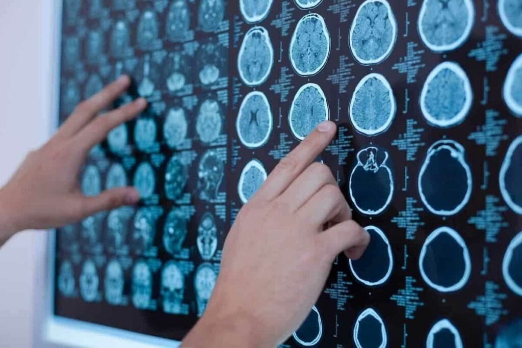
CT scans use X-rays to make detailed pictures of the brain. They move the X-ray source and detectors around the patient. This way, they get images from many angles and make complete pictures.
Key Benefits of CT Scans:
- High-resolution imaging of brain structures
- Rapid scan times, critical for emergencies
- Ability to detect calcifications and bone abnormalities
Tumor Location and Size Assessment
CT scans are great at finding where and how big brain tumors are. Knowing this is key for surgery planning and seeing how far the tumor has spread.
We measure tumor size and how close it is to important brain parts. This helps us make specific treatment plans.
Evaluation of Impact on Surrounding Brain Structures
CT scans also show how tumors affect nearby brain parts. They help us see if tumors push or squeeze important areas. This tells us how the tumor might affect brain function.
CT Contrast Enhancement in Brain Cancer Detection
Using contrast agents in CT scans makes tumors stand out more. These agents show up in abnormal blood flow and tumor blood vessels. This makes it easier to tell tumors apart from other brain issues.
| Feature | Non-Contrast CT | Contrast-Enhanced CT |
| Tumor Visibility | Limited | Enhanced |
| Vascular Detail | Poor | Excellent |
| Diagnostic Accuracy | Moderate | High |
Using CT scans helps us diagnose and treat brain cancer better. The detailed images from CT scans are very helpful in treating brain tumors.
Comparing Brain Cancer X-Ray to MRI and Other Modalities
Many imaging methods are used to find brain cancer. Each has its own good points and not-so-good points. We use these methods to learn about the tumor and how it affects the brain.
X-Ray vs. MRI: Strengths and Weaknesses
X-rays and MRI are two main ways to look at brain cancer. X-rays are great for seeing bone problems, like fractures or calcifications, because they show calcium. But they don’t show soft tissues well.
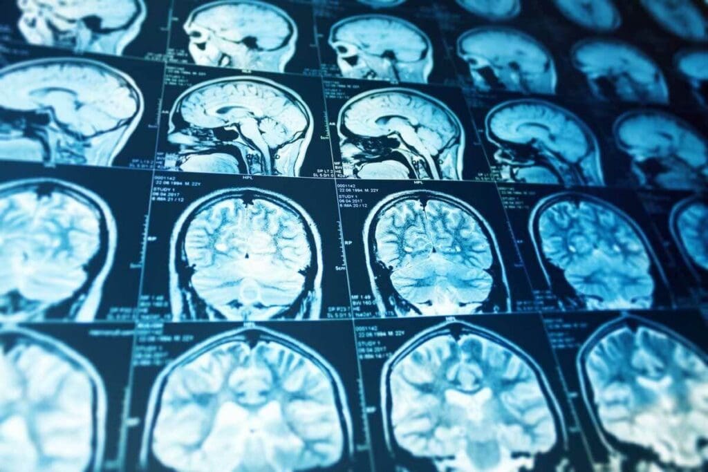
MRI is better at showing soft tissues, making it the top choice for finding brain tumors. MRI shows the tumor’s size, where it is, and how it affects the brain. This info is key for planning surgery and checking treatment.
| Imaging Modality | Strengths | Weaknesses |
| X-Ray | Good for bone abnormalities, quick, and widely available | Poor soft tissue contrast, limited detail |
| MRI | Excellent soft tissue contrast, detailed tumor visualization | More expensive, less available than X-Ray, and claustrophobia issues |
When MRI is Preferred Over X-Ray-Based Imaging
MRI is often the first choice for looking at brain tumors. It’s great for seeing how big the tumor is and how it affects the brain. This info is very important for doctors planning surgery or checking how treatments are working.
Complementary Roles of Different Imaging Techniques
Even though MRI is the best for brain tumors, X-rays and CT scans have their own uses. CT scans, for example, can quickly spot bleeding or calcifications. This is very helpful in urgent situations.
PET and SPECT Scans in Brain Cancer Assessment
PET and SPECT scans show how active brain tumors are. They are very useful for seeing how aggressive the tumor is and how well it’s responding to treatment.
By using different imaging methods together, we get a clearer picture of brain tumors. This helps doctors make more accurate diagnoses and better treatment plans.
The Diagnostic Pathway: When Are Brain Cancer X-Rays Used?
X-rays are used at certain stages in diagnosing brain cancer. We will look at when X-rays are key in finding brain tumors.
Initial Screening and Assessment Protocols
When symptoms suggest brain cancer, doctors start with initial tests. Brain cancer X-rays are often the first imaging test. They are quick and can spot some issues.
These X-rays can show skull bone abnormalities or fractures linked to tumors. They’re not the final say but give early clues.
Emergency Situations and Acute Presentations
In emergencies with severe symptoms like headaches or vomiting, radiographic brain scans are done right away.
X-rays quickly spot fractures or bone shifts, causing symptoms. This fast check is vital in emergencies.
Follow-Up Monitoring After Treatment
After starting treatment for brain cancer, regular checks are needed. X-rays help see if treatment is working and if tumors come back.
- X-rays are used with MRI or CT scans for monitoring.
- They watch for skull changes and new bone issues.
Clinical Decision-Making in Imaging Selection
Choosing the right imaging test depends on many things. This includes symptoms, medical history, and where and what type of tumor is suspected.
“The selection of imaging techniques is a critical aspect of diagnosing brain cancer, requiring a careful balance between diagnostic accuracy and patient safety.” – Expert in Neuro-Oncology..
We weigh these factors to pick between X-rays, MRI, or a CT scan. We s.W e aaimto get a correct diagnosis safely.
Interpreting Brain Cancer X-Ray Results: What to Look For
Understanding X-ray results is key to diagnosing brain cancer. We look for signs and abnormalities in X-ray images. These can show if a tumor is present.
Common Radiological Findings and Their Significance
Brain cancer X-rays often show changes in the skull bones. These changes can mean a tumor is there and how it might affect nearby areas.
Lytic lesions are areas where bone is destroyed, suggesting a tumor. Sclerosis is bone hardening, which can also be a sign. Both are important and need more study.
Signs That Warrant Further Investigation
Some X-ray signs need more tests. These include:
- Unusual calcifications within the brain
- Erosion or destruction of the sella turcica
- Shift of the pineal gland
- Evidence of bone metastasis
These signs might mean brain tumors or other serious issues. More tests, like MRI or CT scans, are needed for a better look.
The Role of Radiologists in Brain Tumor Diagnosis
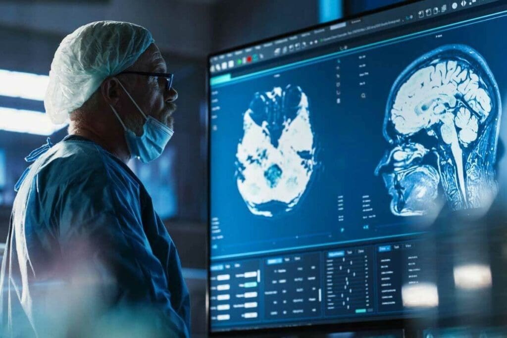
Radiologists are key in reading X-rays and other tests. They spot small changes and understand their importance. They work with doctors to make sure all information is used for a correct diagnosis.
Integration with Clinical Symptoms and History
Just looking at X-rays isn’t enough for brain cancer diagnosis. It’s important to match X-ray findings with symptoms and medical history. This way, doctors can make a precise diagnosis and plan the right treatment.
| Clinical Factor | Relevance to Brain Cancer Diagnosis |
| Headaches and seizures | May indicate increased intracranial pressure or tumor presence |
| Previous cancer history | Could suggest a metastatic brain tumor |
| Neurological deficits | May correlate with tumor location and impact on brain structures |
By combining X-ray results with clinical data, doctors can make better decisions for patient care.
Multidisciplinary Approach to Brain Cancer Imaging
Accurate brain cancer imaging and diagnosis require a team effort. We think combining different medical fields is key to patient care. This way, we get a full picture of a patient’s health, leading to better treatment plans.
Coordination Between Radiology, Neurology, and Oncology
Diagnosing and treating brain cancer is complex. Radiologists look at scans like X-rays and MRIs to spot tumors. Neurologists understand the brain’s symptoms and how tumors affect it. Oncologists then plan treatments based on this info.
Working together, these teams give patients the best care. This teamwork has led to better results by making treatments more effective.
LivHospital’s Integrated Diagnostic Protocols
LivHospital is a leader in brain cancer care. They have a team of experts from radiology, neurology, and oncology. This team works together to give patients the right care for their needs.
Academic and Research-Driven Diagnostic Approaches
Staying up-to-date with research is important for brain cancer imaging. We use the latest tech and research to help our patients. This helps improve treatment outcomes and our understanding of brain cancer.
Research helps us find better ways to diagnose and treat brain cancer. By using new findings in our practice, we can make care more accurate and effective.
Ethical Considerations in Brain Cancer Imaging
Keeping patients safe and respecting their privacy is our top priority. We follow strict ethical standards in our work. This includes being open about the risks and benefits of imaging.
We also focus on being kind and understanding when talking to patients and their families. We aim to care for the whole person, not just their illness.
Patient Experience During Brain Cancer X-Ray Procedures
Brain cancer X-rays are key in finding out what’s wrong. Knowing what to expect can make things easier. We help patients understand what will happen and how safe it is.
What to Expect During Different Imaging Studies
Getting a brain cancer X-ray is usually quick and simple. The X-ray machine takes pictures of your brain and skull. Sometimes, a CT scan is needed for more detailed images.
Preparation is key to good imaging study. You might need to take off metal items or jewelry. A contrast agent might also be used to make certain areas clearer.
Radiation Exposure Concerns and Safety Measures
One big worry is radiation from X-rays. We’re very careful to keep exposure low. This way, we get the images we need without risking too much.
New X-ray tech uses the least amount of radiation needed. Our team makes sure the benefits of the test are worth the risk of radiation.
Preparing for Brain Imaging Procedures
To get ready for a brain imaging test, follow your doctor’s advice. This might mean arriving early, removing metal items, and following diet rules.
Clear communication is key in getting ready. Our team is here to help and support you every step of the way.
Communication of Results and Next Steps
After the test, our experts analyze the results. We know waiting can be hard, so we aim to share the findings and what comes next with care and kindness.
Depending on what we find, you might need more tests or treatment plans. Our team works together to make sure you get all the help you need.
Recent Advances in X-Ray Technology for Brain Cancer Detection
New X-ray technology is changing how we find brain cancer. It makes diagnosis better and safer for patients. This is great news for people all over the world.
Digital Radiography Improvements
Digital radiography has made X-ray images much clearer. High-resolution images help doctors spot tumors and other issues more easily. This is key to catching cancer early and planning treatment.
Switching to digital from film has also made things faster. Patients spend less time in the imaging room. This makes the whole process more efficient.
Reduced Radiation Exposure Techniques
New X-ray tech means less radiation for patients. This is a big deal for those needing many scans during treatment. It keeps them safe without losing image quality.
Low-dose X-ray protocols are now common. They make sure patients get the least amount of radiation needed. This makes brain cancer diagnosis safer for everyone.
AI and Machine Learning in X-Ray Image Analysis
AI and machine learning are changing how we look at X-rays. They can spot things that humans might miss. This makes diathe gnosis more accurate.
AI helps doctors find small changes in X-rays. This can lead to earlier diagnosis and better treatment plans. It’s a big step forward in patient care.
Future Directions in Neurological X-Ray Imaging
We’re expecting even more improvements in X-ray tech soon. Things like better image making and personalized scans will help fight brain cancer. These changes will make diagnosis and treatment even better.
As we look to the future, combining X-rays with other imaging tools will be key. This will help doctors give patients the best care possible. It’s a step towards more complete and effective treatment.
Conclusion: The Evolving Role of X-Rays in Brain Cancer Diagnosis
We’ve looked at how X-rays help diagnose brain cancer, from their basics to their limits. X-rays are useful but have downsides, like not showing soft tissues well. This makes advanced imaging, like CT scans and MR, more important for accurate diagnoses.
Places like LivHospital are leading the way in using the latest neuroimaging. They use these advanced tools to help patients better. This shows how important it is to keep improving our diagnostic methods.
As technology gets better, X-rays’ role in diagnosing brain cancer will change. We might see new tools like AI helping doctors. A team effort from radiologists, neurologists, and oncologists is key to giving patients the best care.
FAQ
What is the primary use of X-rays in brain cancer diagnosis?
X-rays help find problems in the skull, like fractures or bone damage. These signs might mean brain cancer. But, they’re not used seeingsee tumors directly.
How do X-rays work in detecting brain cancer?
X-rays go through the body and show how much each tissue absorbs. For brain cancer, they can spot skull changes that might mean a tumor.
What are the limitations of using X-rays for brain cancer diagnosis?
X-rays can’t show soft tissues well, making tumors hard to see. Also, the skull’s structures can hide important details.
When are CT scans preferred over X-rays for brain cancer diagnosis?
CT scans are better for detailed images. They help see the tumor size, location, and how it affects the brain. They also find tumors more accurately.
How does MRI compare to X-ray in brain cancer diagnosis?
MRI is better for seeing tumors because it shows soft tissues clearly. X-rays are better for finding bone problems.
What is the role of PET and SPECT scans in brain cancer assessment?
PET and SPECT scans show how active tumors are. This helps doctors understand how aggressive the tumor is and how well it’s responding to treatment.
When are X-rays used in the diagnostic pathway for brain cancer?
X-rays might be the first choice for screenings, emergencies, or after treatment checks. The right imaging depends on the patient’s situation.
How are X-ray results interpreted in the context of brain cancer?
Radiologists look for signs like bone damage or fractures in X-rays. They also consider symptoms and medical history.
What can patients expect during X-ray procedures for brain cancer?
X-ray procedures are quick and don’t hurt. Safety from radiation is a big concern. Patients are prepared and told about their results.
How has X-ray technology advanced in recent years for brain cancer detection?
New digital radiography has better images. There are ways to use less radiation. AI and machine learning might help analyze X-rays better.
What is the future direction of neurological X-ray imaging?
The future includes better images and less radiation. AI and machine learning will help doctors make more accurate diagnoses.
References
- National Institute of Biomedical Imaging and Bioengineering. (2023). Computed tomography (CT). https://www.nibib.nih.gov/science-education/science-topics/computed-tomography-ct


