Last Updated on November 27, 2025 by Bilal Hasdemir

At Liv Hospital, we use the latest technology to find and diagnose brain cancer accurately. Magnetic Resonance Imaging (MRI) has changed how we spot and understand brain tumors. It helps doctors see tumors clearly and know what they are.
We use MRI scans to find tiny spots and tell if tumors are harmless or dangerous. This tool is key in helping us care for our patients. It lets us make treatment plans that fit each person’s needs.
Key Takeaways
- Understanding the role of MRI in detecting brain cancer
- Discovering how MRI scans distinguish between benign and malignant tumors
- Learning about the benefits of MRI in patient care
- Exploring the importance of precise diagnosis in treatment planning
- Understanding the advantages of MRI in detecting small lesions
The Critical Role of Brain Imaging in Cancer Detection

Getting a correct diagnosis through brain imaging is key to good cancer treatment and better survival rates. MRI is a top tool for finding and diagnosing brain tumors.
Why Early and Accurate Detection Matters
Finding brain tumors early is very important. Early detection means treatments work better, and patients have a higher chance of recovery. Early and accurate detection lets doctors plan the best treatment, which helps patients do better.
Also, knowing if a tumor is benign or malignant is vital for treatment. Advanced brain imaging helps avoid mistakes in diagnosis. This ensures patients get the right care for their needs.
Evolution of Neuroimaging for Brain Tumors
Neuroimaging has changed a lot over time, making brain tumor diagnosis better. Magnetic Resonance Imaging (MRI) has been a big help. It gives clear pictures of soft tissues, which is great for finding brain tumors.
New imaging methods, like functional MRI and MR spectroscopy, have also helped. They give more details about tumors, helping doctors make better treatment plans.
MRI Scan Brain Cancer: The Gold Standard in Neuroimaging

MRI has changed how we diagnose brain cancer, becoming the top choice in neuroimaging. MRI scans give us detailed brain images. They help us spot and diagnose brain tumors accurately.
How Magnetic Resonance Imaging Works
MRI uses strong magnetic fields and radio waves to create detailed brain images. This method is non-invasive, showing the brain’s soft tissues without surgery or harmful radiation.
It aligns hydrogen nuclei in the body with a strong magnetic field. Radio waves then disturb these nuclei, creating signals for detailed images.
Types of MRI Sequences for Brain Tumor Visualization
There are different MRI sequences for brain tumors, each showing unique tumor details. Some common ones include:
- T1-weighted images: Show detailed anatomy and help see tumor structure.
- T2-weighted images: Are good at showing changes in tissue water, helping spot edema and tumor boundaries.
- FLAIR (Fluid Attenuated Inversion Recovery): Helps find lesions and tumors by suppressing fluid signals.
- Diffusion-weighted imaging: Shows the random motion of water molecules, useful for acute strokes and tumor cellularity.
Contrast Enhancement: What It Reveals About Tumors
Contrast enhancement is key in MRI scans for brain tumor diagnosis. A contrast agent, like gadolinium-based, highlights certain brain areas and tumors. This gives us important tumor information.
Contrast enhancement helps us:
- Identify tumor boundaries: Makes tumor edges clearer.
- Assess tumor vascularity: Shows the tumor’s blood supply and aggressiveness.
- Detect tumor necrosis: It finds dead tissue in the tumor, a sign of aggressive behavior.
By using various MRI sequences and contrast enhancement, we get a full picture of brain tumors. This helps in diagnosis, treatment planning, and patient care.
Fact 1: Exceptional Accuracy in Brain Tumor Detection
MRI technology has changed how we find brain tumors. It gives us clear images of the brain. This helps doctors spot tumors well.
Understanding the 97% Overall Detection Rate
Research shows MRI finds brain tumors 97% of the time. This high success rate is key to early treatment. Finding tumors early can greatly help patients.
Several things help MRI detect tumors so well:
- Advanced MRI sequences that provide detailed images
- High-resolution imaging that helps identify small lesions
- Expert interpretation by radiologists
Detection Capabilities for Small Lesions
MRI is great at finding small tumors that other methods might miss. This is important for treating tumors when they’re small and easier to cure.
MRI’s ability to spot small brain tumors comes from its clear images and many sequences. These help show different parts of the brain.
Comparative Advantage Over Other Imaging Methods
Compared to CT scans, MRI is better at showing soft tissues and tumor types. This makes MRI the top choice for finding brain tumors.
Our experience shows MRI’s detailed images lead to more accurate diagnoses. This means better care for patients.
Fact 2: What Tumors Look Like on MRI Brain Scans
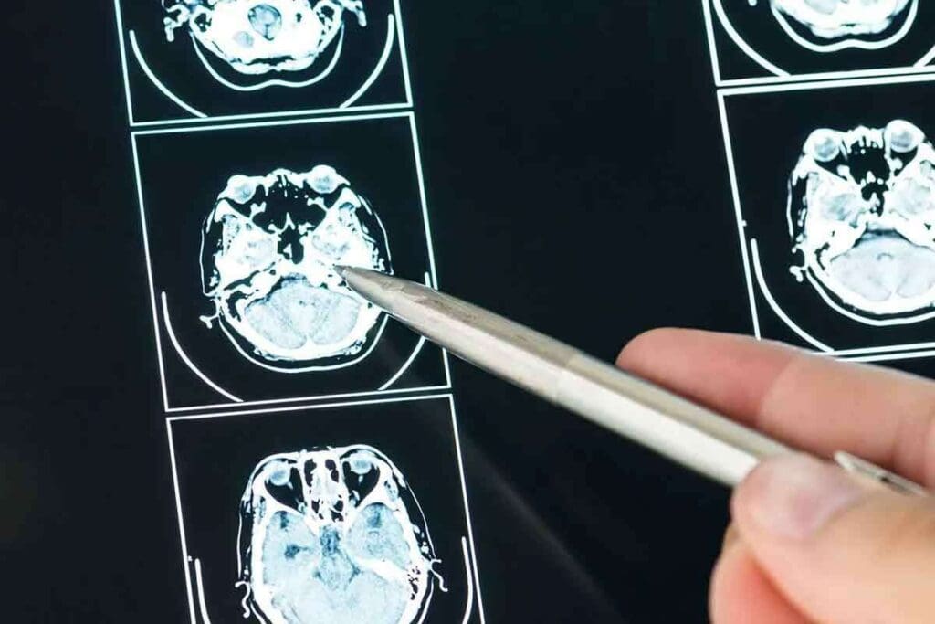
It’s important to know how brain tumors look on MRI scans for accurate diagnosis. MRI scans show the brain’s details well. This helps doctors spot tumors by their looks.
General Appearance of Brain Tumors
On MRI scans, brain tumors look like separate masses from the brain. They can be different sizes, shapes, and places. The general appearance of a tumor can hint at its type, like whether it’s benign or malignant.
Some tumors have clear edges, while others don’t. The signal intensity of a tumor on MRI also matters. Tumors might look brighter or darker than the brain tissue around them, based on their makeup.
Signal Intensity Patterns and Their Meaning
Signal intensity patterns on MRI scans are key to understanding brain tumors. Different MRI sequences, like T1-weighted and T2-weighted images, show different tumor details.
- T1-weighted images show the tumor’s shape and how it fits with nearby structures.
- T2-weighted images are better at showing changes in tissue and swelling around the tumor.
By studying these patterns, doctors can learn about the tumor’s type and how it might affect the brain.
How Radiologists Identify Suspicious Masses
Radiologists look at MRI scans in two ways: by eye and with numbers. They search for unusual areas in the brain and check their features with different MRI types.
The steps are:
- They check the size, shape, and where the mass is.
- They look at the signal intensity patterns on different MRI types.
- They see if the mass is pushing on or moving nearby structures.
By looking at these details, radiologists can spot suspicious masses. These might need more tests or treatment.
Fact 3: Benign vs Malignant Brain Tumor MRI Findings
It’s important to know the difference between benign and malignant brain tumors. MRI scans help doctors tell them apart by showing key features of each.
Characteristics of Benign Tumors
Benign brain tumors have clear edges and look the same on MRI scans. They might show homogeneous enhancement after contrast. These tumors push brain structures aside but don’t invade them.
Benign tumors often have:
- Well-circumscribed margins
- Homogeneous signal intensity
- Limited or no surrounding edema
- Minimal mass effect on adjacent structures
Hallmarks of Malignant Tumors
Malignant brain tumors have irregular borders and look different on MRI. They might show heterogeneous enhancement with necrosis. They also tend to spread into the surrounding brain tissue.
Malignant tumors often have:
- Irregular or infiltrative margins
- Heterogeneous signal intensity
- Presence of necrosis or cystic components
- Significant surrounding edema
- Strong mass effect on adjacent structures
Here’s a table showing the main differences between benign and malignant tumors on MRI:
| Characteristics | Benign Tumors | Malignant Tumors |
| Margins | Well-defined | Irregular or infiltrative |
| Signal Intensity | Homogeneous | Heterogeneous |
| Enhancement Pattern | Homogeneous enhancement | Heterogeneous enhancement with necrosis |
| Surrounding Edema | Limited or none | Significant |
| Mass Effect | Minimal | Strong |
Fact 4: Advanced MRI Techniques Enhancing Diagnostic Precision
We now have advanced MRI methods that give us a clearer view of brain tumors. These new techniques have greatly improved how we diagnose and understand brain cancer.
Diffusion and Perfusion Weighted Imaging
Two advanced MRI techniques, diffusion weighted imaging (DWI) and perfusion weighted imaging (PWI), offer key insights. DWI looks at how water moves in tissues, helping spot high-grade tumors. PWI checks blood flow in tumors, showing how aggressive they might be.
A study in the Journal of Neuro-Oncology showed DWI and PWI. They help tell high-grade from low-grade gliomas, making diagnoses more accurate.
| Technique | Primary Use | Clinical Benefit |
| Diffusion Weighted Imaging (DWI) | Measures water molecule diffusion | Identifies areas of restricted diffusion, often seen in high-grade tumors |
| Perfusion Weighted Imaging (PWI) | Assesses blood flow and perfusion | Provides insights into tumor vascularity and possible aggressiveness |
MR Spectroscopy for Biochemical Analysis
MR spectroscopy (MRS) lets us analyze brain tumors’ biochemistry. It measures metabolic activity, helping tell tumor types and grades apart. For example, high-grade tumors show more choline and less N-acetylaspartate (NAA).
“MR spectroscopy has emerged as a valuable tool in the non-invasive diagnosis and characterization of brain tumors, providing critical information about tumor metabolism and biology.”
Functional MRI Applications in Tumor Assessment
Functional MRI (fMRI) looks at brain function and its link to tumors. It’s key in planning surgery, helping avoid damaging important brain areas.
By using these advanced MRI techniques together, doctors get a better understanding of brain tumors. This leads to more accurate diagnoses and better treatment plans.
Fact 5: Artificial Intelligence Revolution in Tumor Classification
Artificial intelligence is changing how we classify brain tumors, making them more accurate. AI in medical imaging is opening new ways to diagnose and treat brain cancer. This shift in tumor classification is thanks to deep learning models.
Deep Learning Models Achieving 99% Accuracy
Studies show deep learning models can classify brain tumors with up to 99% accuracy. This high accuracy is key to effective treatment and better patient outcomes. AI algorithms help radiologists confidently identify different tumor types.
High accuracy in tumor classification also speeds up diagnosis. AI can quickly assess MRI scans, focusing on urgent cases.
Distinguishing Glioma, Meningioma, and Pituitary Tumors
Distinguishing between brain tumors like glioma, meningioma, and pituitary tumors is a big challenge. AI systems analyze MRI scans to spot each tumor’s unique features. This is vital for choosing the right treatment.
Gliomas come from brain glial cells and vary in aggressiveness. Meningiomas are usually benign and arise from the meninges. Accurate classification is key effective treatment.
How AI Assists Radiologists in Clinical Practice
AI is meant to enhance, not replace, radiologists. It provides initial assessments, helping radiologists make better decisions. This teamwork can lead to better patient care.
In practice, AI points out concerns on MRI scans and suggests diagnoses. It streamlines workflow, allowing radiologists to focus on complex cases and improve accuracy.
Fact 6: The Clinical Pathway from MRI to Diagnosis
When a brain tumor is suspected, the clinical pathway from MRI to diagnosis is key. It helps ensure patients get accurate diagnoses and timely care.
When Physicians Order Brain MRIs
Doctors order brain MRIs when they think there might be a tumor or to check if a tumor is growing. Symptoms like headaches, seizures, or problems with movement might lead to an MRI. MRIs are great when CT scans don’t give clear results or when soft tissue details are needed.
Common reasons for a brain MRI include:
- Suspected brain tumors or metastases
- Monitoring known tumor progression or response to treatment
- Unexplained neurological symptoms
- Abnormal findings on other imaging studies
Interpreting Results: The Multidisciplinary Approach
Reading MRI results is a team effort. Radiologists, neurologists, neurosurgeons, and oncologists work together. They analyze the images, match them with the patient’s symptoms and history, and decide on the next steps.
The process includes:
- Detailed analysis of MRI sequences to identify tumor characteristics
- Correlation with clinical history and symptoms
- Discussion in a multidisciplinary team meeting to reach a consensus on diagnosis and treatment planning
Follow-Up Protocols After Tumor Detection
After finding a tumor, follow-up plans are set to track its growth and how the patient is doing. These plans might include regular MRI scans, changing treatment plans based on new findings, and checking on the patient’s neurological health.
Key parts of follow-up plans include:
- Regular MRI scans to monitor tumor size and characteristics
- Clinical assessments to evaluate neurological function and overall health
- Adjustments to treatment plans as needed based on imaging and clinical findings
By following a clear clinical pathway, healthcare providers can give patients the best care for their needs.
Fact 7: Answering Critical Questions About MRI Capabilities
When we talk about MRI and brain tumors, many questions come up. MRI is key in seeing the brain’s details and finding problems. It’s important to know what it can and can’t do for doctors and patients.
Can MRI Detect All Types of Brain Tumors?
MRI is great at finding many brain tumors because it shows soft tissues well. It can spot tumors by their size, location, and type. But, very small tumors or those in hard-to-see spots might be tough to find. Some tumors might not show up without contrast agents.
Using advanced MRI methods like diffusion-weighted imaging and MR spectroscopy helps more. These methods give extra details about tumors, helping doctors make better choices.
Does MRI Show Brain Cancer in Early Stages?
MRI can find brain cancer early, often before symptoms show. Early detection is key to good treatment and better results. MRI can spot small changes in the brain that might mean a tumor is there.
But MRI’s ability to find early brain cancer depends on the tumor type and where it is. Some tumors might be hard to see or not show up clearly.
Limitations and Possible False Results
Even though MRI is very good, it’s not perfect. It might miss tumors that are small or don’t show up with contrast. It can also say a tumor is there when it’s not, causing worry and more tests.
To avoid these problems, doctors look at MRI results with other tests and symptoms. A team of doctors, including radiologists and oncologists, works together for the best diagnosis and care.
In short, MRI is a big help in finding and diagnosing brain tumors. Knowing its strengths and weaknesses helps us use it better to help patients.
Patient Experience: What to Expect During a Brain MRI
Knowing what to expect during a brain MRI can make you feel less anxious. We want to help you understand the whole process, from start to finish. This includes everything from getting ready to getting your results.
Preparation for the Procedure
Before your brain MRI, you’ll need to take off any metal items like jewelry and glasses. This is important for getting good images and keeping you safe. You might also need to follow special diet rules or skip some medicines. Wear comfy clothes and arrive early to fill out any forms.
Key preparation steps include:
- Removing all metal objects
- Following dietary instructions
- Arriving early to complete paperwork
- Wearing comfortable clothing
During the Scan: Process and Sensations
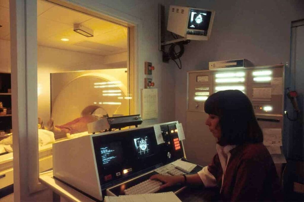
When it’s time for the scan, you’ll lie on a table that moves into the MRI machine. It’s important to stay very quiet to get clear pictures. The scan itself doesn’t hurt, but you might hear loud noises. Don’t worry, you’ll get earplugs or headphones to help block out the sound.
The MRI technician will be with you the whole time to make sure you’re comfortable and safe. They’ll talk to you through an intercom and can help if you have any problems.
After the MRI: Results and Next Steps
After your MRI, you can usually go back to your normal activities unless your doctor says not to. The MRI images are then looked at by a radiologist. They’ll write a report for your doctor to discuss with you.
You might have to wait a few days to a week for your doctor to talk about the results and what to do next. Depending on what the MRI shows, you might need more tests or to see other doctors.
Learning about the brain MRI process can make you feel more ready and less worried. By knowing what to expect, you can handle your diagnostic journey better. This helps you focus on getting better.
Conclusion: The Future of MRI in Brain Cancer Management
MRI will keep being key in finding and managing brain cancer. New MRI tech and AI will make it even better at spotting and understanding brain tumors.
The future looks bright for MRI in brain cancer care. We’ll see better images, more detailed scans, and AI helping doctors. This means doctors can make more precise diagnoses and treatments. Patients will get better care because of it.
As MRI tech gets better, finding and treating brain cancer will get easier. We’ll see more accurate diagnoses and treatments. MRI will keep being a vital tool in the fight against brain cancer, helping patients all over the world.
FAQ
Can MRI detect brain cancer?
Yes, MRI is very good at finding brain cancer. It can spot small lesions too.
What do brain tumors look like on MRI?
On MRI, brain tumors show up as masses. They have specific signal patterns. This helps doctors spot them.
Can MRI distinguish between benign and malignant brain tumors?
Yes, MRI can tell the difference. It looks at the tumor’s borders, how it enhances, and its signal intensity.
What advanced MRI techniques are used for brain tumor assessment?
MRI uses advanced techniques like diffusion and perfusion imaging. It also uses MR spectroscopy and functional MRI. These give important information about the tumor.
How accurate is MRI in detecting brain tumors?
MRI is very accurate in finding brain tumors. It’s better than other imaging methods in many cases.
Can MRI detect brain cancer in early stages?
Yes, MRI can find brain cancer early. This is key to better treatment and outcomes.
What are the limitations of MRI in brain tumor detection?
MRI is very effective but has some limits. It might miss very small tumors or tell different tumor types apart.
How do I prepare for a brain MRI?
To prepare for a brain MRI, remove metal objects. Follow dietary instructions. Arrive early to do paperwork.
What happens during a brain MRI scan?
During a brain MRI, you lie on a table that slides into the machine. You must stay very quiet while it scans.
How long does it take to get the results of a brain MRI?
Getting MRI results can take some time. A radiologist will look at the images and give a report. Your doctor will then talk to you about it.
Can artificial intelligence assist in brain tumor diagnosis with MRI?
Yes, artificial intelligence, like deep learning models, can help doctors classify brain tumors. It can also improve how accurate diagnoses are.
References
- Rethemiotaki, I., et al. (2024). Brain tumour detection from magnetic resonance imaging images using convolutional neural networks. Scientific Reports, 14, 1850. https://www.ncbi.nlm.nih.gov/pmc/articles/PMC10883197/
- Wang, X., et al. (2025). Accurate and real-time brain tumour detection and segmentation with enhanced deep learning techniques. Scientific Reports, 15, 1147. https://www.nature.com/articles/s41598-025-07773-1


