Last Updated on November 27, 2025 by Bilal Hasdemir
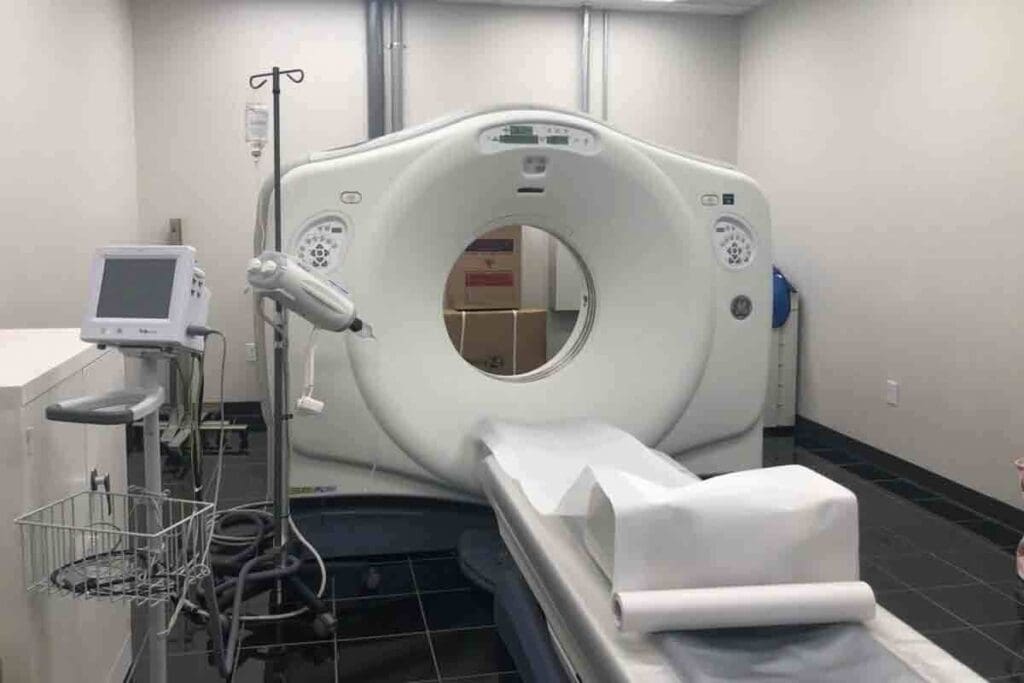
At LivHospital, we use myocardial perfusion imaging (MPI) to check heart health. MPI looks at blood flow to the heart muscle. It helps us find areas where blood flow is low.
We use the latest technology, like the MPI stress test and cardiac PET. This gives us clear views of heart health. Our focus is on safe, effective, and top-notch heart care for our patients.
Key Takeaways
- Understanding myocardial perfusion imaging and its role in diagnosing heart conditions.
- The significance of MPI stress tests in evaluating heart health.
- The benefits of cardiac PET in assessing myocardial perfusion.
- LivHospital’s commitment to patient-centered care and advanced technology.
- The importance of accurate diagnosis in developing effective treatment plans.
Understanding Myocardial Perfusion Imaging: The Foundation of Cardiac Assessment
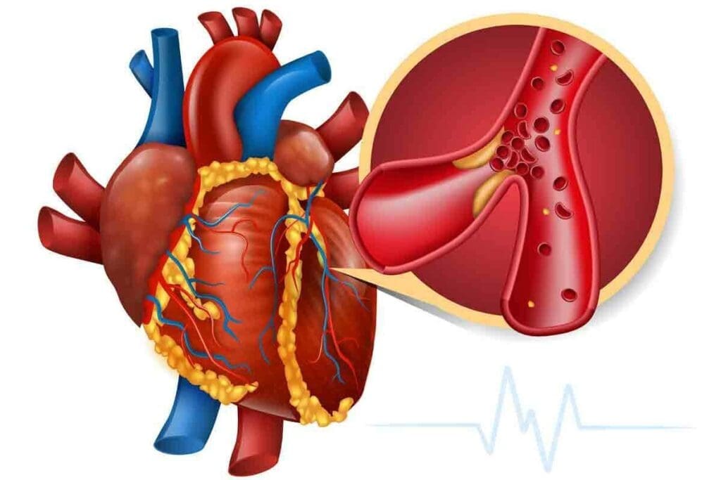
Myocardial perfusion imaging (MPI) is a key tool in cardiology. It helps us see how well blood flows to the heart. This is vital for spotting and treating heart disease.
What Is Myocardial Perfusion and Why Does It Matter
Myocardial perfusion is about blood flow to the heart muscle. This flow is key for the heart to work properly. If it’s blocked, heart problems can occur.
Knowing about myocardial perfusion helps doctors find heart issues. They can see where blood flow is low. This helps them choose the right treatment.
The Evolution of Nuclear Cardiology Techniques
Nuclear cardiology has grown a lot over time. New tech has made MPI more accurate. Old methods were not as good.
New tools like SPECT and PET have made big changes. They help doctors see the heart better. This leads to better care for heart patients.
Common Abbreviations: MPI, Myocardial Perfusion, and More
In cardiology, you’ll see many abbreviations. MPI stands for Myocardial Perfusion Imaging. SPECT and PET are also used for imaging.
Knowing these terms helps everyone talk clearly. It makes it easier for patients to understand their health and treatment.
The Science Behind Ht Muscle Image Spect Mult Technology

Understanding Ht Muscle Image Spect Mult technology is key to its role in heart health checks. We use Single Photon Emission Computed Tomography (SPECT) imaging to see how blood flows to the heart. This is a vital part of checking the heart’s blood flow, or myocardial perfusion imaging (MPI).
Decoding SPECT Imaging for Heart Muscle Evaluation
SPECT imaging is a complex nuclear medicine method. It lets us see how blood moves to the heart muscle. We use tiny amounts of radioactive tracers to get detailed heart images.
“SPECT imaging is a must-have in cardiology,” say experts. It gives us important insights into heart health.
The process starts with the tracer sending out gamma rays. The SPECT camera catches these rays. Then, it makes a 3D image of the heart, showing where blood flow might be low.
Multi-Angle Acquisition: Why Multiple Views Matter
SPECT imaging’s big plus is taking images from many angles. This multi-angle view gives us a full picture of the heart’s work. It lets us spot problems that might not show up from just one angle.
Multiple views are important for accurate diagnoses. For example, a heart muscle problem might only show up from certain angles. So, getting images from many angles is key for a complete check.
Technical Parameters That Ensure Diagnostic Accuracy
Several technical details are vital for SPECT imaging’s accuracy. These include the collimator type, energy window settings, and the algorithms used for image making. Each detail is important for top-notch image quality and reliable data.
- Choosing the right collimator affects image sharpness and sensitivity.
- Setting the energy windows helps cut down scatter radiation, making images clearer.
- The algorithms used are essential for creating accurate 3D images from the data.
By tweaking these technical aspects, we can greatly improve SPECT imaging’s accuracy. This leads to better care for patients.
Key Fact #1: How MPI Stress Tests Detect Coronary Artery Disease
MPI stress tests are key to finding coronary artery disease. They check how well the heart muscle gets blood when stressed and at rest. This test is non-invasive and gives doctors important info about the heart’s blood flow.
Exercise vs. Pharmacological Stress Protocols
There are two main ways to stress the heart in an MPI test: exercise and medicine. Exercise stress uses walking or biking to raise heart rate and blood flow. It’s the best way to test the heart when possible.
Pharmacological stress uses medicine to increase blood flow to the heart. It’s used when exercise is not possible.
A study showed both methods are very good at finding coronary artery disease. The choice depends on the patient’s health.
Interpreting Blood Flow Patterns During Stress and Rest
During an MPI stress test, images of the heart are taken at rest and after stress. These images are compared to see if blood flow changes. Normal blood flow patterns show the same perfusion at rest and stress. But abnormal patterns might show reduced blood flow.
Reversible defects suggest coronary artery disease. Fixed defects might mean scar tissue from a heart attack.
| Blood Flow Pattern | Rest | Stress | Indication |
| Normal | Normal | Normal | No significant coronary artery disease |
| Reversible Defect | Normal | Abnormal | Coronary artery disease is likely |
| Fixed Defect | Abnormal | Abnormal | Possible scar tissue from a previous heart attack |
What Abnormal Results Indicate About Your Heart Health
Abnormal MPI stress test results can show several heart health issues. Cardiologist says, “MPI stress tests are key for understanding the heart’s blood flow. They help us find patients at risk for coronary artery disease and guide treatment.”
“MPI stress tests are invaluable for diagnosing coronary artery disease and assessing the risk of future cardiac events. They help us tailor treatment plans to individual patient needs.”
Cardiologist
These results might lead to more tests or treatment talks. This could include lifestyle changes, medicines, or procedures like angioplasty.
In summary, MPI stress tests are a powerful tool for finding coronary artery disease. They help doctors make better decisions for patient care, which can prevent serious heart problems.
Key Fact #2: SPECT vs. PET in Myocardial Perfusion Imaging
SPECT and PET are key tools for checking how well the heart gets blood. Each has its own benefits. The choice between them depends on how well they work, how much radiation they use, and their cost.
Comparing Imaging Modalities: Resolution and Accuracy
PET scans are better at showing details and accuracy than SPECT. They can measure blood flow to the heart more precisely. This helps doctors see how well the heart is working.
A study found that PET is more accurate than SPECT, which is good for patients with heart disease. PET’s ability to give exact blood flow numbers helps doctors diagnose and treat heart problems better.
SPECT is useful but has some limits. It’s not as detailed as PET. But it’s cheaper and more widely available. Newer SPECT cameras, like CZT, have made it better for diagnosing heart issues.
Radiation Exposure Differences Between Techniques
How much radiation you get from MPI is important. PET scans use more radiation than SPECT, but new PET tech has made it safer. The dose from PET can be higher, but its better accuracy might be worth it for some patients.
SPECT, with newer cameras, can use less radiation. Using stress-only scans and correcting for body parts in the way can also lower radiation. Choosing between SPECT and PET depends on how safe and accurate you need it to be.
Cost-Benefit Analysis for Different Patient Populations
The cost of SPECT vs. PET varies by patient. For those with heart disease, PET’s better accuracy might be worth the extra cost. But for routine checks or in places with less money, SPECT is cheaper and more available.
As healthcare changes, picking between SPECT and PET will depend on the patient’s needs and what’s available. Knowing the good and bad of each helps doctors make the best choice for care.
Key Fact #3: The Complete MPI Procedure: Patient Experience
Understanding the MPI test process is key. Knowing what to expect from start to finish can reduce anxiety. It makes the experience smoother for you.
Preparation Guidelines: Medications, Diet, and Activity
Getting ready for an MPI test means making some changes. Your healthcare provider will give you specific instructions. This might include:
- Avoiding certain medications or foods that could interfere with the test results
- Adjusting your physical activity levels before the test
- Informing your doctor about any allergies or medical conditions
Following these guidelines ensures your MPI test is safe and effective.
During the Test: What Happens in Each Phase
The MPI procedure has several phases, like stress and rest imaging. In the stress phase, you might walk on a treadmill or take medication. This simulates exercise on your heart. The rest phase captures images of your heart at rest. Our team will watch over you to keep you safe and comfortable.
Post-Procedure Care and Results Timeline
After the MPI test, you can usually go back to your normal routine. But always follow any care instructions from your healthcare provider. This ensures your safety and the test’s accuracy.
When you’ll get your MPI test results varies. It usually takes a few days to a week. Your healthcare provider’s specific needs can affect this timeline.
Here’s a summary of what to expect during and after the MPI procedure:
| Procedure Phase | What to Expect |
| Preparation | Adjust medications, diet, and activity levels as instructed |
| Stress Phase | Exercise or receive medication to simulate exercise effects |
| Rest Phase | Imaging of the heart at rest |
| Post-Procedure | Resume normal activities; follow post-procedure care instructions |
| Results Timeline | Receive results within a few days to a week |
Key Fact #4: Cardiac PET Scans: Quantitative Blood Flow Analysis
Cardiac PET scans have changed cardiology by giving exact blood flow numbers. This helps doctors diagnose and treat heart disease better.
How PET Measures Absolute Myocardial Blood Flow
Cardiac PET scans use positron emission tomography (PET) to measure blood flow. They use special tracers like Rubidium-82 or Nitrogen-13 ammonia. These tracers are taken up by the heart muscle based on blood flow.
By counting how much of these tracers are taken up, PET scans can show how well the heart gets blood. This is true whether the heart is at rest or under stress.
Coronary Flow Reserve: A Critical Diagnostic Metric
One key thing PET scans measure is coronary flow reserve (CFR). CFR is the ratio of blood flow when stressed to blood flow at rest. If CFR is low, it means the heart’s blood vessels don’t work well.
This is often a sign of heart disease. Knowing this helps doctors spot patients at risk of heart problems.
PET’s Role in Detecting Balanced Ischemia
Cardiac PET scans are also great at finding balanced ischemia. This is when blood flow to the heart is low all over, often due to many blocked arteries. Unlike other tests, PET scans can show the exact amount of blood flow.
This makes them better at spotting balanced ischemia. It helps doctors understand and treat heart disease more accurately.
In short, cardiac PET scans are a key tool for understanding heart blood flow. They help doctors find and treat heart disease by looking at coronary flow reserve and balanced ischemia.
Key Fact #5: Clinical Applications of Myocardial Perfusion Testing
Myocardial perfusion imaging (MPI) is key in heart care. It helps manage coronary artery disease. It gives insights for treatment plans.
Early Detection of Coronary Artery Disease
Myocardial perfusion testing is vital for spotting coronary artery disease (CAD) early. It finds areas with less blood flow. This helps doctors catch CAD before it gets worse.
Early spotting means:
- Starting treatment sooner
- Stopping big heart problems
- Better health results
Viability Assessment for Revascularization Decisions
MPI checks myocardial viability. This is key for deciding on heart surgeries like CABG or PCI. It shows how much heart muscle can be saved.
Monitoring Treatment Effectiveness Over Time
Also, MPI tracks how well treatments work over time. By doing MPI tests again, doctors see how the heart’s blood flow changes. This helps adjust treatments as needed.
Tracking treatment success includes:
- Seeing changes in heart blood flow
- Checking if treatments are working
- Changing treatments if needed
Key Fact #6: PET Stress Test Advantages in Cardiac Assessment
PET stress tests have changed how we check the heart. They are very good at finding problems. This is key to keeping the heart healthy. They are great for some patients because they are very accurate.
When PET is used with CT angiography, it checks the heart’s function and structure. This gives a full picture of the heart’s health.
Superior Sensitivity and Specificity Rates
PET stress tests are top-notch at finding heart disease. They are more accurate than other tests. This is important for treating heart disease well.
Benefits for Specific Patient Groups
These tests are great for certain patients. They help those with heart disease or who have had heart surgery. They show how much blood the heart is getting. This helps doctors decide the best treatment.
They are also good for patients who can’t do other stress tests.
Integration with CT Angiography in Cardiac PET/CT
Using PET with CT angiography is a big step forward. This combo gives a full view of the heart’s function and structure. It helps doctors understand the heart better and plan treatments.
Here’s a comparison of PET stress tests with other diagnostic tests:
| Diagnostic Test | Sensitivity | Specificity |
| PET Stress Test | 90% | 95% |
| SPECT Stress Test | 80% | 85% |
| Echocardiogram | 75% | 80% |
Key Fact #7: Cardiac PET Scan vs. Echocardiogram Comparison
Cardiac PET scans and echocardiograms are key tools in cardiology. They serve different needs. Echocardiograms show heart structure and function. PET scans look at blood flow and how well the heart works.
Knowing what each test does helps pick the right one for each patient.
Diagnostic Capabilities: What Each Test Reveals
An echocardiogram uses sound waves to show the heart’s details. It checks heart valves and chambers. It also looks at how well the heart moves.
A cardiac PET scan, on the other hand, looks at blood flow and heart health. It shows how active the heart is under stress and at rest.
Comparison of Diagnostic Capabilities:
| Diagnostic Feature | Echocardiogram | Cardiac PET Scan |
| Heart Structure | Detailed images of heart chambers and valves | Limited structural information |
| Myocardial Blood Flow | Limited assessment | Quantitative measurement of blood flow |
| Viability Assessment | Indirect assessment through wall motion | Direct assessment of myocardial viability |
When to Choose PET Over Echocardiography
Choosing between a cardiac PET scan and an echocardiogram depends on the question being asked. For assessing coronary artery disease or assessing heart viability, a PET scan is better. It measures blood flow and checks heart health.
For example, after heart procedures, a PET scan shows if they worked. It helps decide what to do next.
Complementary Roles in Complete Cardiac Evaluation
Cardiac PET scans and echocardiograms work together in heart checks. Echocardiograms look at heart structure and function. PET scans examine blood flow and heart health.
Risks, Limitations, and Considerations in Nuclear Cardiology
Nuclear cardiology is a powerful tool for diagnosing heart issues. Yet, it comes with risks and limitations. We must weigh the benefits against the drawbacks when using tests like myocardial perfusion imaging (MPI).
Radiation Safety in Myocardial Perfusion Imaging
One major concern is radiation exposure from nuclear cardiology tests. MPI uses radioactive tracers to see the heart’s blood flow. The National Center for Biotechnology Information stresses the importance of radiation safety.
To lower radiation risks, several steps can be taken:
- Use the least amount of radioactive tracer needed.
- Optimize imaging to reduce radiation.
- Use advanced tech to cut down radiation doses.
Patient Selection: Who Should Avoid Nuclear Testing
Not everyone is a good candidate for nuclear cardiology tests. Pregnant women and those breastfeeding should talk to their doctor about risks. Radiation could harm the fetus or baby.
Other groups should be cautious too:
- Those allergic to the tracers used in MPI.
- People with severe kidney disease might face complications.
- Individuals who’ve had recent radiation tests, due to added exposure.
Understanding Test Accuracy and Possible False Results
Nuclear cardiology tests are very accurate but not perfect. False positives and negatives can happen. This might lead to wrong treatments or a false sense of security.
Several things can affect test accuracy:
- Patient factors, like body shape or movement during the test.
- Technical factors, like equipment quality and imaging protocol.
- The skill of the doctor in interpreting the results.
By understanding these limitations and taking steps to reduce risks, healthcare providers can use nuclear cardiology tests safely and effectively. As shown in
Conclusion
We’ve looked into how important myocardial perfusion imaging (MPI) is for heart health. It helps find coronary artery disease and helps decide treatment. MPI uses SPECT and PET to give insights into how well the heart works.
The cardiac PET scan is a key tool for diagnosing heart issues. It gives a detailed blood flow analysis and is very accurate. Nuclear cardiology is essential for checking how well blood flows to the heart. This helps doctors make the best choices for their patients.
In short, MPI is a key part of checking the heart’s health. It gives important info about the heart’s function. Knowing about MPI helps both patients and doctors manage heart disease better.
FAQ
What is myocardial perfusion imaging (MPI), and how does it assess heart health?
MPI is a tool to check blood flow to the heart muscle. It helps find heart problems like coronary artery disease. It compares blood flow when you’re stressed and when you’re not.
What is the difference between SPECT and PET imaging in MPI?
SPECT and PET are imaging tools for MPI. SPECT is more common and cheaper. But, PET is better at showing details and is great for finding heart flow issues.
How do I prepare for an MPI stress test?
To get ready for an MPI stress test, talk to your doctor. You might need to change your meds, diet, or activity. Your doctor will give you specific instructions, like avoiding caffeine and certain meds.
What happens during an MPI stress test?
During the test, you’ll have images taken at rest and during stress. This can be exercise or medicine. It shows if there’s less blood flow to the heart, which might mean artery blockages.
What are the benefits of using PET stress tests in cardiac assessment?
PET stress tests are very accurate. They’re great for finding heart problems, like blockages. They can also work with CT scans for a full heart check.
How does a cardiac PET scan compare to an echocardiogram in cardiac evaluation?
PET scans and echocardiograms are used for different things. PET scans look at blood flow and heart health. Echocardiograms check the heart’s shape and how it works. Your doctor will choose the best test for you.
What are the risks associated with nuclear cardiology tests like MPI?
MPI tests use radiation, so it’s important to weigh the risks and benefits. Choosing the right patients for the test is key. Knowing the test’s accuracy and possible false results helps make the right diagnosis and treatment.
What is coronary flow reserve, and how is it measured?
Coronary flow reserve is about how well the heart can increase blood flow under stress. PET imaging measures this, showing how well the heart is working. It helps find heart disease risks.
How is MPI used in the management of heart disease?
MPI helps find heart disease early and check if treatments are working. It helps decide if you need heart procedures. It’s a big part of managing heart disease.
References
- Taylor, A. J., Cerqueira, M., Hodgson, J. M., Mark, D., Min, J. K., Nchimi, A., & Shaw, L. J. (2005). ACCF/ASNC/ACR/AHA/ASE/SCCT/SCMR/SNM 2009 Appropriate Use Criteria for Cardiac Radionuclide Imaging: A Report of the American College of Cardiology Foundation Appropriate Use Criteria Task Force, the American Society of Nuclear Cardiology, and other collaborating organizations. Journal of the American College of Cardiology, 53(23), 2201-2229. https://pmc.ncbi.nlm.nih.gov/articles/PMC3221136/


