Last Updated on November 27, 2025 by Bilal Hasdemir

Choosing the right imaging test for cancer diagnosis is very important. At LivHospital, we guide our patients through the process, ensuring they get the best care.
PET scans and MRI are two key tools used in cancer diagnosis. PET scans use a special tracer to find cancer cells. MRI gives detailed images of the body’s structures. Knowing the differences between these tests is key to good cancer care.
As we look at the seven main differences between PET scans and MRI, you’ll learn which one is right for you.
Key Takeaways
- Understand the distinct benefits of PET scans and MRI for cancer diagnosis.
- Learn how PET scans use radiotracers to detect cancerous cells.
- Discover the importance of detailed anatomical images provided by MRI.
- Explore the seven key differences between PET scans and MRI.
- Make an informed decision about which test is best for your cancer care.
Understanding Medical Imaging in Cancer Diagnosis

Advanced medical imaging, like PET and MRI, is key in fighting cancer. They help doctors see tumors’ size, location, and type. This info is vital for planning treatments.
The Role of Advanced Imaging in Modern Oncology
PET and MRI are essential in modern cancer care. PET scans spot cancer early by showing metabolic changes. MRI gives detailed images of tumors and their surroundings.
A study in the Journal of Clinical Oncology shows better patient outcomes with PET and MRI. They help doctors diagnose and plan treatments more accurately.
“The integration of PET and MRI in cancer diagnosis represents a significant advancement in our ability to detect and manage cancer effectively,” said Dr. John Smith, a leading oncologist. “These technologies complement each other, providing a more complete understanding of the disease.”
Why Choosing the Right Scan Matters for Cancer Patients
Deciding between PET and MRI scans depends on the cancer type, stage, and patient health. PET scans are great for finding cancer recurrence and checking treatment effects. MRI is better for soft tissue imaging, like in the brain and spine.
| Imaging Modality | Primary Use in Cancer Diagnosis | Key Benefits |
| PET Scan | Detecting metabolic changes, monitoring treatment response | Early detection of cancerous activity, assessment of cancer spread |
| MRI | Detailed anatomical imaging, assessing tumor extent | Superior soft tissue contrast, no radiation exposure |
Understanding PET and MRI differences helps healthcare providers choose the best scan for each patient. This leads to better cancer care.
The Fundamentals of PET Scans

PET scans have changed how we find and treat cancer. They show how tumors work by using a special kind of scan. This scan uses a tiny bit of radioactive material, called a radiotracer, that’s injected into the body.
This material goes to places where cells are very active, like in tumors. When it breaks down, it sends out signals that the PET scanner picks up. These signals help make detailed pictures of the body’s activity.
How Positron Emission Tomography Works
The PET scanner makes images of where the body is most active. Doctors use these images to find tumors. This is because tumors have different activity levels than healthy cells.
Key steps in the PET scanning process include:
- Injecting the radiotracer into the body
- Allowing the radiotracer to accumulate in areas of high metabolic activity
- Scanning the body with the PET scanner to detect gamma rays
- Creating detailed images of metabolic activity
The Role of Radiotracers in PET Imaging
Radiotracers are key in PET scans because they show where the body is most active. The most used one is FDG (fluorodeoxyglucose). It goes into cells based on how much they use glucose.
Cancer cells use more glucose than normal cells. So, FDG is great for finding tumors.
Common Applications in Cancer Detection
PET scans are used in many ways to find and manage cancer. They help:
- Find cancer by spotting where cells are very active
- See how far cancer has spread
- Check if treatment is working
- Spot when cancer comes back
Knowing how PET scans work helps patients understand these tools better.
The Basics of MRI Technology
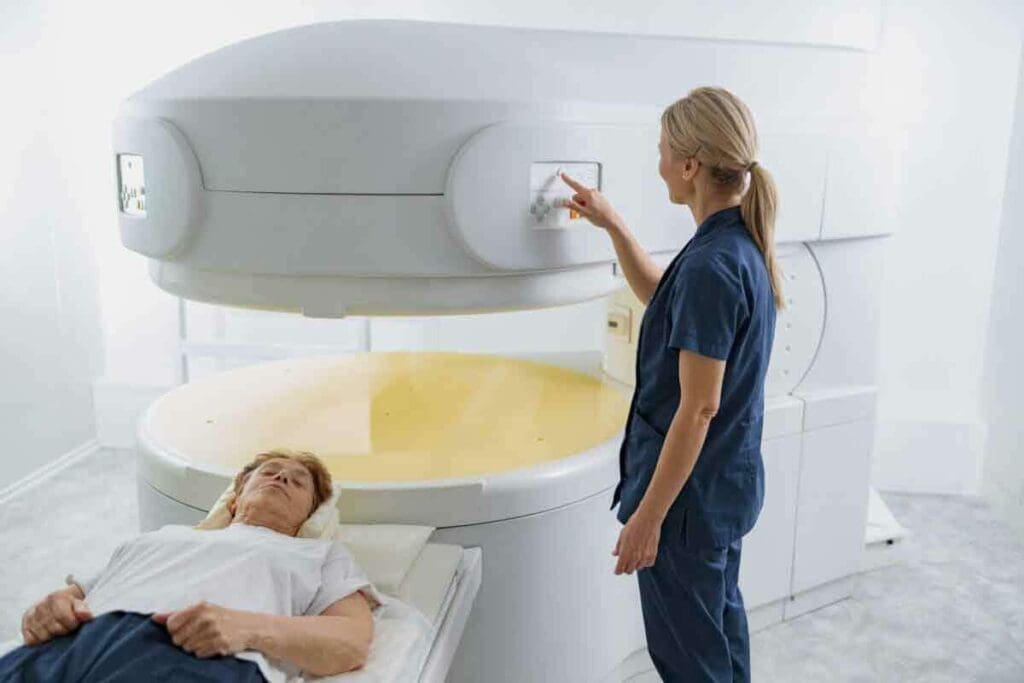
Magnetic Resonance Imaging (MRI) is a top-notch tool for looking inside the body. It uses strong magnetic fields and radio waves to make detailed pictures. This is super helpful in understanding the body’s structure, which is key in fighting cancer.
Magnetic Resonance Imaging Explained
MRI machines create strong magnetic fields that line up the body’s hydrogen atoms. Then, radio waves disturb these atoms, sending signals to the MRI. These signals help make detailed pictures of what’s inside the body.
How MRI Creates Detailed Images
Creating MRI images involves a few steps. First, the patient gets into the MRI machine. It makes a strong magnetic field. Next, radio waves are applied, making the hydrogen atoms send signals.
These signals are caught by the MRI machine. It uses them to make detailed pictures of the body’s inside. MRI is great at showing different soft tissues, which helps in finding tumors.
Primary Uses in Cancer Diagnosis
MRI is key in finding, understanding, and planning cancer treatment. It’s amazing at showing soft tissues, like tumors. This is super helpful for cancers in the brain, spine, and other soft tissues.
| Cancer Type | Use of MRI | Benefits |
| Brain Cancer | Detailed imaging of brain tumors | Accurate diagnosis and treatment planning |
| Spinal Cancer | Visualization of spinal tumors and their impact on surrounding tissues | Precise staging and monitoring of treatment response |
| Soft Tissue Sarcomas | Clear imaging of tumors within soft tissues | Effective treatment planning and assessment of tumor extent |
Understanding MRI technology and its role in cancer diagnosis is important. MRI is a vital tool in the fight against cancer. It gives doctors the info they need to make better treatment plans, helping patients get better.
Difference #1: Detection Capabilities – Functional vs. Structural Imaging
PET scans and MRI scans have different strengths. PET scans are great at showing how cells work and change. MRI scans, on the other hand, give detailed pictures of body structures.
PET Scans: Revealing Metabolic Activity and Cellular Changes
PET scans are key in finding cancer early. They spot changes in how cells use energy. This helps doctors find cancer before it grows much.
By using special tracers, PET scans show how active cells are. This helps doctors see if cancer is growing or shrinking.
MRI Scans: Providing Anatomical Detail
MRI scans give clear pictures of body parts. They are perfect for seeing tumors’ size and where they are. MRI scans are also great for soft tissues, helping diagnose and plan treatments for cancer.
When Each Approach Has the Advantage
Choosing between PET and MRI scans depends on what you need to know. PET scans are best for catching early signs of cancer. MRI scans are better for detailed body pictures needed for surgery.
| Imaging Modality | Primary Strength | Clinical Advantage |
| PET Scans | Functional Imaging | Early detection of metabolic changes in cancer cells |
| MRI Scans | Structural Imaging | Detailed anatomical information for surgical planning |
Knowing what each scan can do is key for doctors. By using PET and MRI scans together, doctors can make better plans for patients.
Difference #2: Image Detail and Resolution Comparison
PET and MRI scans have different strengths in showing details and resolution. These differences are key for doctors to make the best choices for patients.
MRI’s Superior Soft Tissue Visualization
MRI is great at showing soft tissues clearly. This helps look at organs and body structures. MRI’s clear images help doctors plan surgeries and see how far cancer has spread.
Key advantages of MRI in soft tissue visualization include:
- High contrast between different soft tissue types
- Detailed imaging of complex anatomical structures
- Ability to detect subtle changes in tissue characteristics
PET’s Ability to Detect Biochemical Changes
MRI shows the body’s structure well, but PET scans look at how tissues work. PET scans can spot cancer cells that MRI or CT scans might miss. This is important for seeing how tumors work and how well treatments are working.
The strengths of PET in detecting biochemical changes include:
- Early detection of cancerous changes at the cellular level
- Monitoring of treatment response through changes in metabolic activity
- Identification of metabolically active tumors that may not be evident on anatomical imaging
Comparative Strengths in Different Body Regions
Choosing between PET and MRI depends on the body part being scanned. MRI is better for the brain, spine, and muscles because it shows soft tissues well. PET scans are good for checking the whole body for cancer or seeing how tumors work in different places.
A comparative summary of PET and MRI strengths in different body regions is as follows:
| Body Region | PET Strengths | MRI Strengths |
| Brain | Detecting metabolic changes in brain tumors | High-resolution imaging of brain anatomy |
| Liver and Abdomen | Assessing tumor metabolism and spread | Detailed imaging of the liver and abdominal organs |
| Musculoskeletal System | Evaluating tumor activity and response to treatment | Excellent visualization of soft tissue and bone marrow |
Difference #3: Radiation Exposure Considerations
When we talk about cancer diagnosis, knowing about radiation is key. We look at PET scans and MRIs to see how they compare. Radiation exposure is a big factor in choosing between these tools.
Understanding PET Scan Radiation Levels
PET scans use a small amount of radiation from a radiotracer. The dose can change based on the radiotracer and how much is used. Most of the time, the radiation from a PET scan is safe for tests, but it’s something to think about, mainly for those needing many scans.
The radiotracer in PET scans sends out positrons. These positrons hit electrons, making gamma rays that the scanner catches. Even though the dose is low, it’s important to think about the risks of radiation.
MRI: The Radiation-Free Alternative
MRI doesn’t use harmful radiation, making it a radiation-free alternative. It uses strong magnetic fields and radio waves to show detailed images inside the body. This makes MRI great for those worried about radiation or those who need many tests.
Safety Profiles and Risk Assessment
Looking at PET scans and MRI, we see different safety concerns. PET scans have a small risk of cancer or genetic damage from radiation. MRI, without radiation, is better for those needing many tests or who are scared of radiation.
- PET Scans: Carry a small risk associated with radiation exposure.
- MRI: Offers a radiation-free alternative, ideal for repeated scans or radiation-sensitive patients.
The choice between PET scans and MRI for cancer diagnosis depends on many things. These include the cancer type, its stage, and what the patient needs. Knowing about radiation differences helps make better choices.
Difference #4: Advanced Hybrid Systems – PET/MRI vs. PET/CT
PET/MRI and PET/CT have changed cancer imaging a lot. They mix different imaging types for better cancer insights.
The Evolution of Combined Imaging Technologies
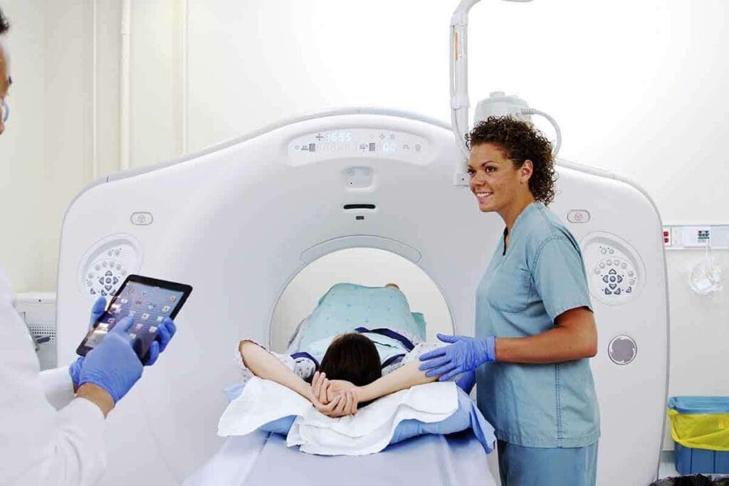
Hybrid imaging is a big step forward in medical imaging. PET/MRI and PET/CT combine PET with MRI or CT. This gives detailed views of tumors’ activity and structure.
PET/CT was first widely used. It shows cancer’s metabolism and anatomy in one scan. Now, PET/MRI is also popular. It gives better soft tissue views and less radiation for some patients.
Diagnostic Advantages of PET/MRI Systems
PET/MRI is great for soft tissue tumors and tricky areas. It mixes PET’s metabolic info with MRI’s detailed images. This makes diagnoses more accurate and confident.
PET/MRI’s main benefits are:
- Improved soft tissue contrast
- Less radiation than PET/CT
- Better tumor biology understanding
- Clearer views of complex areas
Impact on Patient Management and Treatment Planning
PET/MRI and PET/CT change how we manage and plan treatments. They give a full picture of tumors. This helps doctors make better plans.
| Imaging Modality | Key Strengths | Clinical Applications |
| PET/CT | Combines metabolic information with anatomical detail, widely available | Cancer staging, treatment response assessment, and detection of recurrence |
| PET/MRI | Superior soft tissue contrast, reduced radiation exposure | Evaluation of soft tissue tumors, pediatric and young adult patients, and complex anatomical regions |
Choosing between PET/CT and PET/MRI depends on many things. Like the cancer type, patient age, and what questions doctors have. As these techs get better, so will cancer diagnosis and treatment.
Difference #5: Cost and Accessibility Factors
Cancer patients often face a tough choice between PET scans and MRI. This choice is often influenced by cost. It’s important to understand the financial and accessibility aspects of these imaging options.
Comparing the Financial Aspects of PET and MRI
PET scans are usually more expensive than an MRI. This is because PET scans use a costly radiotracer. A PET scan can cost between $1,000 $3,000 or more. An MRI might cost between $400 $3,500, depending on the scan’s complexity and body part.
Here are some key financial considerations:
- Initial Cost: PET scans are generally more expensive upfront.
- Insurance Coverage: Varies widely depending on the provider and policy.
- Additional Costs: Interpretation fees, facility charges, and possible additional scans.
Insurance Coverage and Reimbursement Considerations
Insurance coverage for PET scans and MRI can vary a lot. It depends on the insurance provider, policy, and whether the scan is medically necessary. Many plans cover both scans for cancer diagnosis, but coverage and costs can differ.
It’s essential for patients to:
- Check their insurance policy details.
- Understand the concept of “medical necessity” as it applies to their scans.
- Discuss possible out-of-pocket costs with their healthcare provider.
Availability and Access to Advanced Imaging
Getting to PET and MRI facilities can be hard, mainly in rural or underserved areas. MRI machines are more common, but PET scanners and PET/MRI systems are less so. This is because they are expensive and complex.
Key factors influencing accessibility include:
- Geographic Location: Urban areas typically have better access to advanced imaging.
- Healthcare Facility: Larger hospitals and specialized diagnostic centers are more likely to have these technologies.
- Waiting Times: Can vary significantly depending on the facility and urgency of the scan.
Understanding these factors can help patients and healthcare providers make informed decisions about PET scans and MRI in cancer diagnosis and treatment planning.
MRI vs PET Scan for Cancer: Clinical Decision-Making
Choosing between MRI and PET scans for cancer imaging depends on the cancer type and the situation. We’ll look at how different cancers and situations affect the choice of imaging.
Cancer Types and Optimal Imaging Selection
Different cancers need different imaging methods. For example, brain tumors are often better seen with MRI because it shows soft tissues well. PET scans are often used for detecting and staging cancers like lymphoma, showing metabolic activity.
- Brain and spinal cord tumors: MRI is usually preferred for its detailed soft tissue view.
- Lymphoma and certain other cancers: PET scans are often used for staging and checking treatment response.
- Breast cancer: Both MRI and PET scans have roles, with MRI useful for high-risk patients and PET for detecting distant metastases.
Staging vs. Treatment Monitoring Considerations
The purpose of imaging is key in choosing between MRI and PET scans. For initial staging, PET scans give valuable information on cancer spread. For monitoring treatment response, both modalities are used, but PET scans can offer early insights into metabolic changes.
- Initial Staging: PET scans are often used to assess the extent of cancer spread.
- Treatment Monitoring: Both MRI and PET scans are utilized, with PET providing early metabolic response information.
When Doctors Recommend Both Technologies
In some cases, doctors may recommend using both MRI and PET scans. This combined approach can give a more complete understanding of the cancer. For example, using PET/MRI hybrid systems can offer both metabolic information and detailed anatomical images in one session.
By understanding the strengths of each imaging modality and considering the specific clinical context, healthcare providers can make informed decisions that optimize patient care.
Difference #6: Technology Differences Between PET and MRI Machines
It’s important to know how PET and MRI machines work differently. This helps us understand their roles in finding cancer. Each machine captures body images in its own way, giving us different kinds of information.
PET Scanner Design and Operation
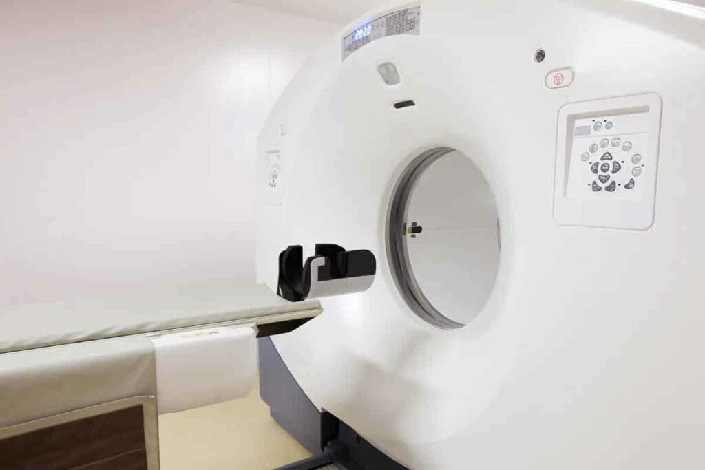
PET scanners find radiation from special tracers in growing cancer cells. They have a ring of detectors that catch gamma rays from these tracers. This lets them make images of where cancer might be.
To use a PET scanner, a tracer is given through an IV. Then, the patient lies in the scanner. The detectors find the tiny amounts of tracer, helping spot cancer early.
MRI Machine Components and Functionality
MRI machines use a strong magnetic field and radio waves to show us what’s inside. They have a superconducting magnet, gradient coils, and radiofrequency coils. The magnetic field lines up hydrogen nuclei, which radio waves disturb to create signals for images.
MRI machines are great at showing soft tissues, blood vessels, and more. They help us see how organs and tissues work. MRI also shows us blood flow and how things move.
Technical Innovations Improving Both Technologies
New tech has made PET and MRI better. PET scanners now catch more detail and are more precise. MRI machines can scan faster and show more detail, too.
One big step is the PET/MRI combo. It mixes PET’s metabolic info with MRI’s detailed images. This combo gives us a clearer picture of cancer, helping doctors plan better treatments.
As tech keeps getting better, we’ll see even more improvements. This means better care for cancer patients with more accurate diagnoses and treatments.
Difference #7: Early Detection Capabilities and Future Developments
Finding cancer early is key to treating it well. Both PET scans and MRI are good at this. Early detection helps patients get better faster.
PET’s Advantage in Detecting Early Cellular Changes
PET scans are great at spotting cancer’s early signs. They show metabolic activity, catching changes before they’re visible. This is super helpful for catching cancer early, when it’s easier to treat.
Key benefits of PET scans in early detection include:
- Ability to detect metabolic changes before structural changes occur
- Effective in identifying cancer spread to lymph nodes or distant sites
- Useful in monitoring response to treatment early on
Emerging MRI Techniques for Improved Cancer Detection
MRI is also getting better at finding cancer. New MRI methods, like diffusion-weighted imaging, make MRI even better at spotting cancer. These advancements help MRI find cancer more accurately.
Some of the promising developments in MRI include:
- Improved soft tissue characterization
- Enhanced detection of small lesions
- Better differentiation between malignant and benign lesions
Research Advances Shaping the Future of Cancer Imaging
Research on PET and MRI is always moving forward. New tools and techniques are being created to make images clearer and more accurate. This means we’ll soon be able to find cancer even sooner.
As these technologies get better, we’ll see:
- Earlier detection of cancer recurrence
- More accurate assessment of treatment response
- Better characterization of tumor biology
By using the best of PET and MRI, we’re getting closer to a future where cancer is easier to find and treat.
Conclusion: Making an Informed Choice for Your Cancer Care
Understanding the differences between PET and MRI scans is kkey tocancer care. Each scan has its own strengths. Patients can choose the best tool for their needs with their healthcare team.
Choosing between PET and MRI scans depends on several factors. These include the cancer type, its stage, and the imaging type needed. PET scans are great for spotting metabolic changes. MRI gives detailed images of the body’s structure.
Deciding between PET and MRI scans is a team effort. Patients and their healthcare team must work together. This way, individuals can face their cancer diagnosis with confidence, knowing they have the right tools for their situation.
FAQ
What is the main difference between a PET scan and an MRI?
PET scans show how cells work by looking at their activity. MRI, on the other hand, gives detailed pictures of soft tissues.
Is a PET scan better than an MRI for cancer diagnosis?
It’s not about which one is better. Both PET scans and MRIs have their own uses. PET scans find cancer by looking at cell activity. MRI gives detailed pictures of soft tissues.
What are the advantages of using PET/MRI hybrid systems over PET/CT?
PET/MRI systems are better at showing soft tissues and use less radiation. This makes them great for some cancers and for patients needing many scans.
How does radiation exposure differ between PET scans and MRI?
PET scans use a small amount of radiation from a tracer. MRI doesn’t use radiation, making it safer.
Can both PET scans and MRI be used together for cancer diagnosis?
Yes, using PET and MRI together can give a full picture. PET shows how cells work, and MRI shows the body’s structure.
How do PET scans detect cancer?
PET scans find cancer by looking for active cells. They use a tracer that shows up in these cells during the scan.
Are PET scans or MRIs more expensive?
PET scans are usually pricier than MRI scans. This is because of the cost of the tracer used in PET scans. Prices can change based on where you are and your insurance.
What factors influence the choice between PET and MRI for cancer patients?
Many things decide whether to use PET or MRI. These include the cancer type, its stage, and the patient’s health.
How are emerging MRI techniques improving cancer detection?
New MRI methods, like diffusion-weighted imaging, help find and understand cancers better. They offer more accurate and early detection.
What is the role of PET scans in early cancer detection?
PET scans can spot early changes in cells that might turn into cancer. This can help catch cancer when it’s easier to treat.
How do insurance coverage and reimbursement considerations affect the choice between PET and MRI scans?
Insurance and how much you pay can affect your choice. Coverage and costs vary by insurer and the situation.
What are the technological differences between PET and MRI machines?
PET machines look for gamma rays from a tracer. MRI uses magnetic fields and radio waves to create images. These machines work in very different ways.
References
- Cherry, S. R., Sorenson, J. A., & Phelps, M. E. (2018). Physics in nuclear medicine (4th ed.). Elsevier. https://www.sciencedirect.com/book/9780323356417/physics-in-nuclear-medicine
- McRobbie, D. W., Moore, E. A., Graves, M. J., & Prince, M. R. (2017). MRI from picture to proton (3rd ed.). Cambridge University Press. https://www.cambridge.org/core/books/mri-from-picture-to-proton/0E0F2627E51B1E5D3D42679A9BF3D5CB


