Last Updated on November 27, 2025 by Bilal Hasdemir
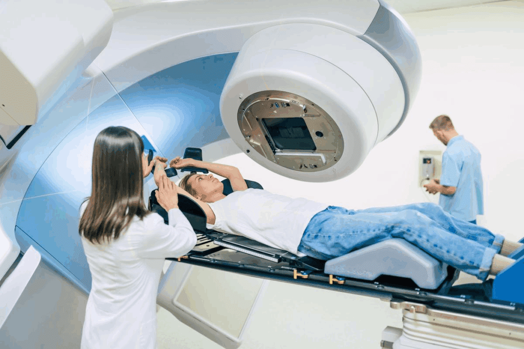
Medical imaging has changed how we find and treat diseases. PET scans are key in seeing how active the body’s cells are.
FDG plays a central role in PET scans, particularly for detecting and monitoring diseases such as cancer. FDG PET scans show how tissues take up glucose. This helps doctors understand what’s happening inside the body.
It’s important for both patients and doctors to know about PET scans and FDG. Knowing what PET scan meaning is can help people understand their health better.
Key Takeaways
- PET scans assess metabolic activity in the body.
- FDG is a tracer used to visualize glucose uptake in tissues.
- FDG PET scans are key in finding and tracking cancer.
- Understanding PET scan results is vital for diagnosis and treatment.
- FDG PET imaging gives insights into metabolic processes.
The Fundamentals of PET Scan Technology
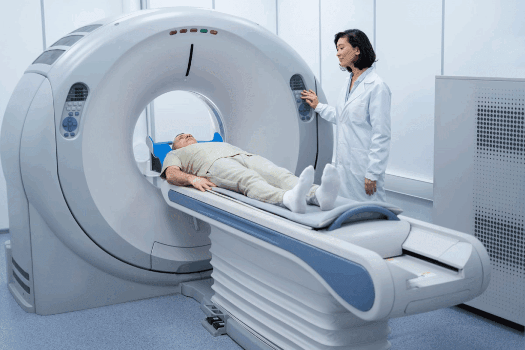
Positron Emission Tomography (PET) scan technology has changed how we diagnose diseases. It shows detailed metabolic activities in the body. This technology is key in nuclear medicine, giving doctors new ways to diagnose.
Basic Principles of Positron Emission Tomography
PET scan technology works by detecting positron emissions from a special drug in the body. The most used drug is Fluorodeoxyglucose (FDG). It builds up in areas that are very active.
When FDG’s positrons meet electrons, they create gamma rays. These rays are caught by the PET scanner. This lets us make detailed maps of metabolic activity.
The steps include giving the drug, the body taking it up, and catching the radiation. This data is turned into images that help doctors diagnose.
Historical Development of PET Scanning
PET scanning started in the 1950s and 1960s. Early scanners were the first steps. Over time, better materials, algorithms, and drugs have made PET scans more useful.
Now, PET scans are vital in many fields like oncology, neurology, and cardiology. As PET tech gets better, we’ll see clearer images and faster scans. New drugs will also help doctors diagnose and treat diseases better.
What is FDG in Medical Imaging?
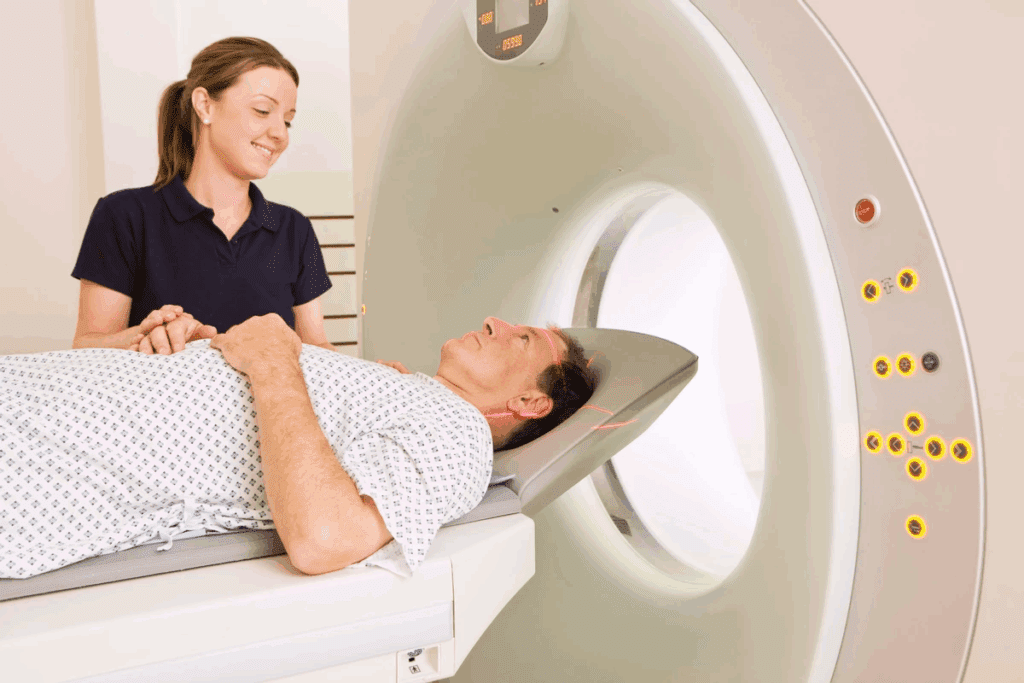
Fluorodeoxyglucose, or FDG, is a glucose-like substance that has changed medical imaging. It’s used in Positron Emission Tomography (PET) scans to see how tissues work. FDG PET imaging is key for finding and treating diseases, like cancer.
Chemical Structure and Properties of Fluorodeoxyglucose
FDG is a made-up glucose molecule with a fluorine-18 atom instead of a hydroxyl group. This makes it fluorodeoxyglucose. It’s perfect for studying how cells use glucose because it gets taken in but not broken down.
How FDG Mimics Glucose in the Body
FDG acts like glucose by being taken into cells the same way. Inside, it gets changed into FDG-6-phosphate by hexokinase. Unlike glucose, this form of FDG stays in the cell, helping us see how cells use glucose.
Production and Synthesis of FDG
Making FDG involves using fluorine-18, a radioactive isotope. This process is complex and needs special places. FDG is made in a cyclotron where fluorine-18 is added to deoxyglucose. This makes sure FDG is pure and ready for medical use.
FDG has greatly improved how we diagnose and track diseases, thanks to PET scans. Its ability to show metabolic activity is very useful in oncology and other areas of medicine.
The Science Behind FDG PET Scans
FDG PET scans work by trapping FDG in cells and detecting positron emissions. This science is key to understanding how PET scans help doctors diagnose and monitor diseases.
Metabolic Trapping of FDG in Cells
FDG, or fluorodeoxyglucose, is a glucose-like substance that cells take up. Inside the cell, it gets stuck as FDG-6-phosphate. This lets doctors see how cells are using glucose.
This method is great for spotting high activity areas, like in some cancers. It helps doctors find and track diseases.
Detection of Positron Emission
PET scans rely on finding positron emissions. When FDG decays, it releases positrons. These positrons then collide with electrons, creating gamma photons.
PET scanners use detectors to find these gamma photons. They figure out where the positrons came from, showing where FDG is.
Image Reconstruction Techniques
Reconstructing images from PET scan data is essential. Special algorithms are used to correct for things like how the body absorbs the signal.
The final images show how active different parts of the body are. This helps doctors diagnose and keep track of many conditions. Improvements in these techniques have made PET scans even more useful.
The Complete FDG PET Scan Procedure
To get accurate results, the FDG PET scan procedure needs precision. It includes patient preparation, FDG administration, and the scanning protocol.
Patient Preparation Requirements
Proper patient preparation is key for quality PET scans. Patients usually fast for hours before the scan. This lowers their blood glucose levels.
High blood glucose can affect FDG uptake. Patients might also avoid strenuous exercise and limit caffeine and sugar intake before the scan.
Patients with diabetes need special care. They might adjust their medication or insulin doses. It’s also important to inform healthcare providers about any medications taken.
FDG Administration Process
Administering FDG is a critical step. It’s injected into a vein in the patient’s arm. The dose is calculated based on the patient’s weight and other factors.
After injection, patients wait about 60 minutes. This allows FDG to be absorbed by tissues. During this time, patients are asked to remain as relaxed as possible.
Scanning Protocol and Duration
The scanning protocol varies based on the application and body region. PET/CT scanners are used, combining PET and CT scans.
The scanning time varies. It can be a few minutes to 30-45 minutes. The time depends on the body region and the specific protocol.
| Scan Type | Typical Scanning Time | Key Considerations |
| Whole-body scan | 30-45 minutes | Requires patient to remain for an extended period |
| Brain scan | 15-30 minutes | May require special positioning and immobilization |
| Cardiac scan | 20-40 minutes | May involve additional steps like ECG gating |
Understanding the FDG PET scan procedure helps patients prepare. This knowledge reduces anxiety and ensures a smooth scan process.
Interpreting FDG Uptake on PET Scans
Understanding FDG PET scan results is key. It helps doctors diagnose and treat many health issues. Knowing what’s normal and what’s not is vital.
Normal Physiologic FDG Distribution
The body’s usual FDG uptake pattern is important to know. The brain, for example, uses a lot of glucose, so it shows high FDG uptake. The heart and liver also have different levels of FDG, based on diet and insulin.
It’s important to understand these patterns. This helps doctors tell the difference between normal and abnormal uptake.
Abnormal FDG Uptake Patterns
Abnormal FDG uptake can mean cancer, infection, or inflammation. In cancer, tumors often take up more FDG because they use a lot of energy. The amount and where FDG is taken up can tell doctors a lot about the disease.
Doctors must look closely at these patterns. They need to match them with what they know about the patient’s health.
Standardized Uptake Value (SUV) Measurements
Standardized Uptake Value (SUV) gives a number for FDG uptake. It compares the activity in a certain area to the body’s weight and the dose of FDG. This helps doctors see how severe a disease is and how well it’s responding to treatment.
But, SUV can be affected by things like how the scanner is set up and how the patient is prepared. Combining SUV with visual checks helps doctors make better decisions. This makes FDG PET scans more useful in treating patients.
FDG Avidity: What It Means for Diagnosis
FDG avidity is key in reading PET scan results for disease diagnosis. It shows how much Fluorodeoxyglucose (FDG) a lesion takes up. This can tell us if a lesion is active or malignant.
Definition of FDG Avid Lesions
FDG avid lesions take up a lot of FDG in a PET scan. They show high metabolic activity, which can mean different diseases. The amount of FDG taken up can tell us a lot about the disease.
Characteristics of FDG Avid Lesions:
- High metabolic rate
- Increased glucose uptake
- Often associated with malignancy or active disease
Correlation Between FDG Avidity and Disease Activity
The level of FDG avidity can show how active a disease is. It helps doctors understand how severe and fast a disease is growing. Studies show that higher FDG avidity means more aggressive diseases.
| Disease State | Typical FDG Avidity | Clinical Implication |
| Malignant Tumors | High | Aggressive disease, potentially requiring intensive treatment |
| Infectious Processes | Variable | May indicate active infection requiring antimicrobial therapy |
| Inflammatory Conditions | Moderate to High | May suggest active inflammation, potentially benefiting from anti-inflammatory treatment |
Differential Diagnosis of FDG Avid Findings
High FDG avidity often means cancer, but it’s not always the case. Other conditions like infections or inflammation can also show high FDG uptake. Doctors must look at the whole picture, including the patient’s symptoms and other tests, to make a diagnosis.
“The interpretation of FDG PET scans requires a broad understanding of the clinical context, as well as the limitations and possible errors of the method.” – Expert in Nuclear Medicine
Understanding FDG avidity helps doctors make better decisions for patients. Using PET scan results with other tests and the patient’s symptoms is key for the best care.
Clinical Applications of FDG PET Scans in Oncology
In oncology, FDG PET scans are key for finding cancer, figuring out how far it has spread, and checking how treatments work. They are very useful in managing many kinds of cancer.
Cancer Detection and Initial Staging
FDG PET scans are great at finding cancer because it uses a lot of energy. They help find the main tumor and any spread, which is important for knowing how serious the cancer is.
- Early Detection: FDG PET scans can spot cancer early, which helps with treatment.
- Accurate Staging: They show how far the cancer has spread, helping plan treatment and predict outcomes.
Treatment Response Monitoring
It’s important to see how well treatment is working in oncology. FDG PET scans show how active tumors are, helping doctors know if treatment is working.
- Early Assessment: Changes in FDG uptake can show if treatment is working early on.
- Treatment Adjustment: Based on FDG PET scan results, treatment plans can be changed to get better results.
Recurrence Surveillance
After treatment, it’s important to watch for cancer coming back. FDG PET scans help find cancer again, often before symptoms show up or other tests find anything.
- Early Detection of Recurrence: Regular FDG PET scans can find cancer coming back early, when it’s easier to treat.
- Guiding Further Treatment: Info from FDG PET scans helps plan the right treatment for cancer that comes back.
Common Cancer Types Evaluated with FDG PET
FDG PET scans help with many cancers, including:
- Lymphoma
- Lung cancer
- Breast cancer
- Colorectal cancer
- Melanoma
These cancers all benefit from the metabolic info FDG PET scans give. It helps with diagnosis, figuring out how serious the cancer is, and planning treatment.
FDG PET Scans in Neurological Disorders
FDG PET scans have changed neurology by showing how the brain works. They help doctors diagnose and treat many brain conditions.
Alzheimer’s Disease and Dementia Evaluation
These scans are key in checking for Alzheimer’s and dementia. They spot changes in brain sugar use that show different brain diseases.
Key Features:
- Reduced glucose metabolism in temporal and parietal lobes
- Characteristic patterns for differential diagnosis
- Early detection of metabolic changes
Epilepsy Focus Localization
For epilepsy, FDG PET scans find where seizures start. This is important for surgery in hard-to-treat cases.
Benefits:
- Identification of hypometabolic regions
- Guiding surgical planning
- Monitoring treatment response
Brain Tumor Assessment
FDG PET scans help with brain tumor checks. They tell if a tumor is fast-growing or slow.
| Tumor Type | FDG Uptake | Clinical Implication |
| High-grade tumor | High | Aggressive, requires intensive treatment |
| Low-grade tumor | Low | Less aggressive, may be monitored |
Parkinson’s Disease and Movement Disorders
FDG PET scans help with Parkinson’s and other movement issues. They look at brain metabolism patterns.
FDG PET scans have greatly helped neurology. They give detailed brain info. This helps doctors make better diagnoses and plans.
Cardiac Applications of FDG PET Imaging
FDG PET imaging is key in diagnosing and managing heart diseases. It helps doctors understand different heart conditions. This information is vital for making treatment plans.
Myocardial Viability Assessment
FDG PET imaging is essential for checking if heart muscle is alive but not working well. This is called hibernating myocardium. Knowing this helps doctors decide if a patient needs heart surgery.
Doctors use FDG PET to see how the heart uses glucose. Alive heart muscle uses more glucose, showing up on the scan. Scar tissue, on the other hand, uses less or no glucose.
| Condition | FDG Uptake | Clinical Implication |
| Viable Myocardium | Preserved or Increased | Potential benefit from revascularization |
| Scar Tissue | Reduced or Absent | Limited benefit from revascularization |
Cardiac Sarcoidosis Evaluation
Cardiac sarcoidosis is a condition where the heart gets inflamed. FDG PET is great for finding and tracking this inflammation. It shows up as increased FDG uptake.
“FDG PET/CT has emerged as a valuable tool for assessing cardiac involvement in sarcoidosis, providing both anatomical and functional information.”
Source: Expert Review in Cardiovascular Imaging
Using FDG PET helps doctors catch cardiac sarcoidosis early. It also helps track how well treatments are working.
Cardiac Inflammation and Infection
FDG PET imaging is also used for heart inflammation and infection. This includes conditions like myocarditis and endocarditis. Increased FDG uptake means there’s active inflammation or infection.
FDG PET’s ability to spot heart inflammation and infection is very helpful. It helps doctors manage and treat heart diseases better.
Limitations and Challenges of FDG PET Scans
FDG PET scans are very useful in medical diagnostics. But, they have their own challenges. Knowing these challenges is key for accurate use and results.
False Positive and False Negative Results
FDG PET scans can sometimes show false positives and negatives. False positives might look like cancer but are not. This can happen when there’s inflammation or infection. False negatives occur when tumors are too small or have low activity.
Experts say the accuracy of FDG PET scans depends on many things. This includes the type of cancer and other diseases present.
To deal with these issues, doctors use other imaging and clinical info. For example, combining PET with CT or MRI helps. This gives both functional and anatomical details.
Physiologic Variants That Mimic Disease
Some normal conditions can look like disease on FDG PET scans. For example, brown fat can take up FDG like cancer. Muscle activity or inflammation in the gut can also confuse scan results.
It’s important for doctors to know about these normal variants. This helps in making accurate diagnoses.
Technical Limitations
Technical issues also affect FDG PET scans. Scanner quality, algorithms, and patient movement can impact results. New technology, like digital PET systems, is improving these areas.
Patient-Related Factors Affecting Scan Quality
Things about the patient can also affect scan quality. Blood sugar levels, body size, and movement can all play a part. High blood sugar, for example, can make scans less sensitive.
Improving patient preparation and scanning methods can help. As the field grows, understanding and fixing these issues is vital for better use of FDG PET scans.
Combining FDG PET with CT or MRI
FDG PET, when paired with CT or MRI, becomes a powerful tool for diagnosis. It combines metabolic and anatomical details. This hybrid imaging is key in clinical practice, giving a deeper look into diseases.
Benefits of Hybrid Imaging
Hybrid imaging, combining FDG PET with CT or MRI, has many benefits. One major advantage is better diagnostic accuracy. It mixes metabolic activity from FDG PET with detailed anatomy from CT or MRI.
This approach helps pinpoint FDG-avid lesions accurately. It’s vital for precise diagnosis and treatment plans. It also helps track treatment success and catch disease return early.
PET/CT vs. PET/MRI Considerations
Choosing between PET/CT and PET/MRI depends on several factors. PET/CT is faster and more common, making it a top pick for many, like in oncology.
PET/MRI, though, shines in soft tissue contrast, which is great for brain scans or soft tissue tumors. It also has less radiation, as MRI doesn’t use ionizing radiation.
Clinical Impact of Integrated Imaging
The impact of FDG PET with CT or MRI on patient care is huge. In oncology, it’s improved cancer staging, monitoring, and catching recurrence. It’s also valuable in neurology and cardiology, giving insights into complex diseases.
As technology advances, hybrid imaging’s role in healthcare will grow. It promises better patient care and outcomes in the future.
Safety and Side Effects of FDG PET Scans
FDG PET scans are mostly safe. But, there are things to know. These include radiation risks, possible side effects from the tracer, and when to avoid them.
Radiation Exposure Concerns
FDG PET scans use a small amount of radiation. This is because the tracer is radioactive. The dose is similar to other scans that use radiation, like CT scans.
It’s important to think about the benefits of the scan against the risks of radiation. This helps make a safe choice.
- The radiation dose from an FDG PET scan is typically in the range of 7-14 mSv.
- Pregnant women and children are more sensitive to radiation, and special precautions are taken.
- The risk of radiation-induced cancer is a consideration, but it’s low.
Potential Adverse Reactions to FDG
Reactions to FDG are rare but can happen. Most are mild. They might include:
- Allergic reactions, such as rash or itching
- Nausea or vomiting
- Headache or dizziness
In rare cases, more serious reactions can happen. Patients are watched closely during and after the scan.
Contraindications and Precautions
Some conditions or situations might mean you can’t have an FDG PET scan. Or you might need extra care:
- Pregnancy and breastfeeding: Special steps are taken to keep risks low.
- Diabetes: Blood sugar levels can affect how the tracer works. Management plans are used.
- Recent chemotherapy or radiation therapy: It can change how results are seen.
It’s key to tell your doctor about any health issues, allergies, or medicines before the scan.
Knowing these details helps patients and doctors make smart choices. This way, the scan’s benefits are maximized, and risks are kept low.
Cost and Insurance Coverage for FDG PET Scans
Knowing the cost of FDG PET scans is key for those thinking about or needing them. The price can change a lot. This depends on where you are, the facility, and if it’s with other tests like CT or MRI.
Average Costs in the United States
In the U.S., FDG PET scan prices vary a lot. They can be between $1,000 to $3,000 or more per scan. It’s best to ask your doctor or the imaging center for a precise quote.
Medicare and Private Insurance Coverage
Usually cover FDG PET scans for some health issues, like cancer. But, the details can differ a lot between plans. Might cover it for certain reasons, based on your health and doctor’s advice.
Has its own rules for FDG PET scans. This includes needing preauthorization, which is a big step before the scan.
Preauthorization Requirements
Preauthorization means the insurance company needs to okay the scan before it happens. Not getting approval can lead to denied claims or higher costs for you. It’s important to work with your doctor to get this approval.
Knowing about preauthorization and your insurance coverage is key to avoid surprise medical bills.
What Patients Should Know About FDG PET Scan Results
Getting your FDG PET scan results can be tough without help. Knowing what your scan says is key to understanding your health. It helps you know what treatments you might need and what to do next.
Understanding Your Radiology Report
Your radiology report is a detailed document from your FDG PET scan. It shows where FDG is in your body, highlighting normal and abnormal areas. The report has sections on the procedure, findings, and impression.
The “findings” section talks about FDG uptake in your body. The “impression” section gives an interpretation of these findings. It might include a diagnosis or suggestions for more tests.
Common Terminology in FDG PET Reporting
FDG PET reports use special terms that might be hard to understand. Terms like “hypermetabolic activity” or “increased FDG avidity” mean higher glucose uptake. This can be a sign of disease.
Knowing these terms helps you understand your report better. For example, “SUV” or Standardized Uptake Value shows how much FDG is taken up. A higher SUV might mean more aggressive disease.
Follow-up Recommendations
Your healthcare provider might suggest more tests or treatments based on your scan. These suggestions are based on your condition and health.
These actions could include more imaging, biopsies, or specialist visits. It’s important to follow these steps for the right care.
Questions to Ask Your Doctor
When talking to your doctor about your scan, have questions ready. Asking the right questions helps you understand and feel better.
- What do my FDG PET scan results indicate about my condition?
- Are there any areas of concern that need further evaluation?
- What are the next steps in my treatment or management plan?
- Are there any additional tests or procedures required?
By asking these questions, you’ll get a clearer picture of your health and what to do next.
A leading oncologist says, “Understanding your PET scan results is key to your care. It’s not just about the numbers or images; it’s about what they mean for your health and well-being.”
Advances in FDG PET Technology and Future Directions
FDG PET technology is changing fast, thanks to new digital PET systems and artificial intelligence. These changes make PET scans more accurate and reliable.
Digital PET Systems
Digital PET systems are a big step up from old PET systems. They use new detector tech for better sensitivity and detail. This means clearer images and spotting small issues easier.
Key benefits of digital PET systems include:
- Improved image quality
- Enhanced sensitivity for detecting small lesions
- Faster scanning times
Artificial Intelligence in PET Interpretation
Artificial intelligence (AI) is being used more in PET imaging. AI can quickly look through lots of data, finding patterns humans might miss.
The role of AI in PET interpretation includes:
- Assisting in the detection of abnormalities
- Providing quantitative analysis of PET data
- Enhancing consistency in image interpretation
Novel Reconstruction Algorithms
New algorithms are also key in making PET images better. They help cut down on noise and improve detail, leading to more accurate diagnoses.
Next-Generation Radiotracers Beyond FDG
Even though FDG is common, scientists are working on new radiotracers. These aim to target specific diseases or biological processes better.
| Technology | Description | Benefits |
| Digital PET Systems | Advanced detector technology for higher sensitivity and resolution | Improved image quality, better detection of small lesions |
| Artificial Intelligence | AI algorithms for analyzing PET data | Enhanced accuracy, quantitative analysis, consistency in interpretation |
| Novel Reconstruction Algorithms | Algorithms to reduce noise and improve resolution | More accurate diagnoses, improved image quality |
| Next-Generation Radiotracers | New radiotracers targeting specific diseases or processes | More effective disease targeting, improved diagnostic specificity |
Conclusion
FDG PET scans have changed how we look at diseases. They give doctors important info for diagnosing and treating many illnesses. This is true for cancers, brain issues, and heart problems.
Knowing how FDG PET scans work is key for doctors and patients. They help find cancers early and check how treatments are working. They also help with brain and heart conditions.
In short, FDG PET scans are a big deal in medicine. They help doctors make better choices by showing how cells work. As tech gets better, FDG PET scans will help even more, making care better for everyone.
FAQ
What does FDG stand for in a PET scan?
FDG stands for Fluorodeoxyglucose. It’s a glucose analog used in PET scans to check metabolic activity in the body.
What is the purpose of a PET scan?
A PET scan helps diagnose and track various medical conditions. It’s mainly used for cancer by looking at metabolic activity in the body.
How does FDG work in a PET scan?
FDG is taken up by cells, like cancer cells, because of their high metabolic activity. The PET scan picks up the positron emissions from FDG. This creates images that show where the activity is high.
What is FDG avidity, and what does it mean for diagnosis?
FDG avidity shows how much a lesion or tissue takes up FDG. High avidity means high metabolic activity. This is often seen in cancer or other diseases.
How is FDG PET used in oncology?
In oncology, FDG PET is used for cancer detection, staging, tracking treatment response, and watching for recurrence.
What are the limitations of FDG PET scans?
FDG PET scans can have false positives and negatives. They can also be affected by normal body functions and patient factors.
How does FDG PET compare to other imaging modalities?
FDG PET gives unique metabolic activity information. It complements CT or MRI anatomical details. Hybrid imaging like PET/CT and PET/MRI combines different modalities’ strengths.
What are the safety concerns associated with FDG PET scans?
Safety concerns include radiation exposure and possible adverse reactions to FDG. There are also contraindications like pregnancy or severe diabetes.
How much does an FDG PET scan cost, and is it covered by insurance?
The cost of an FDG PET scan varies. It’s usually covered approved uses, after preauthorization.
How can patients understand their FDG PET scan results?
Patients should discuss their radiology report with their doctor. They should understand the terms and any follow-up steps.
What are the future directions in FDG PET technology?
Future developments include digital PET systems and artificial intelligence in PET interpretation. There’s also research on new radiotracers beyond FDG.
What is the role of FDG PET in neurological disorders?
In neurology, FDG PET helps evaluate Alzheimer’s disease, find epilepsy foci, assess brain tumors, and manage Parkinson’s disease and other movement disorders.
How is FDG PET used in cardiac applications?
In cardiology, FDG PET assesses myocardial viability, evaluates cardiac sarcoidosis, and diagnoses cardiac inflammation and infection.
Reference
- Love, C., Tomas, M. B., Tronco, G. G., & Palestro, C. J. (2005). FDG PET of infection and inflammation. Radiographics, 25(5), 1357–1368. https://pubmed.ncbi.nlm.nih.gov/16244225/


