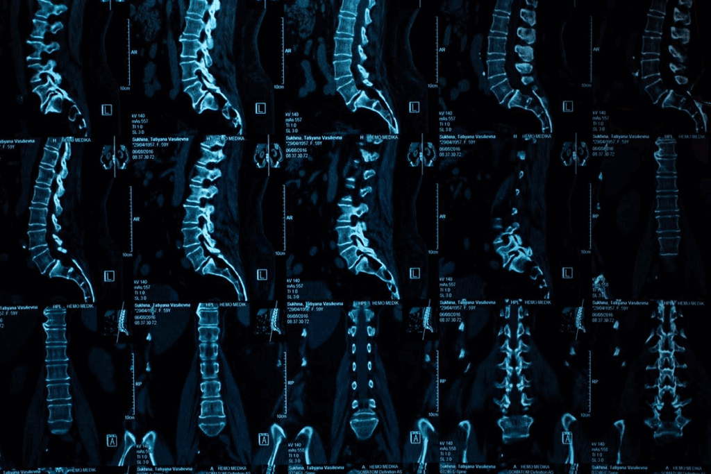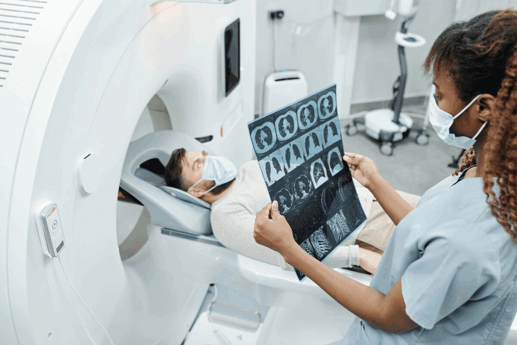
Did you know a herniated disc can cause a lot of pain? It affects your daily life and how you move. This happens when the soft center of the disc leaks out through a tear in the outer layer. It can irritate nearby nerves. Discover how a CT Scan Herniated Disc helps doctors identify disc issues, nerve pressure, and treatment plans.
Medical data shows this condition can lead to pain in areas related to the spine. For example, hip pain can occur if the herniation is in the lower back.
Getting an accurate diagnosis for a herniated disc is very important. Imaging tests are key in this process. We will look at how a CT scan can help diagnose this condition. We will also compare it with other imaging tests.
Key Takeaways
- A herniated disc can cause significant pain and affect mobility.
- The condition can lead to pain in areas related to the affected spine region.
- Imaging tests are critical for diagnosing a herniated disc.
- A CT scan can be used to diagnose a herniated disc.
- The effectiveness of a CT scan is compared with other imaging tests for diagnosis.
- Accurate diagnosis is key to effective treatment of a herniated disc.
Understanding Herniated Discs
It’s important to know about herniated discs to treat back pain well. This condition affects the spine and causes pain for many people.
What is a Herniated Disc?
A herniated disc happens when the soft center of the disc bulges out. This is due to a tear or crack in the outer layer. It can press on nerves, leading to pain, numbness, or weakness in the back or legs.
Common Causes of Disc Herniation
Disc herniation can come from many things, like aging, injury, or genetics. Degenerative changes make discs more likely to herniate. Trauma, like a fall or heavy lifting, can also cause it.
Medical literature says, “the risk of disc herniation goes up with age. This is because discs naturally wear down over time.”
Lifestyle and biomechanical stresses can also play a part. Knowing these causes helps in preventing and treating herniated discs.
Symptoms and Signs of Herniated Discs

It’s important to know the signs and symptoms of herniated discs. These can affect your daily life in different ways. The severity of symptoms can vary.
Pain Patterns and Neurological Symptoms
Pain from herniated discs often starts in the lower back. It can spread to the hips and legs. This pain is called radiculopathy.
It happens when the disc presses on nerves. You might also feel numbness, tingling, or weakness in your limbs. These are neurological symptoms.
The nerves from the spine to the body are affected. The symptoms and how bad they are depend on the disc’s location and the nerves it touches.
When to Seek Medical Attention
Knowing when to see a doctor is key. If your pain is severe and doesn’t get better with rest, seek help. Also, if you have weakness, numbness, or trouble with your bladder or bowels, get medical attention right away.
Seeing a doctor early can help a lot. Here’s a table with symptoms and when to see a doctor:
| Symptom | Description | When to Seek Help |
| Pain | Lower back pain radiating to hip and leg | If severe or persistent |
| Numbness/Tingling | Numbness or tingling sensations in limbs | If progressive or severe |
| Weakness | Muscle weakness in affected limbs | If significant or worsening |
By knowing these symptoms and when to see a doctor, you can manage your condition better.
The Diagnostic Process for Herniated Discs
When someone shows signs of a herniated disc, we follow a few key steps. Finding out if a disc is herniated is key to picking the right treatment.
Initial Clinical Evaluation
The first thing we do is a detailed clinical evaluation. We learn about the patient’s health history and any past injuries. We also check their symptoms to see how the herniated disc might be affecting them.
Physical Examination Techniques
A physical exam is a big part of figuring out what’s going on. We check the patient’s nerves, muscles, and reflexes. This helps us see if the herniated disc is causing nerve problems.
Some common tests we use include:
- Straight leg raise test
- Muscle strength testing
- Reflex testing
When Imaging is Necessary
Even with a thorough exam, sometimes we need imaging tests to confirm a herniated disc. The type of test we choose depends on the patient’s symptoms and health history.
| Imaging Modality | Advantages | Disadvantages |
| X-ray | Quick and widely available | Limited soft tissue visualization |
| CT Scan | Good for bone detail, quick | Radiation exposure, less soft tissue detail |
| MRI | Excellent soft tissue detail, no radiation | More expensive, claustrophobic for some patients |
By looking at the results from exams, tests, and imaging, we can accurately find out if someone has a herniated disc. Then, we can plan the best treatment.
Overview of Spinal Imaging Options
Diagnosing spinal issues, like herniated discs, requires knowing the imaging options. The spine is complex, and imaging it needs techniques that show its details well.
X-rays and their limitations
X-rays are a traditional way to see the spine. They’re good for bones and finding fractures or misalignments. But, they can’t show soft tissues like discs well.
Limitations of X-rays:
- Poor visualization of soft tissues
- Inability to detect disc herniations directly
- Limited detail of spinal anatomy beyond bone structures
Advanced imaging techniques
New imaging methods have changed how we diagnose spinal issues. Computed Tomography (CT) scans and Magnetic Resonance Imaging (MRI) are key today. CT scans show bones well and can spot bone spurs or fractures. MRI is great for soft tissues, like discs, nerves, and the spinal cord.
Choosing the right imaging method
Choosing the right imaging depends on the patient’s condition and symptoms. For herniated discs, MRI is usually best because it shows soft tissues well. But, CT scans are useful for bones or when MRI isn’t possible. The choice between CT and MRI also depends on the patient’s health, what imaging is available, and what’s needed for diagnosis.
In summary, knowing the strengths and weaknesses of spinal imaging is key for accurate diagnosis and treatment. By picking the right imaging, healthcare providers can give patients the best care for their spinal issues.
CT Scan Herniated Disc Detection Capabilities
CT scans use X-rays to create detailed images of the body. They are great at finding herniated discs. This makes them a key tool for diagnosing spinal problems.
CT Scan Technology Explained
CT scans combine X-rays from different angles to show cross-sections of the body. This lets us see the spine’s details, like vertebrae and discs. It’s fast and doesn’t hurt, making it a great tool for doctors.
CT scans are good at showing both bones and soft tissues. This helps doctors spot herniated discs. These discs can press on nerves if they bulge out.
How CT Visualizes Spinal Structures
CT scans are great at showing the spine’s bones, like vertebrae and joints. They also give info on discs, but might miss soft tissue details. The images can show disc herniations and other problems.
When a CT scan looks for disc herniation, a radiologist checks the images. The detailed images help measure the disc and plan treatment.
Knowing how CT scans work helps doctors choose the right test for herniated discs. This ensures patients get the best care.
Accuracy of CT Scans for Diagnosing Herniated Discs

When it comes to diagnosing herniated discs, the accuracy of CT scans is key. CT scans are good for seeing some parts of the spine. But, how well they spot herniated discs is something doctors keep studying.
Detection Capabilities of CT Scans
CT scans use X-rays to make detailed spine images. They can show bone and some soft tissues. But, they’re not as good at showing soft tissues like discs as MRI is.
Accuracy Rates in Clinical Studies
Many studies have looked into how well CT scans diagnose herniated discs. Most say CT scans are not as accurate as MRI for this job. A study comparing CT and MRI found MRI was better at spotting lumbar disc herniation.
| Imaging Modality | Sensitivity | Specificity |
| CT Scan | 70-85% | 80-90% |
| MRI | 90-95% | 95-98% |
The table shows MRI is better at finding herniated discs than CT scans. Yet, CT scans are useful in some cases. This is when MRI is not available or not safe to use.
CT Scan vs MRI for Herniated Disc Diagnosis
Choosing between CT scans and MRI for herniated discs depends on what you need. CT scans are fast and show bones well. MRI is better for soft tissues like discs and nerves. Each has its own strengths for different needs.
Comparative Strengths and Weaknesses
Let’s look at what each can do. MRI is great for soft tissues, like discs and nerves. It spots problems like herniations and nerve compression well. CT scans, on the other hand, are better for bones and are quick, making them good for emergencies.
Key differences between CT scans and MRI include:
- Soft Tissue Visualization: MRI is better for soft tissues, like discs and nerves.
- Bone Imaging: CT scans are better for bones.
- Speed and Accessibility: CT scans are faster and easier to get to, which is good for emergencies.
- Contraindications: MRI might not work for people with metal implants or claustrophobia.
When CT is Preferred Over MRI
There are times when CT scans are better than MRI for herniated discs. In emergencies, CT scans are quick and show the spine well. They’re also good for people who can’t have MRI because of metal implants or claustrophobia.
CT scans are advantageous in these situations:
- Emergency Settings: CT scans are fast for quick spinal injury checks.
- Patients with MRI Contraindications: CT scans work for those who can’t have MRI.
- Bone Detail: CT scans are better for detailed bone images.
In summary, MRI is usually better for soft tissues, but CT scans have their own benefits. The right choice depends on the patient’s needs and the situation. It’s all about what each imaging method is best at.
The CT Scan Procedure for Spinal Imaging
The CT scan procedure for spinal imaging is simple. It includes several steps from preparation to the scan. We know getting a CT scan can make you nervous. So, we’re here to help you know what to expect.
Preparation for the Scan
Before your CT scan, you’ll get instructions on how to prepare. You might need to remove metal objects or jewelry. You’ll also be asked to wear a hospital gown. It’s important to follow these steps to ensure a safe and smooth scan.
Preparation typically involves:
- Removing metal objects and jewelry
- Wearing a hospital gown
- Following any specific dietary instructions if contrast is to be used
What Happens During the Scan
During the CT scan, you’ll lie on a table that slides into a large, doughnut-shaped machine. The machine will rotate around you, taking X-rays from different angles. You’ll need to stay very quiet and might be asked to hold your breath for a bit. The scan is usually very quick, lasting just a few minutes.
The CT scanner uses this data to create detailed cross-sectional images of your spine. These images can help diagnose many spinal conditions, including herniated discs.
Post-Scan Procedures
After the scan, you can usually go back to your normal activities. Unless you’ve been told not to, like if a contrast agent was used. The images from your scan will be reviewed by a radiologist. Then, your doctor will discuss the findings with you and suggest what to do next.
It’s important to know that while CT scans are safe, there is some radiation exposure. But, the benefits of the scan usually outweigh the risks, which is true for diagnosing serious conditions.
CT Scan With Contrast for Herniated Discs
Contrast-enhanced CT scans give a detailed look at the spine. They help find herniated discs. This method uses contrast agents to show specific parts of the spine.
When Contrast Agents Are Used
Contrast agents are not always needed for spine CT scans. But, they help in some cases. For example, if a herniated disc might be pressing on a nerve, the agent makes it easier to see.
Benefits of Contrast Agents:
- They make soft tissues and lesions stand out more
- Help show the spinal cord and nerve roots clearly
- Make it easier to diagnose complex spinal issues
Benefits and Risks of Contrast
Using contrast agents in CT scans has both good and bad sides. They make the scan better by showing important areas. But, there are risks like allergic reactions and concerns about radiation.
Potential Risks:
- Allergic reactions to the contrast
- Problems with the kidneys in those with existing issues
- Radiation exposure, though generally safe
We think about these points for each patient. We make sure contrast agents are used when it’s right and that patients know what’s happening.
In summary, CT scans with contrast are key for diagnosing herniated discs. They offer a clearer view of the spine. While there are risks, choosing the right patients and getting their consent makes this tool safe and effective.
Interpreting CT Scan Results for Disc Herniation
When we look at CT scan results, we search for signs of disc herniation. This includes disc bulging or fragments that have moved out. CT scans give us detailed images of the spine. This helps us see how the discs and surrounding areas are doing.
What Radiologists Look For
Radiologists check CT scans for herniated discs for specific things. They look at the shape and position of the discs. They check if there’s bulging or herniation that might be pressing on nerves.
They also look at the disc’s density to see if there are any fragments that have come out. We use special software to make these details clearer. This helps us make accurate diagnoses.
They also check for nerve root compression or irritation. This is important because it helps decide the best treatment.
Common Findings and Their Significance
CT scans often show disc bulging, protrusion, and extrusion. Disc bulging means the disc goes beyond its usual size but stays together. Protrusion is when the disc bulges but stays inside its outer layer. Extrusion is worse because the disc material breaks through the outer layer.
| Finding | Description | Significance |
| Disc Bulging | The disc extends beyond its normal boundaries. | May cause mild symptoms or be asymptomatic. |
| Disc Protrusion | Disc material bulges out but remains contained. | Can cause nerve compression and pain. |
| Disc Extrusion | Disc material breaks through the outer layer. | Often associated with significant nerve compression and severe symptoms. |
Knowing about these findings helps us figure out the best treatment. By understanding CT scan results, doctors can create treatment plans that fit each patient’s needs.
Limitations of CT Scans in Diagnosing Herniated Discs
CT scans have some big limitations when it comes to diagnosing herniated discs. They are great for seeing some parts of the spine, but they have their limits. These limits can affect how well they can diagnose problems.
Soft Tissue Visualization Challenges
One big problem with CT scans is seeing soft tissues. Soft tissues, like nerves and discs, are harder to see on CT scans than on MRI. This makes it tough to spot issues in these areas. For example, a herniated disc might not show up well on a CT scan, if it’s small or doesn’t press on other parts.
CT scans are better for seeing bones, which is why they’re good for finding bone spurs or breaks. But when it comes to soft tissues, they fall short. This is a big deal for diagnosing herniated discs, where seeing the soft tissues is key.
Radiation Exposure Considerations
Another big issue with CT scans is the radiation they use. CT scans use more radiation than regular X-rays, which is a worry, mainly for people who need lots of scans. We have to think about how much radiation is safe and what benefits it brings.
It’s important to think about how much radiation builds up, which is a bigger worry for younger people or those needing lots of scans. Other tests, like MRI, don’t use radiation and might be better in some cases. But CT scans are useful when the benefits are worth the risks, and other tests aren’t an option.
In short, CT scans are very useful, but knowing their limits is key for good care. By understanding these limits, doctors can choose the best tests for each patient.
Special Applications of CT for Complex Spine Cases
Advanced CT scan technologies are key in managing complex spine conditions. They give us detailed views of spinal structures. This helps us make accurate diagnoses and treatment plans. CT scans are very useful in complex spine cases.
CT Myelography
CT myelography combines CT scanning with myelography. It involves injecting contrast material into the spinal canal. This method gives us detailed images of the spinal cord and nerve roots.
We use CT myelography when other imaging methods don’t give enough information. It’s helpful for diagnosing complex spinal conditions like herniated discs and nerve root compression.
CT for Post-Surgical Evaluation
CT scans are great for checking the spine after surgery. They help see if spinal fusion was successful. They also spot any complications like hardware failure.
CT scans are often recommended after surgery. They show clear images of bone and implanted hardware. This helps us assess how well the surgery went and decide on further treatment.
| Application | CT Myelography | CT for Post-Surgical Evaluation |
| Purpose | Diagnose complex spinal conditions by visualizing the spinal cord and nerve roots. | Assess the spine after surgery, evaluating fusion success and detecting complications. |
| Contrast Use | Requires contrast material injected into the spinal canal. | May or may not use contrast, depending on the specific evaluation needs. |
| Diagnostic Benefits | Detailed images of spinal cord and nerve roots, helpful for diagnosing nerve root compression. | Clear visualization of bone and implanted hardware, aiding in assessing surgical outcomes. |
When Doctors Recommend CT Scans for Suspected Herniated Discs
Doctors look at many things before they suggest a CT scan for a herniated disc. This choice is key to finding out why someone has back pain and what treatment they need.
Patient-Specific Considerations
Doctors think about different things when deciding on a CT scan for herniated discs. They look at if the patient has metal implants, if they get scared in small spaces, and their overall health. For example, if someone has certain metal implants, an MRI might not work, so a CT scan is better.
They also think about how bad the patient’s symptoms are. If someone has really bad pain or neurological problems, they might need a scan right away to figure out what to do next.
| Patient Factor | Consideration for CT Scan |
| Presence of Metal Implants | CT scan is preferred over MRI for patients with certain metal implants. |
| Claustrophobia | CT scan is a more open and less confining option compared to MRI. |
| Severe Neurological Symptoms | Immediate CT scan may be necessary to guide urgent treatment. |
Availability and Accessibility Factors
The location and how easy it is to get a CT scan are also important. Doctors think about how close imaging centers are, how long it takes to get in, and how much it costs.
In emergencies, how fast you can get to a CT scan can really matter. Also, how much insurance covers and what you have to pay out of pocket can affect if you can get the scan.
By looking at these things, doctors can make smart choices about CT scans for herniated discs. This helps make sure patients get the right care.
Cost and Insurance Considerations for Spinal Imaging
The cost of spinal imaging can vary a lot. It’s important to look at insurance coverage and options. When you’re thinking about tests for herniated discs, knowing the costs is key.
Comparing Costs of Different Imaging Methods
Imaging methods have different prices. For example, CT scans and MRI scans cost differently. This is because of the technology and complexity involved.
| Imaging Modality | Average Cost Range (USD) | Typical Use Cases |
| X-ray | $100 – $300 | Initial assessment, bone fractures |
| CT Scan | $300 – $1,000 | Detailed bone imaging, complex diagnoses |
| MRI | $800 – $2,500 | Soft tissue imaging, detailed disc assessment |
Costs can change a lot. This depends on the imaging method, the technology at the center, and where you are.
Insurance Coverage for Diagnostic Imaging
Insurance coverage is key in managing costs for spinal imaging. Most plans cover imaging, but how much can vary.
Key factors influencing insurance coverage include:
- The type of insurance plan
- The specific imaging modality prescribed
- Whether the diagnostic center is in-network or out-of-network
Before getting imaging, check your insurance. This helps avoid surprise bills.
Treatment Options Following Herniated Disc Diagnosis
After finding out you have a herniated disc, you have many ways to treat it. You can choose from non-surgical methods to surgery. The right choice depends on how bad your symptoms are, where the herniation is, and your health.
Conservative Management Approaches
Most people start with non-surgical treatments for herniated discs. This method aims to lessen symptoms and help the body heal without surgery. It includes physical therapy, pain meds, and changes in your daily life to help you get better.
Physical Therapy: A physical therapist will create a special workout plan for you. This plan will help strengthen your spine muscles, improve flexibility, and aid in healing. You might do stretching, strengthening, and manual therapy exercises.
Pain Management: To ease pain and swelling, doctors often suggest over-the-counter pain relievers like NSAIDs. Sometimes, stronger medicines are needed for severe pain.
Surgical Interventions
If non-surgical methods don’t work, or if the herniation is serious, surgery might be needed. Surgery aims to take pressure off nerves and make the spine stable.
- Discectomy: This surgery removes the part of the disc that’s pressing on the nerve. It can be done with traditional surgery or newer, less invasive methods.
- Spinal Fusion: If the herniated disc has made the spine unstable, spinal fusion might be needed. This involves joining the affected vertebrae together to stabilize the spine.
We help patients choose the best treatment plan for them. We consider their unique situation and what they prefer. Our goal is to use the latest medical techniques to help you feel better and live a better life with herniated discs.
Conclusion
Getting a correct diagnosis is key to treating herniated discs well. This article has looked at how CT scans help diagnose herniated discs. We’ve talked about their strengths and what they can’t do.
CT scans give important details for diagnosing herniated discs in some cases. Whether to use CT scans or other imaging like MRI depends on the patient’s situation. It also depends on what the doctor needs to know.
In short, CT scans are important for diagnosing herniated discs. They help doctors make the best choices for each patient. This leads to better treatment plans for herniated disc problems.
FAQ
Can a CT scan detect a herniated disc?
Yes, a CT scan can spot a herniated disc. It uses X-rays to show detailed images of the spine. This includes the discs, vertebrae, and surrounding tissues.
How accurate are CT scans in diagnosing herniated discs?
CT scans are usually good at finding herniated discs. But, how well they work can depend on the disc’s location and how bad the herniation is. Studies show they work well, but MRI is often better.
What is the difference between a CT scan and an MRI for diagnosing herniated discs?
CT scans and MRI are both used to find herniated discs. CT scans are better at showing bones. MRI is better at showing soft tissues like discs. MRI is usually the best choice, but CT scans are used when MRI isn’t available.
Do I need to prepare for a CT scan to diagnose a herniated disc?
Yes, you’ll need to get ready for a CT scan. Your doctor will tell you how. This might include removing jewelry, wearing loose clothes, and avoiding certain foods or meds. You might also need to arrive early to fill out paperwork.
What happens during a CT scan for a herniated disc?
During a CT scan, you’ll lie on a table that slides into a big machine. The machine will take X-ray images from different angles. You might need to hold your breath or stay very quiet during the scan.
Are there any risks associated with CT scans for diagnosing herniated discs?
CT scans use radiation, which has a small risk of causing cancer or other health issues. But, the benefits of a CT scan usually outweigh the risks, which is important for serious conditions like herniated discs.
Can a CT scan with contrast help diagnose a herniated disc?
Sometimes, a CT scan with contrast is used to find herniated discs. Contrast agents highlight certain areas of the spine. But, contrast isn’t always needed, and your doctor will decide based on your situation.
How long does it take to get the results of a CT scan for a herniated disc?
How long it takes to get CT scan results varies. It depends on the facility and how busy the radiologist is. Usually, you’ll get your results in a few hours or days.
What are the treatment options for a herniated disc diagnosed with a CT scan?
Treatment for a herniated disc can include physical therapy, pain management, and lifestyle changes. In some cases, surgery like discectomy or spinal fusion might be needed. This depends on the herniation’s severity and your health.
Will my insurance cover the cost of a CT scan for a herniated disc?
Insurance coverage for a CT scan varies. It depends on your insurance and policy. Always check with your insurance before getting a CT scan to know what’s covered and what you might have to pay for.
Reference
- Chiu, C. C., Chuang, T. Y., Chang, K. H., Wu, C. H., Lin, P. W., & Hsu, W. Y. (2015). The probability of spontaneous regression of lumbar herniated disc: A systematic review. Clinical Rehabilitation, 29(2), 184–195. https://pubmed.ncbi.nlm.nih.gov/24966259/


