Last Updated on November 4, 2025 by mcelik

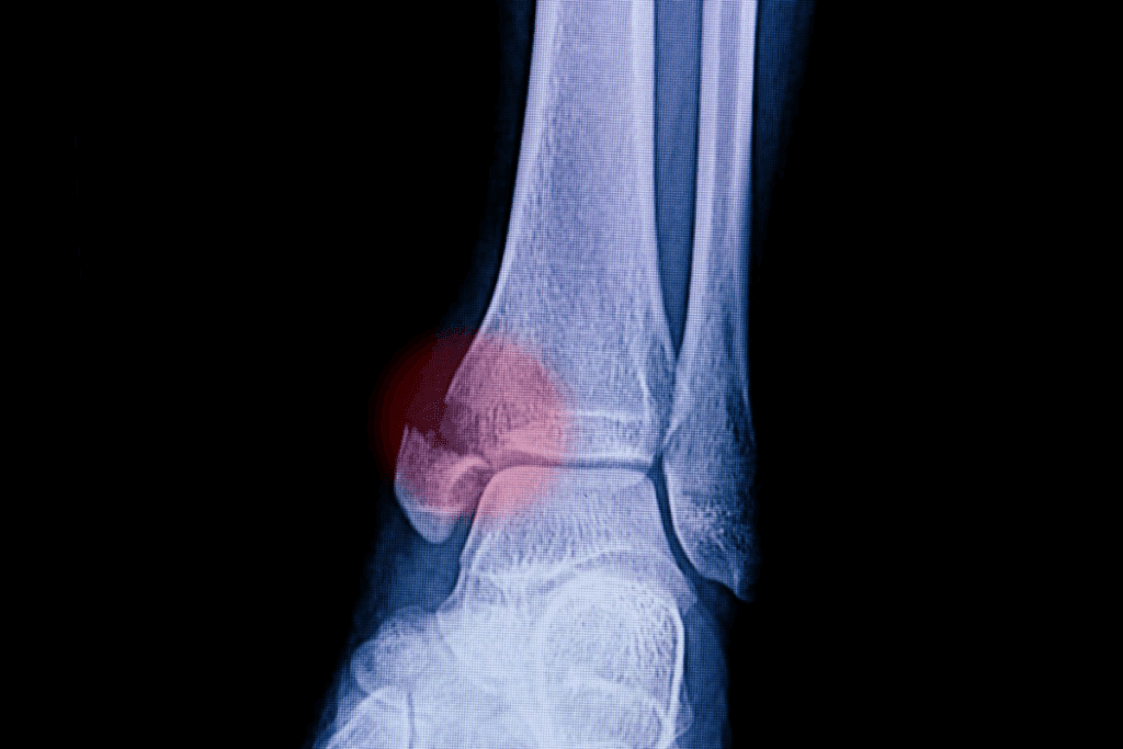
A startling 75% of ankle joint injuries involving displacement also include fractures, according to orthopedic research. This occurs because the body’s connective tissues often withstand trauma better than bone structures. When these high-impact events happen, the results demand urgent expertise to prevent lifelong mobility challenges. Explore the four types of ankle dislocations, their symptoms, and recovery process.
We see these emergencies most frequently in car crashes or sports like basketball, where sudden twists or falls apply extreme force. The talus bone – a critical component of the ankle’s architecture – shifts out of place, tearing soft tissues and destabilizing the entire joint. Patients typically experience immediate swelling, deformity, and an inability to bear weight.
Rapid identification of four directional patterns is prioritized in clinical practice. Posterior shifts dominate clinical practice, making up nearly 60% of incidents globally. However, lateral, medial, and anterior displacements each require unique approaches for safe realignment. Misdiagnosis risks complications like chronic instability or arthritis.
Effective care begins at the accident scene. First responders must stabilize the limb without forcing movement. In trauma centers, imaging studies reveal whether fractures accompany the dislocation, guiding surgical or non-surgical interventions. Recovery timelines vary but consistently emphasize protecting vulnerable tissues during healing.

The ankle’s structure balances remarkable flexibility with load-bearing strength. Three key bones – the tibia, fibula, and talus – form its foundation. These elements work in concert, allowing smooth movement while maintaining critical stability during walking or running.
Your ankle joint functions like a precision hinge. The tibia and fibula create a bony socket (mortise) that cradles the talus from the foot. This arrangement permits up-and-down motion while resisting side-to-side shifts. Strong ligaments reinforce the joint capsule, acting like biological seatbelts during physical stress.
Synovial fluid within the capsule reduces friction between bones. This natural lubricant ensures pain-free motion – until trauma disrupts this delicate system.
High-impact forces overwhelm the ankle’s defenses differently. A sprain stretches or tears ligaments without bone displacement. Fractures involve broken tibia, fibula, or talus. Complete joint separation occurs when bones shift entirely out of alignment.
Most severe injuries happen when the foot twists violently while bearing weight. Car accidents or sports collisions often generate the energy needed to compromise multiple structures simultaneously.
When traumatic forces disrupt the ankle’s intricate architecture, we categorize injuries by the talus bone’s movement relative to its natural position. This systematic approach guides our emergency response and long-term management plans.
Nearly 60% of dislocation cases involve backward talus displacement. These often occur when falls from heights drive the foot into extreme downward flexion. “The body’s weight acts like a hammer pushing the talus out of its socket,” explains our orthopedic team. Associated ligament tears frequently require surgical repair after realignment.
Eversion injuries typically cause inward (medial) shifts, stretching lateral ligaments beyond their limits. Conversely, severe inward twists may force the talus outward – a lateral ankle displacement often accompanied by medial bone fractures.
Anterior movements remain rare but dangerous. High-speed collisions sometimes thrust the talus forward, crushing soft tissues against the tibia. Each type demands specific imaging protocols:
We prioritize rapid categorization because misdiagnosed ankle dislocations increase arthritis risks by 40%. Our trauma surgeons use these classifications to predict recovery timelines and prevent chronic instability.

Severe joint injuries often involve both bone and soft tissue damage, creating complex medical challenges. Our team approaches these cases with dual priorities: restoring structural integrity and preventing long-term complications. Immediate intervention is critical, especially when fractures accompany joint displacement.
We detect bone injuries in over 80% of severe joint trauma cases. High-energy impacts frequently crack the malleoli – the bony protrusions around the ankle. “Open injuries occur when fractured bone pierces the skin,” notes our trauma specialist. These emergencies require urgent surgery to clean the wound and stabilize broken segments.
Advanced imaging guides our decisions. CT scans reveal hidden cracks, while X-rays confirm alignment after manual adjustments. Missed fractures risk improper healing, leading to chronic pain or arthritis.
Stable injuries often respond to non-surgical care. We realign bones under anesthesia, then immobilize the area with fiberglass casts. Patients maintain strict non-weight-bearing status for 3-6 weeks using crutches or walkers.
Complex cases demand surgical precision. Our orthopedic surgeons:
Post-surgery rehabilitation begins within days. Custom physical therapy programs rebuild strength while protecting healing tissues. Regular follow-ups ensure proper recovery and adjust treatment as needed.
Understanding why certain individuals face higher risks requires thorough evaluation of medical history and trauma patterns. We combine this analysis with advanced imaging to create precise treatment roadmaps.
Our team prioritizes patients with connective tissue disorders like Ehlers-Danlos syndrome. These conditions reduce the joint’s natural resistance to traumatic forces. Prior ankle injuries also leave residual instability – 68% of recurrent cases involve individuals with untreated sprains.
| Risk Factor | Impact on Stability | Management Strategy |
| Previous fractures | Altered weight distribution | Custom orthotics |
| Peroneal muscle weakness | Reduced lateral support | Targeted physical therapy |
| Malleolar hypoplasia | Shallow joint socket | Prophylactic bracing |
Initial assessments focus on range motion limitations and swelling patterns. We then deploy a three-tier imaging protocol:
X-rays remain our first-line tool, detecting 92% of bone displacements. For complex cases involving cartilage damage, CT scans map fracture lines in 0.5mm detail. When ligament evaluation becomes critical, MRI reveals soft tissue tears invisible to other methods.
Accurate diagnosis prevents misaligned healing – a key factor in preserving long-term mobility. Our radiologists work alongside orthopedic specialists to interpret results within 90 minutes of imaging completion.
Swift action transforms outcomes in severe foot trauma cases. Our experience shows that 94% of patients regain full movement when treated within six hours of injury. This window matters most when bones shift from their natural position, threatening surrounding tissues.
We combine emergency reduction techniques with personalized rehabilitation plans. Surgical teams focus on restoring stability through precise realignment, while physical therapists prevent range motion loss. This dual approach addresses both immediate damage and long-term complications.
Prevention remains vital. Proper footwear reduces twisting injuries by 37% in high-risk activities. Athletes benefit from balance training that strengthens ankle support structures. For those experiencing trauma, urgent evaluation prevents secondary damage to the fibula or talus.
Our teams prioritize your joint health at every stage – from initial care to final recovery milestones. Immediate intervention paired with expert follow-up ensures optimal results, letting patients reclaim active lifestyles safely.
Posterior dislocations often occur when forceful impacts push the talus backward against the tibia and fibula. This typically happens during high-energy events like falls from height or car accidents, where the foot is fixed in dorsiflexion while sudden pressure shifts the bone position.
While both involve ligament damage, dislocations cause visible joint deformity and complete loss of stability. Sprains may limit movement but don’t displace bones. We use imaging like X-rays to confirm if the talus has shifted from its normal alignment.
Yes, but fractures accompany most dislocations due to the extreme forces involved. Lateral ankle injuries often involve fibula fractures, while medial cases may damage the tibia. We assess stability through stress tests and CT scans to plan appropriate care.
For stable injuries without fractures, we use closed reduction to reposition the joint followed by immobilization in a cast or boot. Physical therapy restores range of motion gradually while monitoring for complications like reduced blood flow or nerve damage.
MRIs reveal soft tissue damage to ligaments, tendons, and cartilage that X-rays can’t detect. This helps us evaluate long-term stability risks and determine if surgical repair is necessary to prevent chronic instability or arthritis.
Weak ligaments from prior sprains, congenital joint abnormalities, or conditions like Ehlers-Danlos syndrome reduce natural stability. Athletes in sports requiring rapid pivoting also face higher risks due to repetitive stress on the ankle’s support structures.
Subscribe to our e-newsletter to stay informed about the latest innovations in the world of health and exclusive offers!