
An 8-week ultrasound, also known as a dating scan, is very important in early pregnancy. It gives a close look at your baby’s growth and important health info.Understand visibility limits for an abdominal ultrasound at 8 weeks pregnant versus TVS. Learn what abdominal ultrasound at 8 weeks pregnant shows.
At this time, your baby is about 0.5 to 1 inch long and looks like a bean. Knowing what’s normal and what’s not is key to watching your baby grow and spotting any problems early.
We will look into the importance of 8-week ultrasound pictures. This will help you feel more confident about your ultrasound.
Key Takeaways
- An 8-week ultrasound confirms pregnancy viability and detects the baby’s heartbeat.
- It establishes an accurate due date, which is important for prenatal care.
- Understanding normal vs abnormal findings helps alleviate concerns.
- Liv Hospital’s patient-centered approach ensures complete support.
- Early imaging appointments are vital for monitoring fetal development.
The Significance of Your First Pregnancy Scan

At 8 weeks pregnant, the excitement is high. The first pregnancy scan gives a peek into the baby’s growth. This time is key as it’s a big step in the baby’s development.
We will look into why this scan matters and what it shows about the pregnancy.
Embryonic Development at 8 Weeks
By the 8th week, the embryo’s main organs start to form. The heartbeat is usually heard.
Medical Expert, “By 8 weeks pregnant, it’s a good time for an early ‘dating and viability scan.'” This scan checks if the pregnancy is viable and gives insights into the embryo’s growth.
Why the 8-Week Mark is Important for Monitoring
The 8-week ultrasound is key for checking if the pregnancy is viable and spotting issues early.
It helps doctors see how the embryo is growing and find any problems.
The main goals of this scan are to check if the pregnancy is viable by finding the fetal heartbeat. It also helps set an accurate due date.
Developmental Milestone | Expected Outcome at 8 Weeks |
Heartbeat Detection | Usually detectable |
Major Organs Formation | Begin to form |
Embryo’s Size | Approximately 1.6 inches |
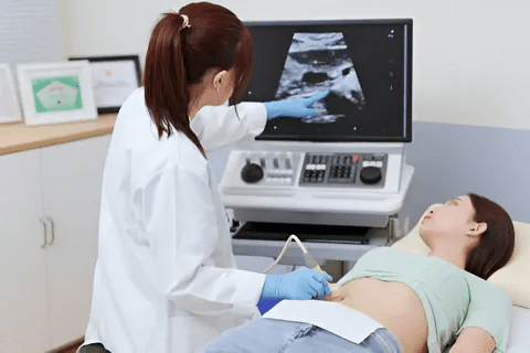
What to Expect During an Abdominal Ultrasound at 8 Weeks Pregnant

As you get closer to your 8-week mark, knowing what to expect at your ultrasound can help. An abdominal ultrasound is a safe way to see your baby. It uses sound waves to make images of your baby inside the womb.
Preparation for Your Ultrasound Appointment
Getting ready for your ultrasound is important. You might need to arrive with a full bladder. This helps get a better view of your uterus. Wear comfy clothes that are easy to move in.
Also, avoid jewelry or clothes with metal. They might get in the way of the ultrasound.
Step-by-Step Ultrasound Procedure
During the ultrasound, you’ll lie on a table. A gel will be put on your belly. This gel helps the sound waves work better.
The sonographer will then move a transducer over your belly. They’ll take pictures of your baby. It’s usually painless and takes 15 to 30 minutes.
Transvaginal vs. Abdominal Approaches: Which Will You Need?
Most ultrasounds are done on the belly. But sometimes, a transvaginal ultrasound is needed, mainly in early pregnancy. This one involves a special transducer in the vagina for clearer images.
Which one you’ll get depends on your pregnancy stage and what your doctor needs to see.
Ultrasound Type | Description | When It’s Used |
Abdominal Ultrasound | Non-invasive; uses a transducer on the abdomen | Later pregnancy stages; when a broader view is needed |
Transvaginal Ultrasound | Involves a transducer inserted into the vagina | Early pregnancy; when detailed, close-up images are required |
Knowing the difference between these can make you feel more ready for your ultrasound. Your doctor will choose the best one for you.
Key Purposes of the 8-Week Dating Scan
At 8 weeks, the dating scan has many important roles. It gives insights into how the pregnancy is going and how the embryo is growing.
Confirming Pregnancy Viability and Heartbeat
The main goal of the 8-week dating scan is to check if the pregnancy is viable. The sonographer looks at the embryo to see if it has a heartbeat. This heartbeat is a good sign that the pregnancy is healthy.
The 8 weeks ultrasound pictures show the heartbeat. It’s a key sign of the embryo’s health.
Establishing Accurate Gestational Age
Another key purpose is to find out the exact age of the embryo. By measuring the crown-rump length, doctors can tell the embryo’s age. This helps predict the due date more accurately.
This info is important for tracking the pregnancy’s progress. It also helps spot any early problems.
Identifying Multiple Pregnancies
The 8-week dating scan also checks for multiple pregnancies. The sonographer looks at the 8 week old ultrasound pictures for signs of twins or triplets. This ensures the pregnancy is monitored and managed correctly.
In summary, the 8-week dating scan is a key part of prenatal care. It gives vital info on the pregnancy’s health, age, and if there are multiple babies. Understanding its importance helps expectant parents appreciate its role in a healthy pregnancy.
Decoding Normal 8-Week Ultrasound Pictures
Understanding 8-week ultrasound images means knowing what to look for. At this time, several important structures should be seen. These signs show a healthy pregnancy.
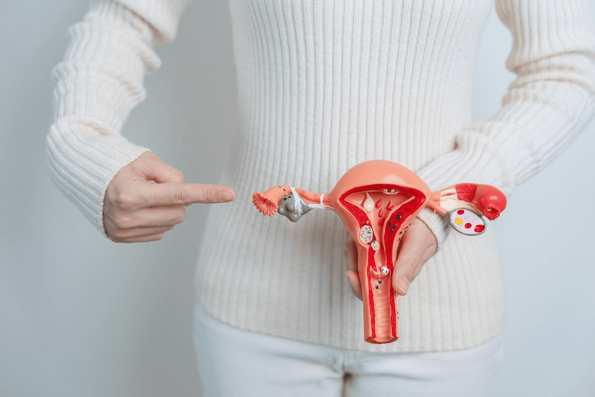
Gestational Sac: The Black Circle
The gestational sac is the first thing seen on an ultrasound. It looks like a black circle-like shape in the uterus. This sac is key because it holds the embryo and other important parts. A normal sac is round and clear, with sharp edges.
Yolk Sac: The Small Bubble Structure
Inside the gestational sac, a small bubble called the yolk sac should be seen. The yolk sac gives the embryo nutrients before the placenta forms. It looks like a small circle attached to the embryo or floating in the sac.
Embryo Appearance: The Bean-Shaped Form
By 8 weeks, the embryo looks like a bean. It’s connected to the yolk sac by the umbilical cord. The embryo’s size and shape might vary, but it should be clear in the sac. You might also see early fetal movements, depending on the ultrasound quality and the sonographer’s skill.
At 8 weeks, seeing the gestational sac, yolk sac, and fetal movements is normal. These signs are key to knowing the pregnancy is healthy. They help parents feel closer to their baby and more informed about their pregnancy.
- The gestational sac should be clearly visible and well-defined.
- A yolk sac should be present, providing nutrients to the embryo.
- The embryo should have a bean-shaped appearance and be actively moving.
These signs mean the 8-week ultrasound is normal. If there are any worries or unusual findings, a healthcare provider can help. They might suggest more tests or close monitoring.
Measurements and Markers in Healthy 8-Week Scans
Understanding the key markers in an 8-week ultrasound can give insights into your baby’s health and growth. At this stage, several important measurements are taken to check on fetal development.
Crown-Rump Length: What’s Standard at 8 Weeks
The crown-rump length (CRL) is a key measurement at 8 weeks. It measures from the top of the head to the bottom of the buttocks. A normal CRL at 8 weeks is usually between 1.6 to 2.2 centimeters. This helps doctors figure out the baby’s age and track its growth.
Heart Rate Expectations (110-160 BPM)
The fetal heart rate is another important marker at 8 weeks. A normal heart rate is between 110 to 160 beats per minute (BPM). This fast heartbeat shows the baby is healthy and growing well. Doctors watch the heart rate to spot any problems early.
Early Movement Detection Possibilities
Some ultrasounds at 8 weeks might show early fetal movements. These first movements show the baby’s muscles are developing. But, seeing these movements depends on the ultrasound quality and the baby’s position.
Key measurements and markers at 8 weeks include:
- Crown-rump length for gestational age assessment
- Fetal heart rate to ensure viability and health
- Early detection of fetal movements, when possible
These measurements and markers give a detailed look at your baby’s development at 8 weeks. They help parents understand their baby’s growth and health.
Visual Guide to Normal Variations in 8-Week Ultrasounds
At 8 weeks, ultrasounds can show different things because of how the baby is positioned and the image quality. Knowing these differences can make parents feel more sure about their baby’s growth.
Position and Angle Differences in Images
The way the embryo is positioned during the ultrasound greatly affects the image. Different angles can make the embryo appear larger or smaller than it actually is. For example, if the embryo’s back is towards the probe, it might look clearer. But if it’s facing the probe, you might see its face better.
It’s key to remember that the sonographer’s skill matters a lot. They adjust the probe and read the images to get the best views.
Individual Development Timing Variations
Just like adults, babies grow at their own pace. Some embryos might be more advanced in their development at 8 weeks, while others might be a bit behind. This difference in growth can change how the embryo looks on the ultrasound.
Things like when the baby was conceived and how fast they grow affect these differences. Doctors look at these things when they check the ultrasound pictures.
How Imaging Quality Affects What You See
The quality of the ultrasound image is very important. High-quality imaging can show more details about the embryo’s growth. But if the image is not as clear, it might be harder to see certain things.
Things that can change image quality include the ultrasound machine, the sonographer’s skill, and the baby’s position. Newer ultrasound technology has made images better, helping doctors see more clearly.
Recognizing Abnormal Signs in 8-Week Ultrasound Pictures
It’s important for expectant parents to understand abnormal signs in 8-week ultrasound pictures. Most ultrasounds at this stage show a healthy embryo. But, some may show signs that need more checking or watching.
During an 8-week ultrasound, many things are checked to see if the pregnancy is healthy. Signs that might need more attention include no heartbeat, an empty sac, or an irregular sac shape. Let’s look at each of these concerns.
Absence of Cardiac Activity When Expected
Seeing a fetal heartbeat is key at 8 weeks. “The absence of cardiac activity when expected can be a concerning sign,” says Medical Expert, a top obstetrician. If the embryo is big enough (around 7-8 mm), a heartbeat should be seen. Not seeing one might mean there’s a problem with the pregnancy.
Empty Gestational Sac (Anembryonic Pregnancy)
An empty gestational sac is when a fertilized egg implants but doesn’t grow into an embryo. This is seen on an ultrasound when there’s an empty sac but no embryo or yolk sac inside.
Medical Expert, “Anembryonic pregnancy is a miscarriage where the embryo doesn’t grow or is absorbed, leaving an empty sac.” This usually means the pregnancy isn’t viable due to chromosomal issues.
Irregular Sac Contours and What They Mean
The shape of the gestational sac can tell us a lot about the pregnancy. An irregularly shaped sac might mean a higher risk of miscarriage or other problems. The exact cause isn’t always clear, but it could be related to how the embryo implants or how the placenta develops.
“Irregular sac contours can be a sign of an underlying issue that may affect the viability of the pregnancy,” warns Medical Expert, a maternal-fetal medicine specialist.
Abnormal Yolk Sac Size or Appearance
The yolk sac is important in early development and is seen first in the gestational sac. An abnormal yolk sac size or shape can signal problems. A yolk sac that’s too big, too small, or irregular might mean a higher risk of miscarriage or chromosomal issues.
Understanding early pregnancy ultrasounds is complex. While these signs might suggest issues, they don’t always mean the pregnancy won’t go on. More tests and monitoring are often needed to check the pregnancy’s health and viability.
Placental Development Visible in 8-Week Ultrasounds
Learning about placental development at 8 weeks is key to understanding the health of mom and baby. The placenta is a vital organ that supports the fetus during pregnancy.
Early Placental Formation Indicators
At 8 weeks, the placenta looks like a grainy area attached to the uterine wall on an ultrasound. This shows early signs of placental formation. The placenta grows from both fetal and maternal tissues. It’s important for exchanging nutrients, gases, and waste between mom and baby.
The process of placental formation is complex. It starts with the blastocyst implanting in the uterine lining. As it grows, the placenta makes hormones that help keep the pregnancy going.
Normal vs. Concerning Placental Appearance
A normal placenta at 8 weeks should be evenly granular and have a clear edge with the uterine wall. Any big changes could mean trouble. For example, an odd-shaped or too small placenta might signal problems.
It’s important to keep an eye on the placenta’s growth during pregnancy. This ensures the health of both mom and baby.
What Isn’t Visible Yet at 8 Weeks
While we can see the placenta at 8 weeks, we can’t see all its details yet. Maturation of the placenta keeps going throughout pregnancy. It reaches full function later on.
- The placenta’s role in hormone production
- Its involvement in waste removal
- The full extent of its nutrient supply to the fetus
These aspects become clearer as the pregnancy goes on.
Next Steps After Your 8-Week Ultrasound
Your 8-week ultrasound is a big deal in your pregnancy. Knowing what comes next can make you feel better. Your doctor will tell you what to do next based on the ultrasound.
Understanding Follow-up Procedures
After an 8-week ultrasound, your next steps depend on the results. If everything looks good, you might have another scan at 12 or 20 weeks. But if there are issues, you might need more tests or closer checks.
Common follow-up procedures include:
- Repeat ultrasounds to monitor growth or address concerns
- Blood tests to check for possible complications
- Consultations with specialists if needed
When Additional Testing May Be Recommended
At times, your doctor might want you to have more tests after the 8-week ultrasound. This could be because of worries about the baby’s growth, if you’re having twins, or other reasons.
Condition | Potential Additional Testing |
Multiple pregnancy | More frequent ultrasounds, specialized prenatal care |
Concerns about embryonic development | Repeat ultrasounds, blood tests for hormonal levels |
History of pregnancy complications | Early screening for gestational diabetes, preeclampsia monitoring |
Questions to Ask Your Healthcare Provider
It’s important to ask questions during your follow-up visits. Some questions to think about include:
- What do the ultrasound results indicate about my pregnancy?
- Are there any specific risks or complications I should be aware of?
- What lifestyle changes or precautions should I take?
- When is my next scheduled appointment or scan?
Emotional Support Resources
Pregnancy can be emotionally challenging. It’s key to have emotional support. Many healthcare providers offer counseling or can suggest support groups.
Remember, you’re not alone in this journey. Talking to family, friends, or online groups can also help.
Conclusion: Making Sense of Your 8-Week Ultrasound Results
Understanding 8-week ultrasound results can be tough, but it’s key for parents-to-be. The report gives a summary of what’s seen, like the heartbeat, age, and any worries.
Looking at your 8-week ultrasound pic, we help you grasp the findings. We check for a heartbeat, the sac’s size and shape, and the embryo’s growth. Knowing these details is vital for your baby’s health and growth.
By carefully looking at your ultrasound results, we can spot any early problems. This lets us take action quickly if needed. It’s all about making sense of the ultrasound to ensure a healthy pregnancy.
We’re here to help you understand your 8-week ultrasound results. We offer clarity and support as you move through this important part of your pregnancy.
FAQ
What is the purpose of an 8-week ultrasound?
An 8-week ultrasound confirms if the pregnancy is viable. It also checks for a fetal heartbeat and determines the gestational age. It can also spot multiple pregnancies.
What can I expect to see during an 8-week ultrasound?
You’ll see the gestational sac, yolk sac, and embryo during an 8-week ultrasound. The embryo looks like a bean. You might see its heartbeat too.
What is a normal crown-rump length at 8 weeks?
At 8 weeks, a normal crown-rump length is between 1.6 and 2.2 centimeters.
What is considered a normal fetal heart rate at 8 weeks?
A normal fetal heart rate at 8 weeks is between 110 and 160 beats per minute.
Can an 8-week ultrasound detect abnormalities?
Yes, an 8-week ultrasound can spot some issues. These include an empty gestational sac, irregular sac shapes, or odd yolk sac sizes.
What does an empty gestational sac mean?
An empty gestational sac means the sac is there but no embryo is inside. It’s also called an anembryonic pregnancy.
How is placental development visible in 8-week ultrasounds?
In 8-week ultrasounds, early signs of placental formation can be seen. The placenta looks like a thick area around the gestational sac.
What are the next steps after an 8-week ultrasound?
After an 8-week ultrasound, your doctor might suggest more tests or follow-ups. They’ll check on the baby’s growth.
What if I have concerns or questions after my 8-week ultrasound?
If you’re worried or have questions after your ultrasound, talk to your doctor. They can offer guidance and support.
Can the position and angle of the ultrasound image affect what I see?
Yes, the ultrasound’s position and angle can change what you see. Different views can show the embryo and its surroundings in different ways.
How does imaging quality affect the ultrasound pictures?
The quality of the ultrasound images matters a lot. Clear images show more details about the baby’s development. Poor images are harder to understand.
References
World Health Organization. 8-Week Ultrasound: Normal and Abnormal Findings in Early Pregnancy. Retrieved from https://www.who.int/publications/i/item/9789241549912

