Last Updated on November 27, 2025 by Bilal Hasdemir

The aortic arch is a key part of the aorta. It’s curved and vital for sending blood from the heart to the upper body. At Liv Hospital, we know how important the aortic arch diagram is for treating heart and vascular issues.
The transverse aortic arch is a big deal in the heart’s system. It starts from the ascending aorta and goes a bit backward and to the left of the trachea. Our team is dedicated to top-notch care. We use the latest info on the aortic arch and its branches to help our patients.
Key Takeaways
- The aortic arch is a critical section of the aorta that distributes oxygenated blood to the upper body.
- Understanding the aortic arch diagram is essential for diagnosing heart and vascular conditions.
- The aortic arch has three main branches that supply blood to the upper body.
- Liv Hospital is committed to providing trusted, patient-focused care using the latest knowledge on the aortic arch.
- Accurate diagnosis and management of cardiovascular conditions rely on a thorough understanding of the aortic arch and its branches.
Understanding the Aortic Arch: Basic Concepts

It’s key to know about the aortic arch to grasp how blood circulates and affects our health. The aortic arch, or the arch of the aorta, is a vital part. It helps spread oxygen-rich blood all over the body.
Definition and Terminology
The aortic arch is the curved part of the aorta. It starts from the ascending aorta and ends as the descending aorta after branching off. It’s a major part of our heart system, making sure blood reaches the head, neck, and upper limbs.
Important terms related to the aortic arch include:
- Aortic arch definition: The curved part of the aorta that gives off branches to the head and upper limbs.
- Branches of the aortic arch: The three main branches are the brachiocephalic trunk, left common carotid artery, and left subclavian artery.
- Aortic arch of the heart: Refers to its origin from the ascending aorta and its role in cardiac circulation.
Role in Cardiovascular Circulation
The aortic arch is vital in cardiovascular circulation. It carries oxygen-rich blood from the heart to the rest of the body. Its shape helps distribute blood through its three main branches:
- Brachiocephalic trunk: Supplies blood to the right arm and head.
- Left common carotid artery: Provides blood to the left side of the head and neck.
- Left subclavian artery: Supplies blood to the left arm.
The aortic arch works well to keep blood pressure right and organs well-supplied. Any problems with it can cause big heart issues. So, knowing how it works is very important.
Anatomical Location and Orientation

The aortic arch is found in the mediastinum, a key area in the chest. It’s surrounded by important structures like the heart, trachea, and esophagus. Its location is vital for its role in the body.
Position Within the Mediastinum
The aortic arch sits in the upper part of the mediastinum, above the left main bronchus. It’s behind the sternum and in front of the trachea. This spot helps it distribute blood well.
Relationship to the Heart
The aortic arch starts from the left ventricle of the heart. It curves backward and to the left, then turns into the descending aorta. This shape is key for the heart’s work.
Transition at T4 Vertebra to Descending Aorta
At the T4 vertebra, the aortic arch changes into the descending aorta. This change is important as the aorta moves from the top to the back of the chest.
| Structure | Relation to Aortic Arch | Significance |
|---|---|---|
| Mediastinum | Contains the aortic arch | Protects and positions the aortic arch |
| Heart | Connected via ascending aorta | Receives blood from the left ventricle |
| T4 Vertebra | Level of transition to descending aorta | Marks the end of the aortic arch |
Knowing where the aortic arch is and how it’s set up is key for health checks and treatments. Its special spot in the mediastinum and tie to the heart show its big role in heart health.
Comprehensive Aortic Arch Diagram Analysis
Aortic arch diagrams give us important insights into the normal and varied anatomy of this key blood vessel. They are vital for both learning and practicing medicine. They help us see the complex anatomy of the aortic arch clearly.
Visual Representation of Normal Anatomy
A aortic arch diagram shows the normal anatomy in detail. It starts from the ascending aorta, curves, and then turns into the descending aorta. It also highlights the three main branches: the brachiocephalic trunk, the left common carotid artery, and the left subclavian artery.
Key Structures Identified in Diagrams
In a typical diagram of the aortic arch, several important structures are shown. These include the aortic arch, its three main branches, and its connection to nearby structures like the trachea and esophagus. Knowing these relationships is key for diagnosing and treating aortic arch-related conditions.
Reading and Interpreting Aortic Arch Imaging
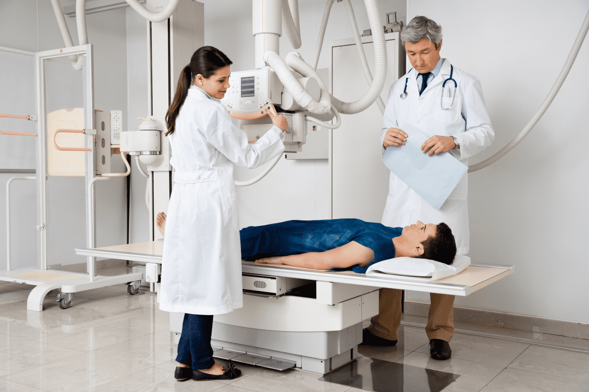
Understanding aortic arch imaging requires knowing the normal anatomy and common variations. Techniques like CT angiography and MRI give detailed views of the aortic arch. This helps doctors assess its structure and spot any issues. Accurate reading of these images is vital for making diagnoses and treatment plans.
By studying aortic arch diagrams and imaging, healthcare experts can better understand the aortic arch’s anatomy and its variations. This knowledge is essential for top-notch patient care and for moving forward in cardiovascular health research.
The Three Main Branches of the Aortic Arch
Knowing the aortic arch’s branches is key to understanding blood flow to the upper body. The aortic arch has three main branches. These branches supply blood to the head, neck, and upper limbs.
Brachiocephalic Trunk (Innominate Artery)
The brachiocephalic trunk, or innominate artery, is the largest branch. It starts at the top of the arch and goes up and to the right. It splits into the right common carotid artery and the right subclavian artery, behind the right sternoclavicular joint.
This artery is vital for blood flow to the right side of the head, neck, and right upper limb.
Left Common Carotid Artery
The left common carotid artery is the second branch. It begins just left of the brachiocephalic trunk. It goes up through the neck, inside the carotid sheath, and splits into the left internal and external carotid arteries.
This artery ensures oxygenated blood reaches the left side of the head and neck.
Left Subclavian Artery
The left subclavian artery is the third branch. It starts at the top of the arch, to the left of the left common carotid artery. It supplies blood to the left upper limb and helps the brain through its vertebral branch.
This artery goes laterally, between the scalene muscles, and into the arm.
To summarize the key characteristics of the three main branches of the aortic arch, we have prepared the following table:
| Branch | Origin | Course | Territory Supplied |
|---|---|---|---|
| Brachiocephalic Trunk | First branch of aortic arch | Upwards and to the right, dividing into right common carotid and right subclavian arteries | Right side of head and neck, right upper limb |
| Left Common Carotid Artery | Second branch of aortic arch | Ascends through neck, dividing into left internal and external carotid arteries | Left side of head and neck |
| Left Subclavian Artery | Third branch of aortic arch | Courses laterally, passing between scalene muscles | Left upper limb, contributes to brain supply via vertebral branch |
Understanding these branches and their distributions is essential for diagnosing and treating vascular conditions affecting the head and upper limbs.
Histological Structure of the Aortic Arch
The aortic arch has three main layers. Each layer has its own role and features. Together, they help the aortic arch do its important job in our heart and blood system.
Tunica Intima Layer
The innermost layer is the tunica intima. It has a single layer of endothelial cells and a thin layer of connective tissue. This layer is key to keeping blood flowing smoothly and preventing blood clots.
The endothelial cells in this layer help control blood flow and clotting. Damage to this layer can cause heart problems.
Tunica Media Layer
The middle layer is the tunica media. It’s made of smooth muscle cells and elastic fibers. This layer helps the aortic arch stretch and return to shape with each heartbeat. It keeps blood pressure steady.
The smooth muscle cells in this layer can change the aortic arch’s size. This helps control blood flow. The elastic fibers give the aortic arch the flexibility needed to handle the heartbeat’s pressure wave.
Tunica Adventitia Layer
The outermost layer is the tunica adventitia. It’s made of connective tissue that wraps around and supports the aortic arch. This layer has nerves, lymphatic vessels, and the vasa vasorum, which supplies blood to the outer layers.
This layer is important for attaching the aortic arch to nearby structures. It also helps nerves and vessels reach the other layers. Its connective tissue helps limit the aortic arch’s expansion during the heartbeat.
Physiological Functions of the Aortic Arch
The aortic arch is key to our heart’s health. It helps spread blood all over our body. This includes the upper parts of our body and our brain.
Blood Distribution Mechanisms
The aortic arch has three main branches. These are the brachiocephalic trunk, left common carotid artery, and left subclavian artery. They carry oxygen-rich blood to our head, neck, and arms.
Its curved shape makes blood flow efficiently. This ensures our brain and upper body get the oxygen and nutrients they need.
Pressure Regulation and Baroreceptors
The aortic arch also helps control blood pressure. It has baroreceptors that sense blood pressure changes. These sensors send signals to our brain to adjust our heart rate and blood vessel size.
This keeps our blood pressure stable. It also makes sure our vital organs get enough blood flow.
Adaptation to Changing Physiological Demands
The aortic arch can adjust to our body’s changing needs. For example, when we exercise, it helps control blood pressure. This ensures our muscles get the oxygen and nutrients they need.
This ability is vital for our heart to work well under different conditions.
In summary, the aortic arch plays a vital role in our body’s blood circulation. It’s essential for our heart’s health. Understanding its functions helps us see how important it is.
Common Anatomical Variations in Aortic Arch Structure
The aortic arch shows many different shapes and sizes. Doctors need to know about these to give the right treatment. These differences can affect how they diagnose and treat patients.
Bovine Arch Configuration
The bovine arch is a common shape of the aortic arch. It has a special way of branching off. This shape is seen in 15-25% of people, more in some ethnic groups.
Clinical significance: This shape can make some medical procedures harder. It might also change the surgery options.
Direct Origin of Left Vertebral Artery
Some people have the left vertebral artery coming straight from the aortic arch. This is true for about 5-6% of the population.
Diagnostic consideration: Knowing this is important for doctors to avoid mistakes in tests.
Aberrant Right Subclavian Artery
The aberrant right subclavian artery is a rare variation. It’s the last branch of the aortic arch. This happens in 0.5-1.5% of people.
Clinical implication: This can cause trouble swallowing because of the way it presses on the esophagus. It might also be linked to other birth defects.
Other Three Vessel Aortic Arch Variants
There are other variations in the three main branches of the aortic arch. These can change how the branches start or connect.
| Variation | Frequency | Clinical Significance |
|---|---|---|
| Bovine Arch | 15-25% | Impacts catheter-based procedures |
| Direct Origin of Left Vertebral Artery | 5-6% | Affects diagnostic imaging interpretation |
| Aberrant Right Subclavian Artery | 0.5-1.5% | Can cause dysphagia and associated congenital anomalies |
It’s key for doctors to understand these variations. This helps them make accurate diagnoses and treatment plans in heart medicine.
Clinical Significance and Pathologies
The aortic arch is key to our heart’s health. Knowing about its problems helps doctors treat them. It’s part of our blood flow system and can face many health issues.
We’ll look at three big problems: aneurysms, dissections, and coarctation. Each one is serious and needs quick medical help.
Aortic Arch Aneurysms
An aortic arch aneurysm is when the aortic arch gets too big. If not treated, it can burst. This is scary because it’s close to the heart and can cause blood clots.
- Risk Factors: High blood pressure, hardening of the arteries, and family history are big risks.
- Symptoms: People might feel chest pain, hoarseness, or trouble swallowing because of the swelling.
- Management: Doctors might watch small ones or operate on bigger or painful ones.
Aortic Dissection
Aortic dissection is when the aorta’s inner layer tears. This lets blood get trapped inside. It’s a serious emergency.
People with dissection often have sharp chest pain that goes to their back. Doctors use CT scans to find it fast.
Coarctation of the Aorta
Coarctation of the aorta is when the aorta narrows. This causes high blood pressure in the arms and weak pulses in the legs. It’s more common in kids but can happen in adults too.
It’s very important to catch and treat this early to avoid heart problems later. Doctors can fix it with surgery or special procedures.
In short, knowing about the aortic arch’s problems is key to helping patients. Spotting these issues early can lead to better health outcomes.
Diagnostic Imaging Techniques for the Aortic Arch
Accurate diagnosis of aortic arch pathologies relies heavily on advanced imaging modalities. These imaging techniques are key in evaluating the aortic arch’s structure and function. They help healthcare professionals identify abnormalities and plan treatments.
CT Angiography
CT angiography is a widely used imaging technique for assessing the aortic arch. It uses computed tomography (CT) scans and contrast agents to visualize blood vessels. CT angiography provides high-resolution images of the aortic arch. This allows for detailed evaluation and detection of pathologies like aneurysms or dissections.
For more information on aortic arch anomalies, you can refer to detailed resources such as aortic arch anomalies on Radiology Key.
MRI Evaluation
Magnetic Resonance Imaging (MRI) is another valuable tool for evaluating the aortic arch. MRI offers detailed images without ionizing radiation. It’s useful for long-term follow-up or when contrast agents are contraindicated.
MRI can provide functional information about blood flow and cardiac function. This makes it a complete diagnostic tool for aortic arch assessment.
Echocardiography and Ultrasound
Echocardiography and ultrasound are non-invasive imaging techniques for the aortic arch. Transthoracic echocardiography (TTE) and transesophageal echocardiography (TEE) are both useful. TEE provides more detailed views due to its proximity to the structure.
Ultrasound technology is used to evaluate blood flow through the aortic arch and its branches. It helps identify stenoses or other abnormalities.
By using these diagnostic imaging techniques, healthcare professionals can understand the aortic arch’s anatomy and function. This facilitates accurate diagnosis and effective treatment planning.
Embryological Development of the Aortic Arch System
Learning about the aortic arch’s start is key to understanding its adult form and any changes. The aortic arch forms from the change of pharyngeal arch arteries during early development.
Formation from Pharyngeal Arch Arteries
The aortic arch system comes from six pairs of pharyngeal arch arteries that change a lot. The fourth pharyngeal arch artery is very important. It helps make the aortic arch on the left side.
The left fourth pharyngeal arch artery turns into the aortic arch. The right fourth arch artery makes the right subclavian artery. This is why the aortic arch is on the left in most people.
Critical Developmental Stages
There are key steps in making the aortic arch:
- The start of the six pairs of pharyngeal arch arteries
- The change and simplification of these arteries
- The left fourth arch artery staying to form the aortic arch
- The growth of the aortic arch’s three main branches
These steps are vital for the aortic arch and its branches to develop right. Problems during these times can cause different shapes and birth defects.
Origins of Common Variations
Many aortic arch variations come from changes in its early development. For example, a bovine arch happens when the left common carotid artery comes from the brachiocephalic trunk. This is because of how the pharyngeal arch arteries change and stay.
Other changes, like an aberrant right subclavian artery, come from how the right fourth pharyngeal arch artery develops. Knowing about these changes helps doctors diagnose and treat problems.
Conclusion
Understanding the aortic arch is key to knowing about heart health. It helps spread blood all over the body. Its shape and how it works are very important for our health.
We’ve looked at the aortic arch’s parts, how it’s made, and what it does. We’ve also talked about its variations and diseases. This helps us see why the aortic arch is so important for heart health.
The aortic arch has three main branches. They carry blood to the head and arms. Its structure is flexible and strong. But, problems like aneurysms and dissections can harm heart health. This shows why finding and treating these issues is so important.
In short, the aortic arch is a vital part of our heart system. Knowing about its structure, function, and diseases helps keep our heart healthy. By understanding its role, we can take care of our heart and fix problems early.
FAQ
What is the aortic arch and its role in the cardiovascular system?
The aortic arch is a key part of the aorta. It starts from the ascending aorta, curves back and to the left, and then becomes the descending aorta. It’s vital for sending oxygen-rich blood to the upper body through its three main branches.
What are the three main branches of the aortic arch?
The aortic arch has three main branches. These are the brachiocephalic trunk (innominate artery), left common carotid artery, and left subclavian artery. They supply blood to the head, neck, and upper limbs.
What is the significance of the aortic arch diagram in understanding its structure and function?
An aortic arch diagram shows the normal anatomy of the aortic arch. It includes its branches and how they relate to other structures. It’s key for understanding its role in blood circulation and spotting any problems.
What are some common anatomical variations of the aortic arch?
There are several common variations of the aortic arch. These include the bovine arch, direct left vertebral artery origin, and aberrant right subclavian artery. These variations can affect diagnosis and treatment of aortic arch issues.
What diagnostic imaging techniques are used to evaluate the aortic arch?
To check the aortic arch, doctors use CT angiography, MRI, and echocardiography with ultrasound. Each method has its own strengths and weaknesses. The choice depends on the patient’s situation and what the doctor needs to know.
What are some common pathologies associated with the aortic arch?
The aortic arch can have problems like aneurysms, dissections, and coarctation. These issues can be serious and need quick diagnosis and treatment to avoid complications.
How does the aortic arch develop embryologically?
The aortic arch forms from the pharyngeal arch arteries during development. Its formation goes through many stages. Any issues during these stages can lead to congenital variations and problems.
What is the histological structure of the aortic arch?
The aortic arch is made of three layers: the tunica intima, tunica media, and tunica adventitia. Each layer has its own role. They work together to keep the aortic arch strong and functional.
What is the clinical significance of the aortic arch’s physiological functions?
The aortic arch’s functions are vital for heart health. It helps distribute blood, regulate pressure, and adapt to body needs. Any issues with the aortic arch can be serious and need immediate attention.
References
- Kenhub. (n.d.). Aorta: Anatomy, branches, supply. Retrieved October 23, 2025, from https://www.kenhub.com/en/library/anatomy/aorta
- AnatomyZone. (n.d.). Aortic arch. Retrieved October 23, 2025, from https://anatomyzone.com/articles/aortic-arch/
- Radiopaedia. (n.d.). Aortic arch. Retrieved October 23, 2025, from https://radiopaedia.org/articles/aortic-arch
- National Center for Biotechnology Information. (2023). Aortic arch. In StatPearls. Retrieved October 23, 2025, from https://www.ncbi.nlm.nih.gov/books/NBK499911/
- MedScience Group. (2018, August 1). Aortic arch morphometry and its clinical implication – A computed tomography study. Archives of Anatomy and Physiology, 3(1), 5–8. https://www.medsciencegroup.us/articles/AAP-3-111.php


