Last Updated on November 26, 2025 by Bilal Hasdemir

Breast calcifications are common on mammograms. While most are harmless, some patterns might show breast cancer. At Liv Hospital, we take suspicious findings seriously, like clustered microcalcifications or microcalcification clusters in the breast.
We follow advanced protocols to make safe decisions. A biopsy is often needed to check these calcifications. This article will cover important facts about biopsy for calcification and clustered microcalcifications.
Key Takeaways
- Breast calcifications can be benign or a sign of breast cancer.
- Clustered microcalcifications may indicate cancer and require further evaluation.
- A biopsy is often necessary to determine the nature of calcifications.
- Liv Hospital uses advanced protocols for safe and effective diagnosis.
- Early detection and optimal outcomes are critical in breast cancer treatment.
Understanding Breast Calcifications and Their Clinical Significance

It’s key to know the different types of breast calcifications. This helps figure out their importance and if more tests are needed. Calcifications in the breast are calcium deposits found through mammograms and ultrasounds.
Types of Breast Calcifications
Breast calcifications fall into two main groups: macrocalcifications and microcalcifications. Macrocalcifications are big and usually harmless, linked to aging. They rarely need a biopsy. Microcalcifications, being smaller, can be harmless or show cancer signs. If they cluster or look unusual, more tests might be needed.
Differentiating Between Benign and Suspicious Calcifications
Telling apart harmless and possibly cancerous calcifications is vital. Harmless ones are bigger and more spread out. Suspicious ones are smaller, clustered, and irregular. Mammography and breast ultrasound help figure out what they are. If microcalcifications cluster, a biopsy might be required to check for cancer.
Also, microcalcification in breast cluster might hint at early cancer. So, a detailed check and possibly a biopsy are important for breast health.
The Clinical Importance of Clustered Microcalcifications

Microcalcification clusters are small calcium deposits in the breast. They are key for checking breast health. These clusters might show early signs of breast cancer.
Definition of Microcalcification Clusters in the Breast
A microcalcification cluster is a group of at least five calcifications in a small area of breast tissue, usually 1 cm. They can point to different breast issues, from benign changes to cancer.
The look of microcalcifications matters a lot. For example, pleomorphic calcifications, which are different in size and shape, are more worrying than uniform ones.
Risk Assessment of Grouped Microcalcifications
Figuring out the risk of grouped microcalcifications involves looking at their imaging and clinical findings. The Breast Imaging Reporting and Data System (BI-RADS) helps by sorting calcifications by how likely they are to be cancerous.
- BI-RADS 3 lesions are probably benign, with a <2% chance of malignancy.
- BI-RADS 4 lesions are suspicious and warrant biopsy, with a 2-95% chance of malignancy.
- BI-RADS 5 lesions are highly suggestive of malignancy, with a >95% chance of cancer.
Getting the risk right is key for deciding what to do next. This could be watching them, getting a biopsy, or other steps.
Biopsy for Calcification: When and Why It’s Necessary
Deciding on a breast biopsy for calcification depends on several factors. These include the pattern and characteristics of the calcifications. We use these factors to decide if a biopsy is needed.
Indications for Breast Calcification Biopsy
A biopsy is often suggested when calcifications show suspicious patterns or are linked to other concerning imaging features. These patterns include:
- Clustered or grouped microcalcifications
- Pleomorphic calcifications
- Calcifications with a segmental or linear distribution
Our team looks at these findings to decide on a biopsy. The presence of pleomorphic calcifications of the breast often concerns us. It may mean a biopsy is needed to check for cancer.
Correlation Between Calcifications and Breast Cancer Risk
Some calcifications are linked to a higher risk of breast cancer. For example, pleomorphic calcifications can signal cancer. The link between calcifications and cancer risk is complex. It depends on the calcification’s type and how it’s spread.
| Calcification Type | Characteristics | Risk Association |
| Benign | Typically large, coarse, or scattered | Low risk |
| Suspicious | Clustered, pleomorphic, or segmental | Moderate to high risk |
| Malignant | Fine, linear, or branching | High risk |
Knowing how calcifications relate to cancer risk helps us decide on a breast biopsy for calcification. By looking at the calcification’s characteristics and how it’s spread, we can better judge the risk. This helps us suggest the right diagnostic steps.
Diagnostic Imaging Techniques for Breast Calcifications
Diagnostic imaging greatly helps in finding and understanding breast calcifications. It’s key in telling the difference between harmless and possibly dangerous calcifications.
Mammography as the Gold Standard
Mammography is the top choice for spotting and studying breast calcifications. It gives clear images that let us see the shape, spread, and type of calcifications. We use mammography to find calcifications that need more checking.
Key advantages of mammography include:
- High sensitivity for detecting calcifications
- Ability to characterize calcification morphology
- Effective for monitoring changes over time
Microcalcifications Breast Ultrasound: Applications and Limitations
Even though mammography is the top choice, breast ultrasound is useful for certain cases, like dense breasts. Ultrasound helps check for any masses or lesions nearby.
| Imaging Modality | Strengths | Limitations |
| Mammography | High-resolution imaging of calcifications | May not be as effective in dense breast tissue |
| Ultrasound | Useful for assessing associated masses or lesions | Limited in detecting microcalcifications |
We use ultrasound to look closely at calcifications found on mammograms, if we think there might be a mass. But, ultrasound can’t find tiny calcifications as well as mammography can.
By using both mammography and ultrasound, we can fully check out breast calcifications. This helps us decide if a biopsy or other treatments are needed.
Pleomorphic Calcifications in Breast Tissue: Special Concerns
Pleomorphic calcifications in breast tissue need careful attention. These calcifications vary in size, shape, and density. This makes them stand out in breast imaging.
Characteristics of Pleomorphic Microcalcifications
Pleomorphic microcalcifications look different from each other. They are small, irregular, and fragmented. They often appear in a specific area of the breast.
Because of their irregular shape, they are linked to a higher risk of cancer. Their varied appearance suggests a possible underlying disease. This calls for a closer look.
Why Pleomorphic Calcifications Raise Red Flags
Pleomorphic calcifications are concerning because they might be linked to early breast cancer. Their irregular and varied nature is common in cancerous lesions. This makes them a key finding in breast imaging.
When these calcifications are found, more tests are usually needed. A biopsy might be done to check their nature and rule out cancer.
Key Features of Pleomorphic Calcifications:
- Variability in size and shape
- Irregular morphology
- Association with malignancy
- Necessity for further diagnostic evaluation
| Characteristics | Implications |
| Varied size and density | Increased suspicion for malignancy |
| Irregular shape | Potential indicator of pathological process |
| Fragmented appearance | May require a biopsy for diagnosis |
Types of Biopsy Procedures for Breast Calcification Clusters
There are several biopsy procedures for diagnosing breast calcification clusters. Each has its own benefits and uses. The right technique depends on the calcification’s size, location, and the patient’s health.
Core Needle Biopsy Techniques
Core needle biopsy is a common method for sampling breast calcifications. It uses a needle to take a small tissue sample from the area of concern. Core needle biopsy is great for evaluating calcification clusters because it gives a bigger tissue sample than fine-needle aspiration.
The American Cancer Society says core needle biopsy is a common way to diagnose breast abnormalities, including calcifications. This procedure is done under imaging, like ultrasound or stereotactic mammography. It helps target the calcification cluster accurately.
Stereotactic Breast Biopsy for Precise Targeting
Stereotactic breast biopsy uses mammography to guide the needle to the calcification cluster. It’s good for targeting small calcifications not seen on ultrasound. Stereotactic biopsy offers precise sampling of the calcification cluster, lowering the chance of error.
A study in the Journal of the American College of Radiology shows that stereotactic breast biopsy is very accurate. It has a diagnostic accuracy rate of over 95%. This makes it a reliable choice for checking suspicious calcification clusters.
Vacuum-Assisted Biopsy Benefits for Calcifications
Vacuum-assisted biopsy (VAB) is another method for sampling breast calcifications. It uses a vacuum device to collect multiple tissue samples through one site. VAB is good for evaluating calcification clusters because it gets larger tissue samples, often avoiding the need for more procedures.
A leading breast radiologist says VAB has changed how we diagnose breast calcifications. It offers a more detailed tissue sampling. This is helpful for pleomorphic calcifications or other suspicious features.
In summary, there are many biopsy procedures for breast calcification clusters, each with its own benefits. Understanding these techniques helps healthcare providers choose the best one for each patient. This ensures accurate diagnosis and effective treatment planning.
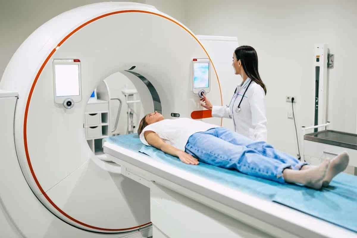
The Biopsy Process: What Patients Should Expect
Getting a breast calcification biopsy can feel scary, but knowing what to expect can help. We’ll walk you through each step, from getting ready to healing. This way, you’ll feel more in control and supported.
Pre-Procedure Preparation and Considerations
Before your breast calcifications biopsy, our team will give you clear instructions. You might need to stop taking certain medicines that can make bleeding more likely. Also, wear comfy clothes and avoid jewelry that could get in the way.
“Following these instructions is key for a smooth and safe biopsy,” says our lead radiologist.
Step-by-Step Procedure for Calcification Biopsy
On biopsy day, you’ll lie on a special table. Our team can easily reach the area we need to check. Here’s what happens next:
- Local anesthesia is given to make you comfortable
- Imaging like mammography or ultrasound helps find the calcification
- A biopsy needle is used to take tissue samples
- The needle is removed, and pressure is applied to stop bleeding
Post-Biopsy Care and Recovery
After the biopsy procedure, you might feel some soreness or bruising. This is usually mild and goes away quickly. We’ll give you detailed instructions on how to care for the area, manage any pain, and watch for infection signs. It’s important to follow these steps to heal right.
“Most patients can go back to their usual activities in a day or two. But, it’s vital to follow our care instructions to avoid problems,” says our patient care coordinator.
Knowing what to expect during your breast calcifications biopsy can help you prepare. Our team is here to offer caring support every step of the way.
Interpreting Biopsy Results for Microcalcification Clusters
Understanding biopsy results for microcalcification clusters is key. Results can be benign, malignant, or indeterminate. Each type has its own meaning for patient care.
Understanding Benign Findings
Benign findings mean the calcifications are not cancerous. This can include fibrocystic changes, fibroadenomas, or calcified debris. Patients with benign results usually don’t need immediate treatment but should be monitored.
A study in a medical journal found most biopsies for microcalcifications are benign. This means many patients avoid further invasive procedures.
“The significance of benign biopsy results cannot be overstated, as they provide reassurance to both patients and healthcare providers,” notes a leading expert in breast health.
| Benign Condition | Description | Typical Management |
| Fibrocystic Changes | Common, non-cancerous changes in the breast tissue | Monitoring, symptom management |
| Fibroadenoma | A benign tumor | Observation, possible removal if symptomatic |
| Calcified Debris | Calcium deposits within the breast tissue | Monitoring |
Malignant Findings and Their Implications
Malignant findings mean cancer is present. This often includes ductal carcinoma in situ (DCIS) or invasive breast cancer. Such findings require immediate treatment, which can include surgery, radiation, or chemotherapy.
The implications of malignant findings are significant. Knowing the type and extent of cancer is vital for treatment planning.
Indeterminate Results: Next Steps and Follow-up
Indeterminate results mean the findings are unclear. This can happen when the sample is not enough or the pathology is complex. Such results can cause anxiety and require careful thought from healthcare providers.
For indeterminate results, more tests or a repeat biopsy might be needed. The goal is to get a clear diagnosis for proper management.
- Additional imaging or diagnostic tests
- Repeat biopsy
- Close surveillance
In conclusion, interpreting biopsy results for microcalcification clusters is complex. Understanding the different types of results helps patients and healthcare providers make informed decisions.
Single Small Cluster of Microcalcification: Management Approaches
When a single small cluster of microcalcification is found, doctors must be very careful. They look at many things to decide how to manage it.
Diagnostic Challenges with Isolated Clusters
Isolated microcalcification clusters are hard to diagnose. The main worry is telling if they are harmless or could be cancer. Advanced imaging techniques help a lot in making this decision.
We use high-resolution mammography and sometimes other imaging to check the microcalcifications. We look at their shape, how they spread, and if they change over time. These details help us understand if they are serious.
Risk Stratification and Decision-Making
It’s important to figure out the risk of a single small cluster of microcalcification. We use doctor’s opinion, imaging results, and sometimes biopsy to judge the risk.
Deciding on a biopsy or more imaging depends on a detailed risk assessment. We consider the patient’s history, family history of breast cancer, and the microcalcifications’ features.
| Risk Factor | Low Risk | High Risk |
| Calcification Morphology | Benign appearance | Suspicious or pleomorphic |
| Distribution | Scattered or diffuse | Clustered or segmental |
| Patient History | No family history of breast cancer | Family history of breast cancer |
By looking at these factors carefully, we can make smart choices about managing single small clusters of microcalcification. This way, we make sure patients get the right care without doing too much.
Advances in Detecting and Evaluating Breast Cancer Calcification Clusters
New imaging and analysis methods are changing how we find and check breast cancer calcification clusters. These changes help make diagnoses more accurate and improve patient care.
New Imaging Technologies and Their Impact
New imaging tools have made it easier to spot calcification clusters. Digital mammography and tomosynthesis give clearer images of tiny calcifications.
Contrast-enhanced mammography also shows more about the blood flow in calcified areas. This can help tell if a calcification is cancerous.
| Imaging Technology | Key Features | Impact on Calcification Detection |
| Digital Mammography | High-resolution images, improved contrast | Enhanced detection of microcalcifications |
| Tomosynthesis | 3D imaging, reduced overlap of tissue | Better visualization of calcification clusters |
| Contrast-Enhanced Mammography | Highlights vascularity of lesions | Improved differentiation between benign and malignant calcifications |
Artificial Intelligence in Calcification Analysis
Artificial intelligence (AI) is now used to study calcification patterns. AI can spot patterns that humans might miss, making diagnoses more accurate.
AI can also sort patients by risk based on calcification patterns and other data. This helps doctors plan better care.
Future Directions in Non-Invasive Assessment
The future of breast cancer diagnosis is non-invasive. Advanced imaging modalities and AI-driven analysis will be key.
Studies are looking into diffusion-weighted imaging and magnetic resonance imaging (MRI) for non-invasive calcification checks.
As these technologies get better, we’ll see better early detection and treatment of breast cancer linked to calcification clusters.
Conclusion: Making Informed Decisions About Breast Calcification Biopsies
We’ve looked into the details of breast calcifications and how biopsies help diagnose them. Making smart choices is key when it comes to breast calcification biopsies.
Deciding to get a biopsy should be a well-thought-out decision. It’s important to know the risks and benefits. Patients and doctors can make better choices by looking at the latest research and guidelines.
Breast calcification biopsies are important for finding and diagnosing breast cancer early. Knowing about different calcifications and biopsy methods helps us feel more confident in our choices.
Choosing the right path for breast calcification biopsies needs teamwork between patients and doctors. By working together, we can make sure patients get the best care and results.
FAQ
What are breast calcifications, and are they a cause for concern?
Breast calcifications are calcium deposits in the breast tissue. They are common and usually not harmful. But, some types, like clustered microcalcifications, might mean a higher risk of breast cancer and need more tests.
What is the difference between macrocalcifications and microcalcifications?
Macrocalcifications are big calcium deposits that are usually harmless. Microcalcifications are small and can be either harmless or cancerous. The small, varied microcalcifications are most concerning.
Why is a biopsy necessary for calcification clusters?
A biopsy is needed to check on calcification clusters, mainly if they look suspicious. It helps find out if they are cancer or something else.
What is the role of mammography in detecting breast calcifications?
Mammography is key for spotting breast calcifications. It shows the size, shape, and where they are, helping to figure out if they are important.
Can ultrasound detect breast calcifications?
Ultrasound can find calcifications, but mammography is better at it. Ultrasound is more useful for looking at soft tissue or when mammography results are unclear.
What are pleomorphic calcifications, and why are they concerning?
Pleomorphic calcifications are small, varied calcium deposits. They are worrisome because they might be cancerous, so a biopsy is needed to confirm.
What types of biopsy procedures are used for breast calcification clusters?
There are core needle biopsy, stereotactic breast biopsy, and vacuum-assisted biopsy. Each is chosen based on the calcification type and the patient’s situation.
How are biopsy results for microcalcification clusters interpreted?
Results can be benign, malignant, or unclear. Benign means no cancer, malignant means cancer, and unclear might need more tests or another biopsy.
What are the management approaches for a single small cluster of microcalcification?
Management depends on the microcalcification type, patient risk, and doctor’s judgment. This might include watching them, doing a biopsy, or more tests.
Are there advances in detecting and evaluating breast cancer calcification clusters?
Yes, new imaging and artificial intelligence are helping. These aim to better diagnose breast cancer linked to calcification clusters.
How can patients prepare for a calcification biopsy?
Follow your doctor’s instructions. This might include avoiding certain meds, bringing someone with you, and understanding the procedure risks.
What is the significance of grouped microcalcifications in breast cancer diagnosis?
Grouped microcalcifications can signal breast cancer, like if they’re new or changing. Their look and behavior guide if more tests or a biopsy are needed.
References:
Leconte, S. et al. (2024). Stereotactic breast biopsy. RadiologyInfo.org.


