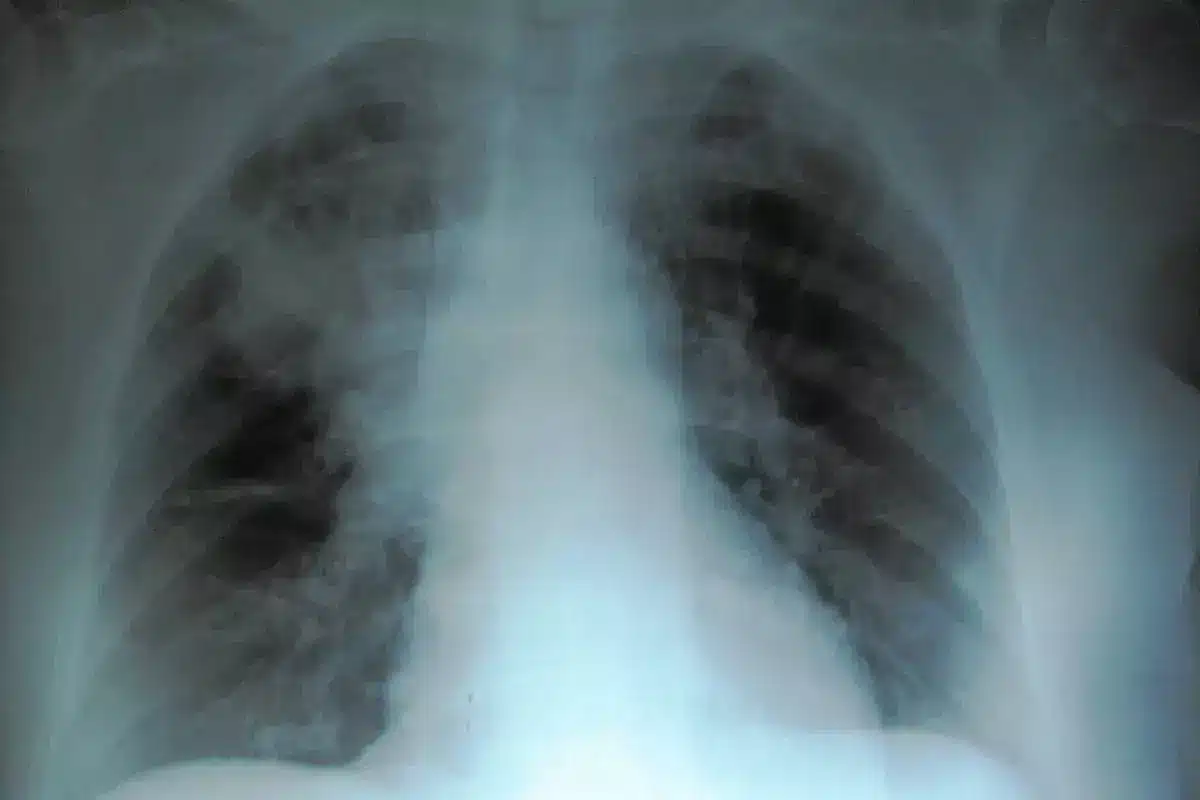
Choosing between a bone scan vs PET scan is key for finding cancer accurately. At Liv Hospital, our team is known worldwide for top-notch nuclear scans. We make sure each scan fits perfectly into your treatment plan, helping doctors choose the best approach for accurate diagnosis and effective treatment.
We use both diagnostic tools to find and track cancer well. Bone scans help spot bone metastases in cancers like breast, lung, and prostate. But PET scans are better at finding metastases because they’re more sensitive and specific.
It’s important to know how these tools differ. This knowledge helps in treating cancer better.
Key Takeaways
- Both bone scans and PET scans are vital for cancer detection.
- Liv Hospital is known worldwide for its nuclear medicine expertise.
- The right choice between a bone scan and a PET scan depends on the cancer type and stage.
- PET scans are better at finding metastases than bone scans.
- Good cancer treatment starts with accurate diagnosis using the best tool.
The Evolution of Nuclear Medicine Imaging
Nuclear medicine imaging has made huge strides in medical technology. It has greatly improved our ability to diagnose and treat diseases. From its early days, nuclear medicine has seen many advancements in scanning technologies.
The Science Behind Nuclear Medicine
Nuclear medicine uses tiny amounts of radioactive materials to help diagnose and treat diseases. These materials, called radiopharmaceuticals or tracers, target specific areas in the body. They give us important information about organs and tissues.
The science of nuclear medicine is about how these tracers work with the body. It uses advanced imaging to show where and how much they are present. This helps doctors diagnose many conditions, like cancer and heart diseases.
Development of Nuclear Scanning Technologies
Nuclear scanning technologies have improved a lot over time. Now, they can take clearer images faster and are more comfortable for patients. Modern scanners, like PET and SPECT, give doctors detailed images for better diagnoses.
Hybrid imaging systems combine nuclear medicine with CT or MRI. They give both functional and anatomical information. This helps doctors understand diseases better.
Impact on Early Disease Detection
Nuclear medicine imaging has greatly helped in finding diseases early. It lets doctors see disease processes early, leading to better treatment outcomes. For example, in cancer, early detection through nuclear scans can greatly improve treatment success and survival rates.
It also helps in tracking how diseases progress and how well treatments work. This is key in managing chronic conditions and ensuring patients get the best care.
What is a Bone Scan?
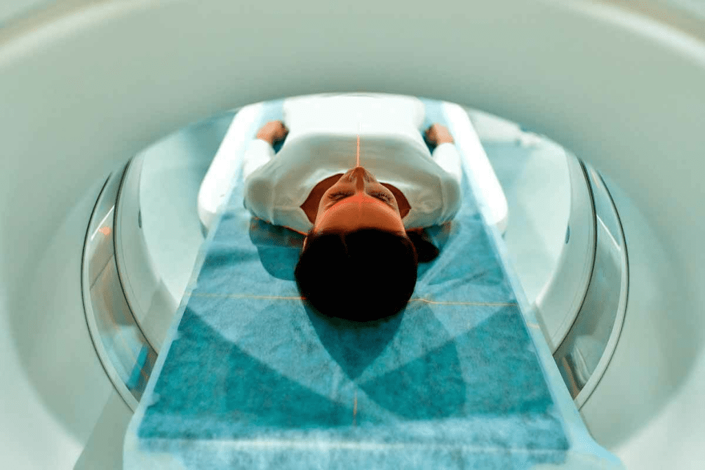
We use bone scans to see the skeletal system and find abnormal bone activity. A bone scan is a nuclear medicine test that diagnoses and monitors bone conditions. This includes cancer metastases.
Definition and Basic Principles
A bone scan injects a radioactive tracer into the blood. This tracer goes to the bones. A gamma camera then picks up signals from the tracer, showing bone images.
The main idea is that sick or damaged bones take more tracer than healthy ones. This helps spot areas of abnormal bone activity. This can be cancer, fractures, or infections.
NM Whole Body Bone Scan Technology
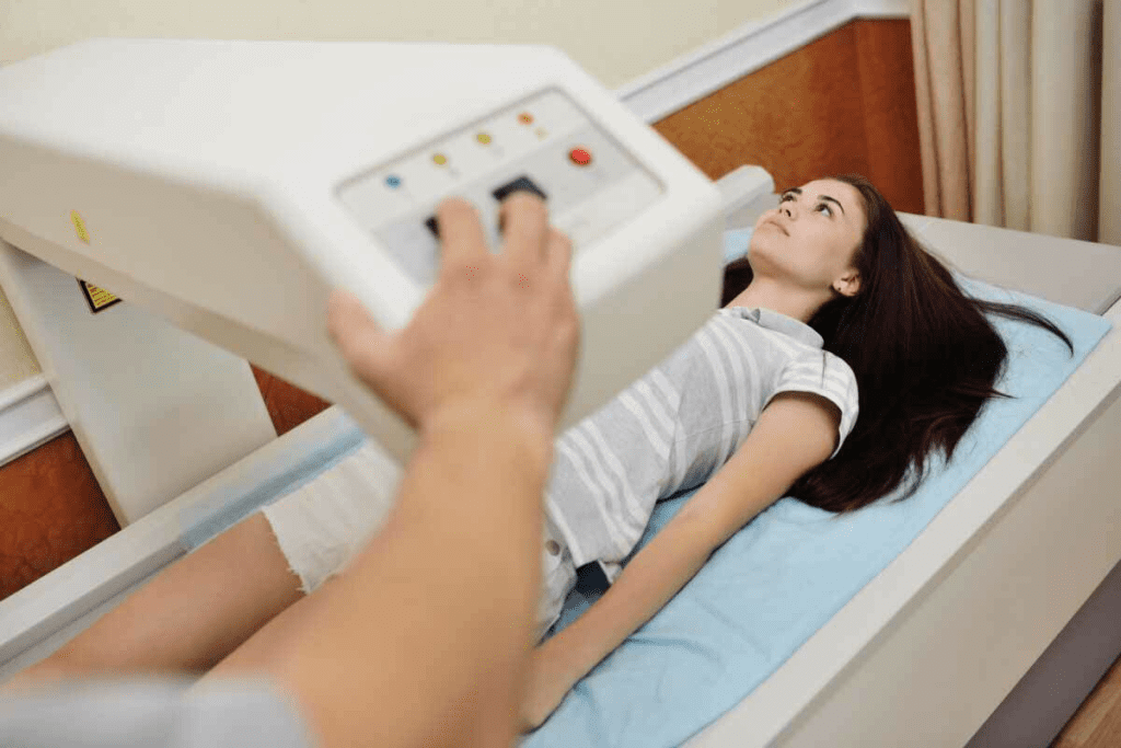
NM whole body bone scan technology uses a gamma camera to see the whole skeletal system. The camera moves around the body, catching signals from the tracer. These signals help make detailed bone images.
This technology is great for finding bone metastases. It gives a full view of the bones. This helps catch bone diseases early and track them.
Gamma Camera Imaging Process
The gamma camera imaging process has a few steps:
- The patient gets a radioactive tracer injection.
- The tracer builds up in the bones over time.
- The gamma camera picks up signals from the tracer.
- These signals make images of the bones.
These images are key for checking bone health. They help doctors diagnose and keep track of bone conditions.
| Characteristics | Bone Scan | Other Imaging Tests |
| Tracer Used | Radioactive tracer (e.g., Technetium-99m) | Varies (e.g., X-rays, CT scans) |
| Imaging Focus | Skeletal system | Varies (e.g., specific organs or tissues) |
| Detection Capability | Detects bone metastases, fractures, infections | Varies (e.g., detects tumors, injuries) |
What is a PET Scan?
A PET scan is a high-tech imaging method used to find and manage diseases, like cancer. It shows how tissues and organs work by looking at their metabolic activity. This helps doctors diagnose, plan treatments, and check how well treatments are working.
Definition and Fundamental Concepts
PET scans detect gamma rays from a special tracer in the body. The most used tracer is Fluorodeoxyglucose (FDG), a glucose molecule with a radioactive tag. Cancer cells, with their high metabolic rates, take up more FDG. This lets PET scans spot areas of high activity.
Research on PubMed Central shows PET scans are very useful in cancer care. They help see how cancer spreads and how well treatments are working.
PET-CT Integration and Benefits
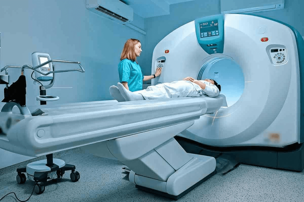
PET-CT combines PET scans with CT scans. This gives a detailed look at both the metabolic activity and the body’s structure. It offers a better understanding of diseases.
The benefits of PET-CT include:
- Improved diagnostic accuracy
- Enhanced ability to stage cancer
- Better treatment planning
- More accurate assessment of treatment response
Nuclear CT Scan Components
Nuclear CT scans are part of PET-CT. They use a CT scanner for detailed body images. The main parts are:
| Component | Description | Function |
| CT Scanner | Device that uses X-rays to create detailed images of the body’s internal structures | Provides anatomical information |
| PET Scanner | Device that detects gamma rays emitted by the tracer | Provides metabolic activity information |
| Computer System | Software and hardware that reconstructs images from PET and CT data | Combines PET and CT images for a complete analysis |
By mixing PET and CT, doctors get a full view of a patient’s health. This leads to better treatment plans.
Bone Scan vs PET Scan: 7 Key Differences
It’s important to know how bone scans and PET scans differ. They are both key tools in finding and treating cancer. Each has its own role and features.
Difference #1: Tracer Types and Mechanisms
Bone scans use Technetium-99m methylene diphosphonate (Tc-99m MDP). This tracer shows how bones are working. PET scans, on the other hand, use Fluorodeoxyglucose (FDG). This is a sugar-like substance that cancer cells take up.
“The choice of tracer is key to the scan’s success,” say nuclear medicine experts. The tracer affects what the scan can show.
Difference #2: Biological Function vs Anatomical Imaging
Bone scans look at bone activity. They help find cancer, fractures, or infections. PET scans, though, show how active tissues are. They help spot cancer by its high sugar use.
Difference #3: Sensitivity Rates (Up to 93.8% for PET)
PET scans are better at finding cancer than bone scans. They can spot cancer up to 93.8% of the time. This makes them very useful in diagnosing cancer.
Difference #4: Specificity Rates (Up to 98.8% for PET)
PET scans are also very specific. They can confirm cancer is not there up to 98.8% of the time. This means they’re great at both finding and ruling out cancer.
Looking at these differences, it’s clear PET scans have an edge in accuracy and detail. Both scans are important in nuclear medicine, but PET scans have unique benefits.
Cancer-Specific Applications of Nuclear Scanning Tests
Nuclear scanning tests have changed how we detect and manage cancer. They give us precise tools to find, stage, and watch different cancers. This includes breast, lung, and prostate cancer.
Breast Cancer Detection and Staging
In breast cancer, nuclear medicine is key. PET scans help see how far cancer has spread and if treatments are working. We use PET scans to:
- Detect primary breast tumors
- Identify lymph node involvement
- Detect distant metastases
PET scans are very good at finding breast cancer. Studies show they are very accurate in spotting cancer cells.
Lung Cancer Evaluation Protocols
Nuclear scanning tests are essential for lung cancer. We use PET-CT scans to get detailed images. This helps us plan treatments better. The steps include:
- Initial PET-CT scan to check the main tumor and find any spread
- Follow-up scans to see how treatments are working
- Helping decide where to take biopsies
Prostate Cancer Monitoring Strategies
Nuclear medicine helps a lot in prostate cancer care. We use bone scans and PET scans with special tracers. This helps us find and track cancer cells. The methods are:
- Using bone scans to find cancer in bones
- Using PET scans with PSMA to find prostate cancer cells
- Watching how tumors change to see if treatments are working
These tests help us make treatment plans that fit each patient. This makes care better for prostate cancer patients.
Nuclear Body Scan Procedures and Patient Experience
Exploring nuclear body scans, we must focus on the patient’s journey. From start to finish, knowing what to expect can ease worries. This knowledge makes the diagnostic process smoother.
Preparing for Nuclear Medicine Examinations
Getting ready for a nuclear medicine test is key. Patients usually arrive 30-60 minutes early. This allows time for the special medicine needed for the scan. Following your doctor’s instructions is very important, including what to eat and drink, and which medicines to skip.
For example, if you’re getting a bone scan, you might need to avoid certain foods. Drinking lots of water is also important to help your body get rid of the medicine.
“Proper patient preparation is key to obtaining high-quality images and accurate diagnostic results.”
Nuclear Medicine Practitioner
During the Procedure: What to Expect
During the scan, you’ll lie on a table. The scan uses a special camera or PET scanner. The scan can last from 30 minutes to several hours, depending on the type and how it’s done.
You’ll need to stay very quiet and not move during the scan. The room is made to be comfortable, with things like music or TV to make the time pass.
| Scan Type | Typical Duration | Patient Requirements |
| Bone Scan | 1-2 hours | Remain silent, may need to drink water |
| PET Scan | 30-60 minutes | Avoid talking, remain silent |
Post-Scan Care and Radiation Safety
After the scan, you’ll be watched for a bit to see if you have any reactions. Drinking lots of water is a good idea to help get rid of the medicine.
Nuclear scans do involve some radiation. But the benefits of these tests are usually worth it, like finding and managing cancer.
Knowing what happens during a nuclear body scan helps patients prepare. This leads to better diagnoses and treatment plans.
Nuclear Medicine Scanners: Technology and Equipment
Nuclear medicine scanners have changed how we diagnose diseases. These scanners let doctors see inside the body in great detail. This helps them understand how the body works better than ever before.
Gamma Camera Systems for Bone Scans
Gamma camera systems are key in nuclear medicine, mainly for bone scans. They find the gamma rays from special medicines in bones. This gives doctors clues about bone health and problems.
Key Features of Gamma Camera Systems:
- Detects gamma radiation
- Can image the whole body
- Very good at finding bone cancer
PET Scanner Technology
PET scanner technology is a big step forward in nuclear medicine. PET scanners find special radiation from positron-electron interactions. This shows how tissues work and what’s happening inside the body.
Advantages of PET Scanner Technology:
- Very sensitive and specific for cancer
- Shows how tissues are working
- Works well with CT or MRI for full images
Hybrid Imaging Systems (PET-CT, PET-MRI)
Hybrid systems like PET-CT and PET-MRI mix different imaging types. They give both how the body works and its structure. This makes diagnosis and treatment planning more accurate.
| Hybrid System | Features | Benefits |
| PET-CT | Combines PET metabolic activity with CT anatomical detail | Improved cancer staging and treatment monitoring |
| PET-MRI | Integrates PET metabolic information with MRI soft tissue contrast | Enhanced visualization of soft tissue tumors and neurological disorders |
Advancements in nuclear medicine scanners keep coming. Research is always looking for new ways to improve diagnosis and care for patients.
Interpreting Nuclear Imaging Results
Understanding nuclear scans needs a mix of technical skills and medical knowledge. We look at many factors to make sure diagnoses are right. This helps us decide the best treatment.
Reading Bone Scan Images: Hotspots and Cold Spots
Bone scan images show us where the tracer is more or less active. Hotspots mean high activity, often from bone problems like fractures. On the other hand, cold spots show low activity, which can be from bone damage or death.
- Hotspots are usually found in areas with lots of bone activity.
- Cold spots might mean bone destruction or death.
- It’s important to match the scan with the patient’s health history to tell if it’s a problem.
Analyzing PET Scan Metabolic Activity
PET scans show how active tissues are, helping find cancer that’s hard to see. We check the metabolic activity by looking at how much FDG (fluorodeoxyglucose) is taken up by tissues.
- High activity usually means cancer.
- Low activity might be from non-cancerous or dead tissues.
- Using Standardized Uptake Values (SUV) helps measure how serious the disease is.
Standardized Uptake Values (SUV) in PET Interpretation
SUV is a way to measure how much tracer is taken up by tissues. It’s calculated by comparing the activity in a certain area to the dose given and the patient’s weight. SUV helps us compare different scans and patients.
| SUV Value | Interpretation |
| Low SUV (<2.5) | Usually means it’s not cancer or has low activity |
| High SUV (>5) | Often means high activity, possibly cancer |
By using both bone scans and PET scans, and looking at SUV, we can make more accurate diagnoses. This helps us choose the best treatment.
Multidisciplinary Approach in Oncology Care
Cancer care has changed a lot with a new care model. This model combines many medical fields for better care. It makes sure patients get the best treatment.
Integration of Nuclear Medicine in Treatment Planning
Nuclear medicine is key in planning cancer treatments. It gives detailed info on tumors, helping doctors plan better. For example, PET scans show how active tumors are. This helps in precise treatment planning.
We use nuclear medicine for:
- Checking how far cancer has spread
- Finding the best treatment options
- Watching how well treatment works
Tumor Boards and Collaborative Decision-Making
Tumor boards are vital in cancer care. They gather experts from different fields to plan treatments. Nuclear medicine experts share insights from scans.
Collaborative decision-making considers all patient details. This leads to better, more personal treatments.
Measuring Treatment Response with Nuclear Scans
Nuclear scans help see how well treatments work. They compare scans before, during, and after treatment. This helps doctors decide if treatment should change.
PET-CT scans show tumor activity. This is key for cancers like lung or pancreatic cancer.
Limitations and Challenges of Nuclear Medicine Scans
Nuclear medicine scans are powerful tools for doctors. But, they also have their own set of challenges. These can affect how accurate diagnoses are and how well treatments work.
False Positives and False Negatives
One big issue with nuclear medicine scans is false positives and false negatives. False positives can cause patients a lot of worry. They might need more tests that are not needed. False negatives can mean patients don’t get treated on time. This can make their health worse.
Why do these mistakes happen? It depends on the tracer used, the scan technology, and the patient’s health. Things like body type and how fast they metabolize can play a part.
Patient-Specific Considerations
Every patient is different, and this affects how well scans work. For example, diabetes can change how tracers work in the body. This can lead to wrong results. How well a patient prepares for the scan, like what they eat and drink, also matters a lot.
Another thing to think about is radiation exposure. This is a big concern for pregnant women, kids, and people who need to have scans often. Doctors have to think carefully about the benefits and risks for these patients.
Accessibility and Resource Limitations
Getting a nuclear medicine scan can be hard for some people. It’s expensive, needs special equipment, and requires trained staff. These resource limitations can make it hard for people in poor or rural areas to get the care they need.
Also, reading these scans needs experts. If there aren’t enough skilled people, it can slow down getting a diagnosis and treatment.
Emerging Trends in Nuclear Medicine Imaging
Recent changes in nuclear medicine are changing the game, opening new doors for cancer detection and treatment. As we explore new frontiers in medical imaging, several trends are set to make a big difference.
Next-Generation Tracers and Radiopharmaceuticals
New tracers and radiopharmaceuticals are key areas of research. They allow for more precise and sensitive imaging. This is vital for catching diseases early and tracking how treatments work.
For example, new tracers are being made to find specific cancer biomarkers. This makes diagnosis more accurate.
There’s also big progress in radiopharmaceuticals. New compounds are being created to improve imaging. These next-generation radiopharmaceuticals aim to be more specific and sensitive, cutting down on errors.
| Tracer/Radiopharmaceutical | Application | Benefits |
| Fluorodeoxyglucose (FDG) | Cancer metabolism imaging | High sensitivity for tumor detection |
| Fluciclovine | Prostate cancer imaging | Improved detection of recurrence |
| Gallium-68 PSMA | Prostate cancer imaging | High specificity for prostate cancer cells |
Artificial Intelligence in Image Interpretation
Artificial intelligence (AI) is being used more in nuclear medicine imaging. AI can quickly and accurately analyze complex data. This helps doctors spot patterns they might miss.
AI in nuclear medicine goes beyond just analyzing images. It also helps predict patient outcomes and tailor treatments. By looking at large datasets, AI can suggest the best treatment plans for each patient.
Theranostics: Combining Diagnosis and Therapy
Theranostics is a big change in nuclear medicine. It combines diagnosis and treatment in one step. This means doctors can diagnose and treat diseases with the same probe.
Theranostics is very promising in fighting cancer. It allows for personalized treatment plans and real-time monitoring. This way, doctors can choose the best treatment for each patient, improving results and reducing side effects.
As we look ahead, trends like new tracers, AI, and theranostics will keep changing nuclear medicine imaging. These advancements are promising for better patient care and fighting cancer.
Conclusion: Choosing the Right Nuclear Scan for Cancer Detection
Nuclear medicine is key in finding and managing cancer. When picking between a bone scan and a PET scan, it depends on the cancer type and stage. Knowing the differences between these tools is important for good diagnosis and treatment plans.
The choice between bone scans and PET scans depends on the cancer type, stage, and patient health. PET scans are better at finding some cancers, while bone scans are great for checking bone metastases. Healthcare providers use these details to choose the best scan for each patient.
Choosing a nuclear scan should be based on a full understanding of the patient’s condition and the scan’s abilities. As nuclear medicine grows, we’ll see better ways to detect and treat cancer.
FAQ
What is the difference between a bone scan and a PET scan?
A bone scan uses a small amount of radioactive material to highlight areas of bone activity. On the other hand, a PET scan uses a different type of radioactive material. It shows how tissues and organs are functioning.
What is a nuclear medicine scan?
A nuclear medicine scan, also known as a nuclear scan or radionuclide scan, is a diagnostic imaging test. It uses small amounts of radioactive material to diagnose and determine the severity of various diseases. This includes many types of cancers, heart disease, and more.
How does a bone scan work?
A bone scan works by injecting a small amount of radioactive tracer into the bloodstream. This tracer accumulates in the bones. A gamma camera then detects the radiation emitted by the tracer. It creates images of the bones to help diagnose bone-related conditions.
What is a PET-CT scan?
A PET-CT scan is a diagnostic imaging test that combines a PET scan and a CT scan. It provides both functional and anatomical information. This allows for more accurate diagnosis and staging of cancer, as well as monitoring of treatment response.
How do I prepare for a nuclear medicine scan?
To prepare for a nuclear medicine scan, avoid certain foods or medications. Also, arrive at the scanning facility at the scheduled time. Specific instructions will be provided by the healthcare provider or scanning facility.
What are the benefits of using PET scans in cancer detection?
PET scans offer high sensitivity and specificity in detecting cancer. They can help identify cancer at an early stage. They are also useful in monitoring treatment response and detecting cancer recurrence.
What are the limitations of nuclear medicine scans?
Nuclear medicine scans have limitations. They may have false positives or false negatives. They are not suitable for all patients. They also involve exposure to small amounts of radiation.
How are nuclear medicine scans used in oncology care?
Nuclear medicine scans are used in oncology care to diagnose and stage cancer. They monitor treatment response and detect cancer recurrence. They are often used with other diagnostic tests and treatments.
What is theranostics?
Theranostics is a concept that combines diagnosis and therapy using nuclear medicine. It involves using a radioactive tracer to diagnose a condition. Then, a similar tracer is used to deliver targeted therapy.
What are the emerging trends in nuclear medicine imaging?
Emerging trends in nuclear medicine imaging include next-generation tracers and radiopharmaceuticals. Artificial intelligence is being used in image interpretation. Theranostics is also being explored.
How do I interpret nuclear imaging results?
Interpreting nuclear imaging results requires expertise in nuclear medicine. It should be done by a qualified healthcare professional. They will consider the results in the context of the patient’s medical history and other diagnostic tests.
What is the role of nuclear medicine in treatment planning?
Nuclear medicine plays a critical role in treatment planning. It provides information on the extent and location of disease. This information informs treatment decisions.
What is a nuclear CT scan?
A nuclear CT scan is not a standard term. It may refer to a CT scan used in conjunction with nuclear medicine, such as a PET-CT scan.
What is the difference between a nuclear scan and a CT scan?
A nuclear scan uses small amounts of radioactive material to diagnose disease. A CT scan uses X-rays to create detailed images of the body’s internal structures.
References
- Cook, G. J. R., et al. (2008). Efficacy comparison between 18F-FDG PET/CT and bone scintigraphy in detecting bony metastases of non-small-cell lung cancer. Lung Cancer, 65(3), 333-338. https://www.sciencedirect.com/science/article/abs/pii/S0169500208006600
- Müller, J., et al. (2014). Comparison of the diagnostic accuracy of 99m-Tc-MDP bone scintigraphy and 18-F-FDG PET/CT for the detection of skeletal metastases. Acta Radiologica, 55(2), etc. https://pubmed.ncbi.nlm.nih.gov/25533313


