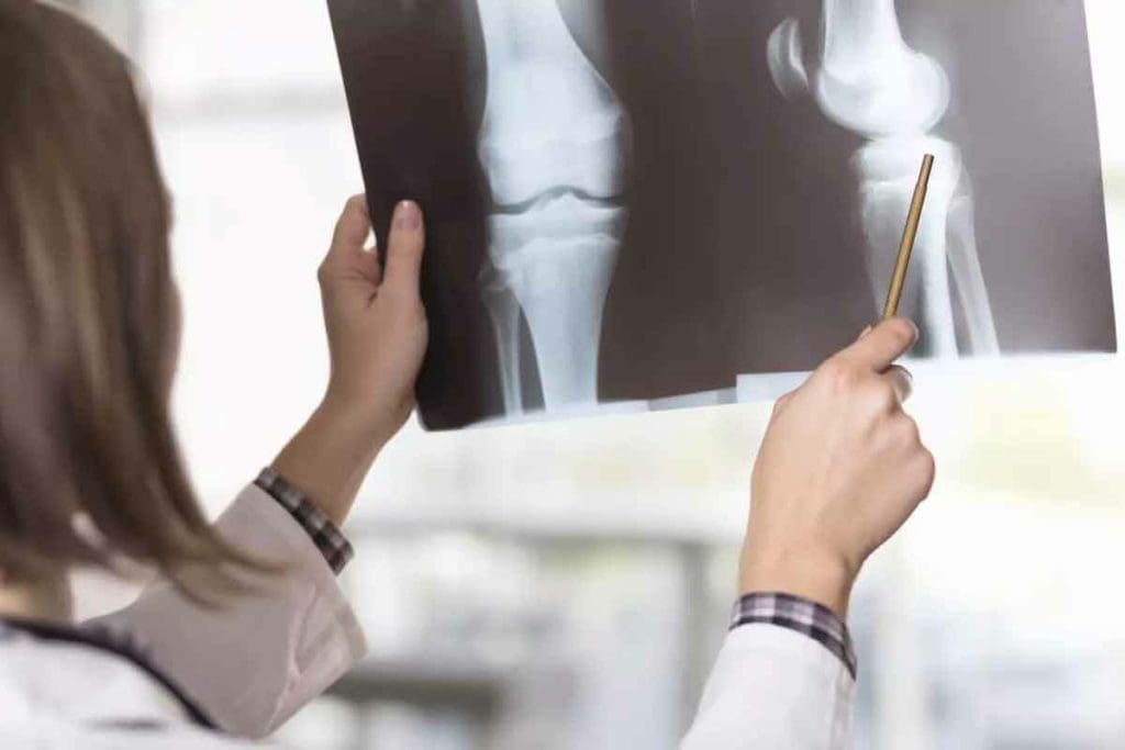
Bone scintigraphy is a detailed nuclear medicine imaging method. It helps find and diagnose different bone diseases. This includes fractures, infections, and cancer.
A bone scan uses a special radioactive tracer. It looks for bone damage or disease. The scan is usually done in a hospital’s nuclear medicine unit. First, a radiopharmaceutical, often technetium-99m (99mTc), is injected into the patient. Then, it gets absorbed by the bones.
The scan can take up to four hours. This includes the time for the tracer to circulate and the imaging process. This tool is key for spotting bone-related issues early. It helps start treatment quickly and effectively.
Key Takeaways
- Bone scintigraphy is a nuclear medicine imaging technique for diagnosing bone diseases.
- The procedure involves injecting a radioactive tracer and scanning with a gamma camera.
- Images are typically acquired 2-5 hours after the injection.
- Bone scintigraphy is useful for detecting fractures, infections, and cancer.
- The technique is considered safe and effective for diagnosing bone-related conditions.
Understanding Bone Scintigraphy
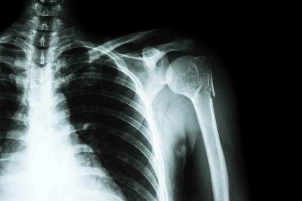
Bone scintigraphy, also known as skeletal scintigraphy, is a way to see bone damage or disease. It uses a small amount of radioactive material injected into the vein. This material goes to the bones, showing where there’s abnormal activity.
Definition and Basic Principles
Bone scintigraphy is a method that uses nuclear medicine to look at the bones. It works by using radiotracers that go to areas of bone problems. The most common radiotracer is Technetium-99m methylene diphosphonate (99mTc-MDP), which sticks to bone, showing where there’s more activity.
This technique is great for seeing the whole skeleton. It helps find and track many bone diseases. It’s good at showing where bone metabolism is higher, which can mean there’s a problem.
Terminology: Scintigrafie Osoasa and Skeletal Scintigraphy
“Scintigrafie osoasa” and “skeletal scintigraphy” mean the same as bone scintigraphy. “Scintigrafie osoasa” is the Romanian term, and “skeletal scintigraphy” is another English term. They all talk about the same imaging technique for checking bone health.
Knowing these terms is important for doctors and patients. It helps with clear communication and accurate diagnosis.
Historical Development of Bone Scanning
The first bone-seeking radiopharmaceuticals were introduced in the 1960s. Over time, better technology and new radiotracers have made bone scintigraphy more accurate.
Today, bone scintigraphy is a key tool in medicine. It helps doctors understand bone health and diseases. Research keeps improving it, making it even more useful for bone-related issues.
The Science Behind Bone Scintigraphy
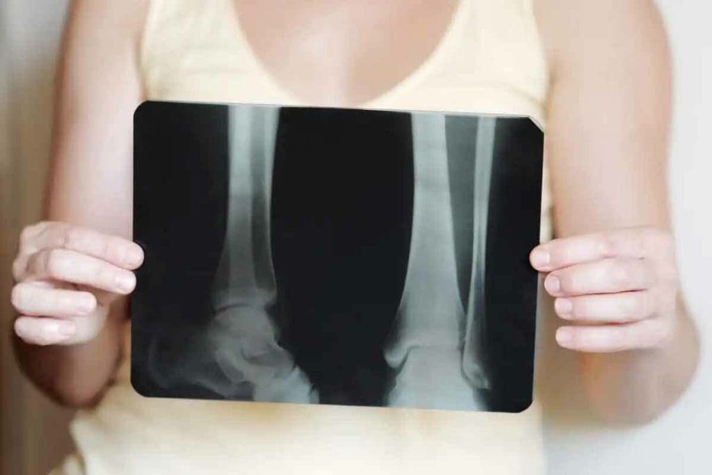
Bone scintigraphy uses nuclear medicine and special radiotracers. It’s a way to see the skeletal system through imaging. This technique relies on nuclear medicine principles.
Nuclear Medicine Fundamentals
Nuclear medicine uses tiny amounts of radioactive materials. It helps diagnose and treat diseases like cancer and heart disease. In bone scintigraphy, it helps see how bones work and their structure.
Radiopharmaceuticals are key in this process. They are compounds with a radioactive element. These are made to target specific areas in the body, like bone tissue in bone scintigraphy.
Radiotracers in Bone Imaging
Radiotracers are substances with a radioactive element. They help track biological processes. In bone scintigraphy, Tc-99m labeled phosphonates are used. They build up in bone tissue based on how active it is.
Choosing the right radiotracer is important. It affects how accurate and detailed the scan is. The best one should stick well to bone, be safe, and emit gamma radiation that cameras can detect.
The Role of Technetium-99m (99mTc)
Technetium-99m (99mTc) is a key isomer in nuclear medicine. It has a short half-life of about 6 hours. This makes it safe for medical use because it decays quickly.
In bone scintigraphy, 99mTc is linked to phosphonates like methylene diphosphonate (MDP). This creates a radiopharmaceutical that bone tissue takes up. The images from this help doctors understand bone health and diagnose bone disorders.
How a Bone Scan Works
To understand a bone scan, let’s look at its main parts. A bone scan, or bone scintigraphy, is a way to see how bones work and find bone problems. It has several steps to do this.
Patient Preparation
Before a bone scan, patients need to get ready. They might need to take off jewelry or metal things that could mess with the scan. Drinking lots of water is also important to get rid of the radiotracer after the scan. For more info, check RadiologyInfo.org.
Radiotracer Injection Process
The scan starts with a tiny amount of radioactive material, called a radiotracer, being injected into a vein. Technetium-99m (99mTc) methyl diphosphonate (MDP) or hydroxymethylene diphosphonate (HDP) is often used. This material goes to active bone areas, like fractures or tumors.
Waiting Period and Tracer Distribution
After the injection, the patient waits for 2 to 4 hours. This lets the radiotracer spread and build up in bones. During this time, the patient might drink water to help the tracer move around.
Image Acquisition with Gamma Cameras
After waiting, the patient lies under a gamma camera. This camera picks up the gamma rays from the radiotracer. It shows where the bone activity is. The whole scan takes about an hour, and the patient must stay very quiet.
The whole bone scan, from start to finish, can take four hours. New tech, like SPECT/CT hybrid imaging, makes bone scans better. It helps find and understand bone issues more accurately.
Types of Bone Scintigraphy Procedures
There are many types of bone scintigraphy procedures. Each one is designed for different needs and gives important insights. At Liv Hospital, they use the latest methods and focus on the patient.
Whole-Body Skeletal Surveys
Whole-body skeletal surveys scan the whole skeleton. They are great for finding widespread bone diseases or cancer. This scan is key for checking bone health in patients with cancer or bone disorders.
Three-Phase Bone Scans
Three-phase bone scans have three parts: blood flow, soft tissue, and bone metabolism. They help find issues like osteomyelitis or bone trauma. This makes them perfect for checking bone health and grafts.
Targeted Regional Scans
Targeted regional scans look at specific areas of the body. They are very good at finding small problems. These scans help with bone pain, checking bone lesions, or seeing how tumors respond to treatment.
Bone scintigraphy is vital for diagnosing many bone issues. It includes whole-body scans, three-phase scans, and targeted scans. The right scan depends on the patient’s needs and what doctors want to find out.
Clinical Applications of Bone Scintigraphy
Bone scintigraphy is used in many ways, like finding fractures, infections, and cancers. It’s key in today’s medicine because it shows how well bones are doing.
Detecting Bone Fractures and Trauma
Bone scintigraphy is great for spotting fractures that X-rays can’t see. It catches changes in bone activity, helping find hidden fractures.
Diagnosing Bone Infections and Inflammatory Conditions
It’s also good for finding bone infections and inflammatory diseases. It helps figure out what kind of infection and how big it is, helping doctors decide the best treatment.
Cancer Detection and Metastasis Evaluation
Bone scintigraphy is key in finding and checking cancer spread in bones. It helps doctors know how far the cancer has spread and if treatments are working.
Other Clinical Applications
Beyond finding fractures and infections, bone scintigraphy is used in other ways. It checks bone health, watches bone grafts, and looks at some metabolic bone diseases. Its many uses make it a valuable tool in many medical situations.
Interpreting Bone Scan Results
Understanding bone scan results is key for good patient care. Bone scintigraphy shows how bones are working. But, it’s not always easy to get the meaning right.
Normal vs. Abnormal Findings
A normal bone scan looks even across the skeleton. No spots stand out too much. But, an abnormal scan shows hot spots or cold spots. These spots mean the bone is acting differently.
Hot spots mean the bone is more active. This could be because of a fracture, infection, or tumor. A study on the National Center for Biotechnology Information (NCBI) website says so (PMC4932135).
Hot Spots and Cold Spots: What They Mean
Hot spots on a bone scan mean the bone is working hard. This could be because of a fracture, infection, or tumor. Or, it could be because of joint disease.
- Fractures or trauma
- Infections or inflammatory conditions
- Tumors or metastatic disease
- Degenerative joint disease
Cold spots, on the other hand, mean the bone is not working right. This can happen in avascular necrosis or some tumors.
| Condition | Typical Bone Scan Finding |
| Fracture | Hot spot |
| Avascular Necrosis | Cold spot |
| Tumor or Metastasis | Hot spot |
Correlation with Other Diagnostic Tests
It’s hard to understand bone scan results alone. But, when you compare them with X-rays, CT scans, or MRI, you get a clearer picture. This helps doctors make better choices for patients.
“The integration of bone scan results with clinical findings and other diagnostic tests is vital for the best patient care.”
” Clinical Nuclear Medicine
By looking closely at bone scan results and comparing them with other tests, doctors can make better decisions. This helps patients get the care they need.
Advantages of Bone Scintigraphy Over Other Imaging Techniques
Bone scintigraphy stands out among other imaging methods. It’s great for spotting problems early and checking the whole body. This technique is very good at catching changes in bone activity early on.
Early Detection Capabilities
Bone scintigraphy is very sensitive. It can find even small changes in bone activity. This is key for catching issues like bone infections, fractures, and cancer early.
Early treatment is often the best way to manage bone diseases. Finding problems early lets doctors start treatment sooner. This can lead to better results for patients.
Whole-Body Assessment Benefits
Bone scintigraphy gives a whole-skeleton overview with low radiation. This is great for seeing how systemic conditions affect the skeleton. It’s very useful for checking on diseases that can spread to many bones.
Seeing the whole skeleton at once helps find many problems or areas of abnormal activity. This is very important for tracking cancer or spotting widespread bone disease.
Functional vs. Anatomical Imaging
Bone scintigraphy focuses on functional information about bone activity. This is different from some other imaging that looks at structures. It lets doctors see how active bone lesions are, which can be more helpful than just looking at structures.
By mixing functional info with anatomical images, doctors get a better picture of what’s going on. This helps a lot with diagnosing and planning treatment.
Limitations and Safety Considerations
It’s important to know the limits and safety of bone scintigraphy. This test is very useful but has its own set of rules and risks.
Radiation Exposure and Risk Assessment
Bone scintigraphy uses tiny amounts of radioactive materials. These materials are injected into your vein. They leave your body in 2 to 3 days.
The radiation is very low. It’s much less than what you get from a CT scan.
Radiation Exposure Comparison
| Imaging Technique | Typical Effective Dose (mSv) |
| Bone Scintigraphy | 4-6 |
| CT Scan (Abdomen and Pelvis) | 10-20 |
Specificity Challenges and False Positives
Bone scintigraphy can sometimes show false positives. This means it might show activity that’s not related to the condition. It’s important to check the results with other tests and symptoms.
Contraindications and Special Populations
While safe, bone scintigraphy has some limits. For example, it’s not recommended for pregnant women because of the radiation. Breastfeeding women should also stop for a short time after the test.
In summary, bone scintigraphy is a great tool but knowing its limits and safety is key. This helps use it effectively and safely.
Advanced Developments in Bone Scintigraphy
Recent advancements in bone scintigraphy have changed the field of nuclear medicine. New technologies have made bone scans more accurate and useful. A key development is SPECT/CT hybrid imaging technology. It combines SPECT’s functional data with CT’s detailed images.
SPECT/CT Hybrid Imaging Technology
SPECT/CT hybrid imaging is a big step forward in bone scintigraphy. It mixes SPECT’s functional data with CT’s detailed images. This gives a better understanding of bone problems.
A study on NCBI shows SPECT/CT improves diagnosis in many cases.
SPECT/CT helps find and understand bone issues better. It can tell the difference between harmless and serious problems. This technology is key in modern nuclear medicine, helping with complex bone disorders.
New Radiotracer Developments
New radiotracers are being researched for bone scintigraphy. Traditional agents like Technetium-99m (99mTc) are used but new ones are being tested. For example, Fluorine-18 Fluoride (18F-NaF) PET/CT is promising for finding bone metastases.
New radiotracers could lead to earlier and more accurate bone disease detection. As research goes on, bone scintigraphy will get even better.
Standardization Guidelines and Protocol Improvements
As bone scintigraphy evolves, standardization and protocol improvements are needed. Standardization ensures consistency and helps with large studies. Work is being done to standardize image acquisition, processing, and interpretation.
“Standardization of imaging protocols is essential for ensuring high-quality images and accurate diagnoses in bone scintigraphy.”
Advances in SPECT/CT, new radiotracers, and standardization will keep improving bone scintigraphy. These changes will help patients and make managing bone disorders more effective.
Patient Experience During a Bone Scan
Knowing what to expect during a bone scan can make you feel less anxious and more comfortable. At Liv Hospital, we focus on our patients. We make sure you get the best care and support during your bone scan.
What to Expect Before, During, and After the Procedure
Before your bone scan, you might not need to do much. “You may be asked to drink extra water after you receive the radiotracer to help flush the material from your bladder,” says the prep steps. Drinking lots of water is key to getting rid of the radiotracer after the scan.
During the scan, you’ll lie on a table and stay very quiet. The gamma camera will take pictures of your bones. The scan is usually painless and can last from 30 minutes to a few hours, depending on the scan’s details.
After the scan, you can usually go back to your normal day unless your doctor says not to. The radiotracer leaves your body through urine and feces.
Managing Anxiety and Ensuring Comfort
It’s important to manage your anxiety during a bone scan. Talking openly with your healthcare team can really help. Don’t be afraid to ask questions or share your worries.
Try breathing exercises and relaxation techniques to calm down. Some places might even offer things like TV or music to help you relax during the scan.
“The patient-centered approach at Liv Hospital ensures comfort and care during the procedure,” highlighting the importance of a supportive environment in managing patient anxiety.
Understanding the bone scan process and how we ensure your comfort can help you feel more at ease. You’ll be better prepared to handle the experience with less worry.
Conclusion
Bone scintigraphy is a key tool for checking bone health and finding bone problems. It’s safe and helps find many bone issues. This makes it very important for doctors and patient care.
This imaging method helps spot bone problems early and checks the whole body. It’s vital for diagnosing and managing bone diseases. Knowing how it works helps doctors give the right treatment.
Using bone scintigraphy, doctors can improve patient care. It’s a reliable way to diagnose and watch bone conditions. This helps patients get better care.
FAQ
What is bone scintigraphy?
Bone scintigraphy, also known as a bone scan, is a way to see inside bones. It uses tiny amounts of radioactive material to find and track bone problems.
What is a bone scan used for?
A bone scan helps find bone fractures, infections, and cancer spread. It also checks how bone diseases are doing.
How does a bone scan work?
A bone scan injects a tiny bit of radioactive material into your blood. This material goes to your bones. Then, a special camera takes pictures of your skeleton.
What is the role of technetium-99m (99mTc) in bone scintigraphy?
Technetium-99m (99mTc) is a special tracer used in bone scans. It shows up in active bones, helping doctors see bone problems.
What are the different types of bone scintigraphy procedures?
There are many types of bone scans. These include whole-body scans, three-phase scans, and targeted scans. Each has its own use and benefits.
How do I prepare for a bone scan?
Before a bone scan, remove any metal items. You might also need to drink lots of water to clear the tracer.
Is a bone scan safe?
Bone scans are usually safe. But, they do involve some radiation. Pregnant or breastfeeding women should talk to their doctor first.
What are the advantages of bone scintigraphy over other imaging techniques?
Bone scans are good at finding problems early. They can look at the whole body and show how bones are working.
What are the limitations of bone scintigraphy?
Bone scans have some downsides. They involve radiation, can be tricky to interpret, and might not be right for everyone.
How are bone scan results interpreted?
Doctors look at the scan images for any unusual activity. They then match these findings with other tests and your medical history.
References
- Adams, C. (2023). Bone Scan – StatPearls. National Center for Biotechnology Information. https://www.ncbi.nlm.nih.gov/books/NBK531486/
- Radiopaedia. (2024). Bone scintigraphy. https://radiopaedia.org/articles/bone-scintigraphy-1
- Van den Wyngaert, T. et al. (2016). The EANM practice guidelines for bone scintigraphy. PMC. https://www.ncbi.nlm.nih.gov/pmc/articles/PMC4932135/
- Stokkel, M. (2025). A bone scan with technetium-99m: Early detection, better treatment. Nuclear Medicine News. https://www.nrgpallas.com/news/a-bone-scan-with-technetium-99m-early-detection-better-treatment
- RadiologyInfo.org. (2024). Bone Scan. https://www.radiologyinfo.org/en/info/bone-scan


