Last Updated on November 27, 2025 by Bilal Hasdemir

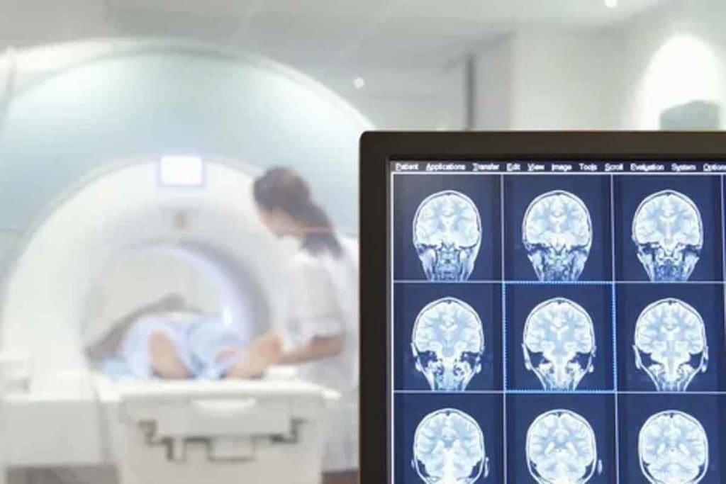
Getting a correct diagnosis and treatment plan for brain tumors depends a lot on advanced imaging. Brain tumor MRI is key for spotting and figuring out brain masses. It gives clear images of soft tissues without using harmful radiation.
At Liv Hospital, we use the latest MRI tech to find and tell apart brain tumors. This lets our team make good treatment plans. We focus on our patients and follow global medical standards to give them the best care.
Key Takeaways
- MRI is the main way to find and understand brain masses.
- Advanced MRI methods help tell brain tumors apart.
- Getting the right diagnosis is key for good treatment.
- Liv Hospital puts patients first and follows global medical standards.
- Our team uses top-notch MRI tech to diagnose and treat brain tumors.
The Fundamentals of Neuroimaging for Brain Masses
Neuroimaging is key for spotting brain masses. We use it to check for tumors and plan treatments. MRI is top-notch for this because it shows brain details well.
Why MRI is the Gold Standard for Brain Tumor Detection
MRI is the best for finding brain tumors. It’s very good at showing soft tissues. Doctors use MRI to see where, how big, and how tumors look.
MRI is a must for brain tumor care. It’s safe and doesn’t use harmful radiation. This makes it perfect for checking patients often.
High Spatial Resolution and Tissue Contrast Advantages
MRI shines because it shows things clearly and in detail. This helps doctors tell different tumors apart. Tumors can be inside or outside the brain, affecting it differently.
| Characteristics | Intra-Axial Tumors | Extra-Axial Tumors |
| Origin | Within brain parenchyma | Outside brain parenchyma |
| Common Types | Gliomas, astrocytomas | Meningiomas, schwannomas |
| MRI Features | Variable enhancement, edema | Often strong enhancement, dural tail |
Knowing these differences helps doctors make better diagnoses. MRI’s clear images are vital for good care.
Key Finding 1: Distinguishing Intra-Axial vs Extra-Axial Tumors
It’s key to tell intra-axial from extra-axial tumors for right diagnosis and treatment. Intra-axial tumors grow inside the brain, while extra-axial ones come from outside, like the meninges or skull. Knowing this helps us read brain MRI scans well.
Characteristics of Intra-Axial Tumors
Diffuse gliomas are common primary brain tumors in adults. They start in the brain and can spread to nearby tissue. MRI shows intra-axial tumors as:
- Ill-defined edges because they spread into brain tissue
- Variable brightness on T1 and T2 images
- Varied contrast enhancement
Extra-Axial Masses and Their MRI Signatures
Extra-axial tumors grow outside the brain, often from the meninges, nerves, or skull. Examples are meningiomas and schwannomas. MRI shows them as:
- A wide dural base or dural tail sign
- Clear edges with the brain
- Even enhancement after contrast
Distinguishing intra-axial from extra-axial tumors matters a lot. It affects treatment planning and how well a tumor will do. Radiologists and doctors need to know these differences to give the right diagnosis and treatment.
Key Finding 2: Enhancement Patterns in Brain Tumor MRI
MRI enhancement patterns help us understand brain tumors. These patterns give us clues about the tumor’s nature. This information is key for diagnosis and treatment planning.
Homogeneous vs. Heterogeneous Enhancement
There are two main types of enhancement patterns on MRI: homogeneous and heterogeneous. Homogeneous enhancement means the tumor takes up contrast evenly. This suggests a simpler tumor biology. Heterogeneous enhancement shows uneven contrast uptake. It might mean the tumor has different parts, like necrosis or cysts.
Some tumors show specific enhancement patterns. For example, low-grade gliomas might not show much enhancement. But high-grade gliomas often have uneven enhancement because they are aggressive.
Ring Enhancement and Its Diagnostic Significance
Ring enhancement is when the tumor’s edge shows up on the scan, but the center doesn’t. This pattern is important because it often means the tumor is aggressive or an abscess. The edge shows where the tumor is alive, and the center shows where it’s dead.
Ring enhancement is a sign of aggressive tumors and a poor prognosis. Doctors use this to decide on treatments, like more surgery or other therapies.
| Enhancement Pattern | Tumor Characteristics | Diagnostic Implication |
| Homogeneous | Uniform tumor biology | Suggests less aggressive tumors |
| Heterogeneous | Complex tumor composition | May indicate high-grade tumors |
| Ring Enhancement | Viable tumor periphery with necrotic center | Often associated with aggressive tumors or abscesses |
By looking at MRI enhancement patterns, we can learn more about tumors. This helps us make better decisions for patient care. New imaging and AI models help us spot more details and predict tumor behavior.
Key Finding 3: Signal Intensity Variations Across Tumor Types
Understanding how signal intensity changes is key to identifying different tumors on MRI. Signal intensity shows how bright or dark a tissue looks on an MRI. It depends on the tissue’s magnetic properties and the MRI sequence used.
T1-Weighted Image Characteristics
T1-weighted images are great for seeing brain anatomy. Most tumors look darker than brain tissue on these images. But, some tumors might look the same or brighter, depending on their makeup.
T2-Weighted Image Characteristics
T2-weighted images show water content differences in tissues. This makes them good for spotting edema and tumor spread. Tumors usually look brighter on T2 images because they have more water. The brightness on T2 images helps tell different tumors apart.
FLAIR Sequence Findings and Their Importance
The FLAIR sequence is excellent for finding brain lesions. It reduces free water signal, making it great for spotting issues near CSF spaces. Tumors and swelling appear bright on FLAIR images, helping doctors see how big a tumor is and if there’s swelling around it.
Recent advances in deep learning, like CNNs, have made MRI analysis better for tumor identification. These technologies use signal changes across MRI sequences to improve accuracy.
| Tumor Type | T1-Weighted Signal | T2-Weighted Signal | FLAIR Signal |
| Low-Grade Glioma | Hypointense | Hyperintense | Hyperintense |
| High-Grade Glioma | Hypointense | Hyperintense | Hyperintense with heterogeneity |
| Meningioma | Isointense/Hypointense | Hyperintense | Hyperintense |
By looking at signal intensity changes on T1, T2, and FLAIR images, we can better understand brain tumors. This helps doctors make better decisions.
Key Finding 4: Peritumoral Edema as a Diagnostic Clue
Peritumoral edema on MRI scans is a key sign in brain tumor diagnosis. It shows swelling around a tumor. This swelling gives important clues about the tumor and its effect on the brain.
Vasogenic vs. Cytotoxic Edema on MRI
There are two main types of peritumoral edema: vasogenic and cytotoxic. Vasogenic edema happens when the blood-brain barrier breaks down. This leads to fluid buildup outside cells. On MRI, it looks like bright spots on T2-weighted images around the tumor.
Cytotoxic edema is caused by cell damage, often from lack of blood flow or certain tumors. It makes cells swell. But, it’s harder to spot on MRI than vasogenic edema.
Edema Patterns Associated with Different Tumor Types
Brain tumors show different edema patterns. For example, glioblastomas have a lot of vasogenic edema because they grow fast and damage the blood-brain barrier. But, some low-grade gliomas might not show much edema at all.
Knowing these patterns helps doctors and radiologists diagnose and treat brain tumors better. By looking at the edema on MRI, we can learn more about the tumor and its effects on the patient.
Key Finding 5: Advanced MRI Techniques for Tumor Characterization
Advanced MRI techniques have changed neuro-oncology a lot. They give us detailed views of brain tumors. This helps us tell different tumors apart and see their edges clearly. This is key for planning treatments.
Diffusion-Weighted Imaging Applications
Diffusion-weighted imaging (DWI) is great for looking at tumor cells. It checks how water moves in the tumor. This tells us about the tumor’s cell density and health.
Table 1: Comparison of Diffusion Characteristics in Different Tumor Types
| Tumor Type | Diffusion Characteristics | DWI Appearance |
| High-Grade Gliomas | Restricted Diffusion | Hyperintense |
| Low-Grade Gliomas | Facilitated Diffusion | Hypointense |
| Metastatic Tumors | Variable Diffusion | Variable |
Perfusion Imaging for Vascularity Assessment
Perfusion imaging looks at how tumors get blood flow and how open their blood vessels are. It shows how well tumors can grow blood vessels. This is important for knowing how aggressive a tumor is and how it will react to treatment.
MR Spectroscopy for Metabolic Profiling
MR spectroscopy checks the tumor’s metabolism by looking at certain chemicals. It finds out how much choline, creatine, and N-acetylaspartate there are. High choline levels mean the tumor is growing fast, which is a sign of cancer.
By using these MRI methods together, we get a full picture of brain tumors. This helps us make better diagnoses and treatment plans for each patient.
Brain Tumor MRI: Identifying Non-Enhancing Lesions
Non-enhancing brain tumors are hard to spot on MRI because they look similar to normal brain tissue. Doctors need to look closely to figure out what they are and if they’re serious.
Low-Grade Gliomas and Their Subtle MRI Appearance
Low-grade gliomas are a kind of brain tumor that shows up as non-enhancing lesions on MRI. They look the same on T1-weighted images and are brighter on T2-weighted images. They don’t show up after contrast is added, which is a key sign, but not the only one.
We use special MRI techniques to learn more about these tumors. Diagnosing brain tumors often means using different imaging methods and talking to the patient’s doctor.
| MRI Sequence | Typical Appearance of Low-Grade Gliomas |
| T1-Weighted | Hypointense or isointense |
| T2-Weighted | Hyperintense |
| FLAIR | Hyperintense |
| Post-Contrast T1 | No enhancement |
Differential Diagnosis of Non-Enhancing Brain Masses
When we see non-enhancing brain masses, we have to think of many possibilities. These include low-grade gliomas, benign growths, inflammation, and other tumors.
Key considerations in differential diagnosis:
- Clinical presentation and history
- Imaging characteristics on various MRI sequences
- Presence of mass effect or edema
Getting a correct diagnosis means looking at all the imaging and clinical details. We have to think about all the possible causes when we see non-enhancing brain masses on MRI.
Key Finding 7: Calcifications in Brain Tumors and Their Imaging Correlates
Understanding calcifications in brain tumors is key for good treatment plans. These calcifications can change how we diagnose and treat tumors.
Imaging Characteristics of Calcified Brain Tumors on MRI
Calcified brain tumors show clear signs on MRI. On T1-weighted images, they might look brighter. But on T2-weighted images, they can appear darker. The look can change based on the type and amount of calcification.
MRI Features: MRI’s susceptibility-weighted imaging (SWI) can hint at calcifications. It’s good at spotting calcifications and bleeding.
Correlation Between CT and MRI Findings for Calcified Masses
While MRI is great for soft tissues, CT scans are better at showing calcifications. So, it’s important to compare CT and MRI scans to fully understand calcified brain tumors.
| Imaging Modality | Characteristics of Calcified Tumors |
| CT | Hyperdense areas indicating calcifications |
| MRI (T1-weighted) | Variable signal intensity, sometimes hyperintense |
| MRI (T2-weighted) | Often hypointense due to calcifications |
| MRI (SWI) | Susceptibility effects highlighting calcifications |
By using both CT and MRI, we can get a clearer picture of calcified brain tumors. This leads to more accurate diagnoses and better treatment plans.
Key Finding 8: Small Brain Tumor Detection Strategies
Small brain tumors are hard to find, making early detection key. This is important for good treatment and outcomes.
Contrast Enhancement Protocols for Small Lesion Visualization
Contrast enhancement is vital in MRI for spotting small brain tumors. It uses agents like gadolinium to make tumors stand out.
To enhance contrast, a contrast agent is given through an IV before the MRI. The right amount and timing are important for clear images.
| Contrast Enhancement Protocol | Description | Benefits |
| Gadolinium-based Contrast | Administered intravenously before MRI | Enhances tumor visibility, improves boundary delineation |
| Dosage Optimization | Tailored to patient weight and scan parameters | Maximizes contrast effect, minimizes side effects |
| Timing of Contrast Administration | Optimized for best image quality | Ensures optimal tumor visualization |
High-Resolution Techniques for Detecting Minute Tumors
High-resolution MRI is essential for finding small brain tumors. It uses advanced sequences for detailed brain images.
Some high-resolution techniques include:
- 3D Imaging Sequences: Give detailed volume data for better tumor assessment.
- Thin Slice Thickness: Helps spot small tumors by reducing partial volume effects.
- High-Field MRI: Offers clearer images of small brain structures with better signal-to-noise ratio.
Combining contrast enhancement with high-resolution techniques boosts small brain tumor detection. This approach improves diagnosis and treatment planning.
Key Finding 9: Tumor Boundaries and Infiltration Assessment
MRI helps doctors understand tumor boundaries and how they spread. This info is key for making treatment plans. Knowing how far a tumor has spread helps doctors plan the best course of action.
Sharp vs. Ill-Defined Margins and Their Clinical Implications
The clarity of tumor margins on MRI is very important. Tumors with sharp margins are usually easier to remove surgically. Tumors with ill-defined margins are more aggressive and harder to plan surgery for.
How clear the tumor margins are affects treatment choices. For example, a tumor with sharp margins might be removed completely. But a tumor with ill-defined margins might need a more careful approach, like removing part of it or using other treatments.
| Margin Type | Clinical Implication | Treatment Approach |
| Sharp Margins | Potential for gross total resection | Surgical resection |
| Ill-Defined Margins | Infiltrative nature, complicating surgery | Conservative approach, subtotal resection, or alternative treatments |
Detecting Tumor Infiltration with Advanced MRI Sequences
Advanced MRI sequences are key in finding where tumors spread. Diffusion-weighted imaging and MR spectroscopy show how active and dense tumors are. This helps spot where tumors might be spreading.
We use these advanced scans to see how far tumors have spread. For example, diffusion-weighted imaging can tell the difference between tumor spread and swelling. MR spectroscopy shows the tumor’s metabolic activity, helping to judge how aggressive it is.
By using MRI and advanced sequences together, we get a clearer picture of tumors. This helps us create treatment plans that really work for each patient.
Key Finding 10: Differentiating Tumor Recurrence from Treatment Effects
It’s very important to tell the difference between tumor recurrence and treatment effects. This is a big challenge in neuro-oncology because they can look similar on MRI. We will look into the complexities of MRI changes after treatment, the issue of pseudoprogression, and new ways to improve diagnosis.
Post-Treatment MRI Changes and Pseudoprogression
After treatment, MRI scans can show changes that might mean the tumor is back or it’s just a treatment effect. Sometimes, these effects can look like the tumor is getting worse, known as pseudoprogression. A study in the American Journal of Neuroradiology found that about 20-30% of patients on chemoradiation therapy experience pseudoprogression (ACR).
It’s key to understand when and how these changes happen. Pseudoprogression usually shows up in the first few months after radiation therapy, often in patients with glioblastoma. It looks like the tumor is growing, but it’s not.
“Pseudoprogression is a treatment-related reaction that mimics tumor progression on imaging, making it a diagnostic challenge.”
Advanced Techniques for Recurrence Detection
New imaging methods are being used to tell tumor recurrence apart from treatment effects. These include diffusion-weighted imaging, perfusion-weighted imaging, and MR spectroscopy.
| Technique | Application | Benefit |
| Diffusion-Weighted Imaging | Assesses water diffusion in tissues | Helps differentiate between tumor recurrence and necrosis |
| Perfusion-Weighted Imaging | Evaluates blood flow and vascularity | Identifies areas of high vascularity indicative of tumor recurrence |
| MR Spectroscopy | Analyzes metabolic changes in tissues | Detects metabolic signatures of tumor recurrence |
These new methods give us important information to help make a correct diagnosis. Experts say, “Advanced MRI techniques help us tell tumor recurrence from treatment effects, which improves patient care.”
Conclusion: Integrating MRI Findings for Comprehensive Brain Tumor Diagnosis
Integrating MRI findings is key for a full brain tumor diagnosis. We’ve talked about MRI’s big role in finding and understanding brain tumors. It’s great at showing details and differences in tissues.
MRI is very important for diagnosing and planning treatment for brain tumors. It helps us tell apart different types of tumors and see how they affect the brain. It also shows how the tumor is growing and if it’s causing swelling.
Using MRI, we can make a better diagnosis and plan a good treatment. MRI’s role in diagnosing brain tumors is huge. It helps doctors make the right choices and improves how patients do. As MRI technology gets better, we’ll get even more accurate diagnoses and treatments.
FAQ
What is the role of MRI in brain tumor diagnosis?
MRI is key in finding and understanding brain tumors. It shows detailed images of soft tissues. This helps doctors plan treatments.
Why is MRI considered the gold standard for brain tumor detection?
MRI is top-notch for spotting brain tumors. It offers clear images of brain structures and tumors.
What is the difference between intra-axial and extra-axial tumors?
Intra-axial tumors start inside the brain. Extra-axial tumors start outside. Their looks and where they are on MRI are different.
What are the different enhancement patterns seen in brain tumor MRI?
MRI shows tumors in three ways: all the same, mixed, or with a ring. Each pattern tells doctors something about the tumor.
How do T1-weighted, T2-weighted, and FLAIR sequences help in characterizing tumors?
These MRI types show how tumors look. They help doctors understand what kind of tumor it is.
What is the significance of peritumoral edema in brain tumor diagnosis?
Edema around tumors is important. It helps doctors figure out what kind of tumor it is.
How do advanced MRI techniques aid in tumor characterization?
New MRI methods like diffusion-weighted imaging help. They show how tumors work and what they’re made of.
How are non-enhancing lesions identified on brain tumor MRI?
MRI finds tumors that don’t show up well. Doctors have to be careful to make the right diagnosis.
What are the imaging correlates of calcifications in brain tumors on MRI and CT?
MRI and CT show tumors with calcium differently. Knowing this helps doctors make accurate diagnoses.
How are small brain tumors detected on MRI?
MRI uses special techniques to find small tumors. This makes it easier to see tiny lesions.
How is tumor infiltration assessed on MRI?
MRI checks how tumors spread. It uses advanced methods to see if tumors are growing.
What are the challenges in differentiating tumor recurrence from treatment effects on MRI?
It’s hard to tell if a tumor is coming back or if it’s just a reaction to treatment. But new MRI methods help.
References
- Chong, A., Song, C., & Shin, H. (2014). Application of bone scans for prostate cancer staging. Journal of Cancer Research and Therapeutics, 10(3), 605-610. https://www.ncbi.nlm.nih.gov/pmc/articles/PMC4137016/


