Last Updated on November 27, 2025 by Bilal Hasdemir

Neuro-oncology is moving forward thanks to top-notch datasets, like MRI scans of brain tumors. New tools in deep learning are changing how we analyze these scans. This is helping both doctors and artificial intelligence.
Having many and well-annotated MRI datasets is key for research and better care. As the field grows, finding reliable and easy-to-get datasets is more important than ever.
Key Takeaways
- High-quality MRI scan datasets are essential for advancing neuro-oncology research.
- Diverse and annotated datasets support the development of AI tools.
- Reliable datasets drive innovation and improve patient outcomes.
- Access to extensive datasets remains a significant challenge.
- Advancements in deep learning are transforming brain tumor analysis.
The Importance of Quality Brain Tumor MRI Scan Datasets

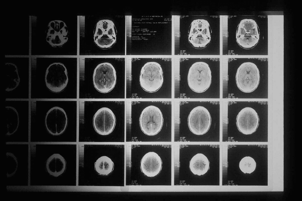
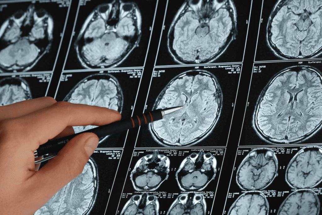
High-quality brain tumor MRI images are key in neuro-oncology research. They help improve clinical studies and AI in medical imaging.
Large datasets like BRISC offer 6,000 T1-weighted MRI scans with various tumor types. Public datasets like BraTS are also vital for brain tumor segmentation.
Advancing Clinical Neuro-Oncology Research
Brain tumor MRI scan datasets are vital for neuro-oncology research. They help in creating new diagnostic and treatment methods. Annotated brain tumour images MRI lead to better studies on tumors.
| Dataset | Description | Size |
| BRISC | Contrast-enhanced T1-weighted MRI scans | 6,000 |
| BraTS | Multimodal brain tumor segmentation dataset | Varies |
Fueling AI Development in Medical Imaging
Brain tumor MRI images are essential for AI in medical imaging. They help train and test AI models. This makes AI better at tumor segmentation and classification.
Using large, annotated datasets, researchers can create advanced AI models. These models improve diagnosis and support personalized medicine.
Understanding MRI Modalities in Brain Tumor Imaging

Knowing about MRI modalities is key for spotting brain tumors right and planning treatments well. MRI shows grayscale images that show different tissue types. These include proton density, FLAIR, T1-weighted, and T2-weighted images. They help doctors understand tumors and make decisions.
T1-Weighted vs. T2-Weighted Sequences
T1-weighted and T2-weighted sequences are basic in brain tumor scans. T1-weighted images show body parts clearly, and they’re better after contrast. This shows where the contrast goes, marking tumor edges. On the other hand, T2-weighted images spot water changes well, helping find swelling and lesions.
Knowing the difference between T1 and T2 images is important. Tumors look darker on T1 and brighter on T2. This helps see how big a tumor is, where it is, and if it’s spreading.
FLAIR and Contrast-Enhanced Protocols
FLAIR and contrast-enhanced scans add more to MRI’s power in brain tumor imaging. FLAIR sequences help see lesions near CSF spaces better. This is great for spotting swelling around tumors and telling it apart from fluid.
Contrast-enhanced MRI uses gadolinium to show where the blood-brain barrier is broken. This is common in brain tumors. It’s key for seeing tumor edges and how well treatments are working.
| MRI Modality | Primary Use in Brain Tumor Imaging | Key Features |
| T1-Weighted | Anatomical detail, contrast enhancement | Tumor appears hypointense, good for assessing tumor boundaries post-contrast |
| T2-Weighted | Detecting edema and lesions | Tumor appears hyperintense, sensitive to changes in tissue water content |
| FLAIR | Suppressing free water signal | Useful for detecting peritumoral edema and lesions near CSF spaces |
| Contrast-Enhanced | Delineating tumor margins, assessing treatment response | Highlights areas of blood-brain barrier disruption |
In summary, using T1, T2, FLAIR, and contrast-enhanced MRI gives a full view of brain tumors. Each type gives special info, helping doctors diagnose and plan treatments better.
Comprehensive Brain Tumor MRI Scan Collections
Comprehensive brain tumor MRI scan collections are key for neuro-oncology research. They give researchers the tools to create and test new ways to diagnose and treat brain tumors.
BRISC Dataset: 6,000 Annotated T1-Weighted Scans
The BRISC dataset is a big help in brain tumor research. It has 6,000 annotated T1-weighted MRI scans. This large set helps researchers train and test AI for better tumor detection and segmentation.
BraTS Challenge Dataset: Multimodal Segmentation Standard
The BraTS Challenge dataset is a top choice for testing brain tumor segmentation algorithms. It has MRI scans from different angles, like T1, T2, and FLAIR. This gives a full view of brain tumors. It also includes other CNS tumors and has datasets for both kids and adults with glioma.
TCIA Glioblastoma Collection (TCGA-GBM)
The TCIA Glioblastoma Collection, linked to TCGA-GBM, is a big help for glioblastoma research. It has lots of MRI scans with detailed clinical info. This makes it easier to study how tumors grow and how they respond to treatment.
RIDER Neuro MRI Dataset
The RIDER Neuro MRI dataset is also key for understanding brain tumors. It offers MRI scans with annotations. This helps improve automated analysis tools.
| Dataset | Description | Modalities |
| BRISC | 6,000 annotated T1-weighted scans | T1-weighted |
| BraTS | Multimodal segmentation standard | T1, T2, FLAIR |
| TCIA Glioblastoma | Large collection with clinical data | Multiple |
| RIDER Neuro MRI | Annotated MRI scans | Multiple |
These brain tumor MRI scan collections are vital for research. They offer large, detailed datasets. This helps create more accurate diagnostic tools and treatments.
Longitudinal Brain Tumor Imaging Resources
Studies using brain tumor MRI scans give us key insights. They show how tumors grow and how well treatments work. This is important for understanding tumor behavior over time.
Yale-Brain-Mets-Longitudinal: 11,892 Studies Over Two Decades
The Yale-Brain-Mets-Longitudinal dataset has 11,892 studies from 20 years. Researchers say it’s a great chance to see how brain metastases change. Check out the Yale-Brain-Mets-Longitudinal dataset for detailed research.
TCIA Collections with Temporal Progression Data
The The Cancer Imaging Archive (TCIA) has many collections. They include data on how brain tumors grow over time. These are great for those studying tumor growth and treatment effects on brain tumor images.
Harvard’s Neuroimaging Longitudinal Analysis Dataset
Harvard’s dataset is another important resource. It has MRI pictures of brain cancer taken at different times. This helps us learn more about tumor growth and improve diagnosis.
In summary, resources like Yale-Brain-Mets-Longitudinal and others are key for neuro-oncology research. They offer images for brain tumor studies. These are essential for finding better treatments.
Specialized Datasets for Tumor Classification
Specialized datasets are key in brain tumor classification research. They help create and test AI models for MRI scans. These models can accurately identify brain tumors.
Figshare Brain Tumor Classification Dataset
The Figshare Brain Tumor Classification Dataset is a great resource. It has a big collection of MRI images, all labeled and sorted. This makes it perfect for training AI to spot different brain tumors.
Key Features:
- Large collection of MRI images
- Meticulously labeled and categorized
- Useful for training machine learning models
Radiopaedia’s Curated Case Collection
Radiopaedia’s Curated Case Collection is also vital. It has a wide variety of cases with detailed notes. It’s great for learning and research.
Notable aspects include:
- Diverse range of cases
- Detailed annotations
- Useful for educational purposes and research
MICCAI Brain Tumor Classification Challenge Dataset
The MICCAI Brain Tumor Classification Challenge Dataset is a benchmark. It’s used in challenges to test algorithms. This helps improve brain tumor classification.
Key Benefits:
- Standardized dataset for evaluation
- Promotes advancements in brain tumor classification
- Used in various challenges and competitions
In conclusion, these datasets are essential for brain tumor research. They offer high-quality MRI images for better classification models.
Kaggle-Hosted Brain Tumor MRI Resources
Kaggle is a key platform for sharing brain tumor MRI datasets. It offers a wide range of resources for training AI models in brain tumor detection and classification.
7,023 Classified Images in Brain Tumor MRI Dataset
The Brain Tumor MRI Dataset on Kaggle has 7,023 classified images. It’s a big help for researchers. This dataset is key for training models to spot and classify brain tumors accurately.
Brain Tumor Detection 2020 Dataset
The Brain Tumor Detection 2020 Dataset is another great resource on Kaggle. It’s made to help develop algorithms for finding brain tumors in MRI scans.
Brain MRI Images for Brain Tumor Detection
Kaggle also has a set of Brain MRI Images for Brain Tumor Detection. This collection offers a wide range of images for training and testing brain tumor detection models.
The table below shows the main features of the Kaggle-hosted brain tumor MRI datasets:
| Dataset Name | Number of Images | Primary Use |
| Brain Tumor MRI Dataset | 7,023 | Classification |
| Brain Tumor Detection 2020 Dataset | Not specified | Detection |
| Brain MRI Images for Brain Tumor Detection | Not specified | Detection |
These datasets on Kaggle are vital for brain tumor imaging research. They give researchers access to lots of MRI scans. This helps in making AI models for brain tumor diagnosis more accurate and reliable.
AI-Optimized Brain Tumor Imaging Collections
AI-optimized brain tumor imaging collections are changing neuro-oncology. They help artificial intelligence understand and analyze complex medical images better.
Advanced datasets like DeepLesion and Medical Segmentation Decathlon (MSD) lead this change. They offer large, detailed clinical data for AI model training.
DeepLesion: Large-Scale Annotated Clinical Dataset
DeepLesion has 32,735 lesions from different body parts, including the brain. It’s a great source for training AI algorithms with annotated images.
“The DeepLesion dataset has been instrumental in advancing research in medical imaging analysis, providing a large-scale resource for lesion detection and segmentation.”
MSD (Medical Segmentation Decathlon) Brain Tumor Dataset
The MSD Brain Tumor Dataset is part of a bigger challenge. It tests segmentation algorithms on various medical imaging tasks. It includes 484 multi-modal MRI scans with detailed annotations.
The MSD dataset is key for developing AI models that can accurately segment brain tumors. This is vital for treatment planning and patient care.
- Large-scale annotated datasets for AI training
- Multi-modal MRI scans for thorough analysis
- Detailed annotations for precise segmentation
These AI-optimized brain tumor imaging collections are not just advancing research. They’re also making clinical practices in neuro-oncology more accurate and efficient.
Practical Considerations When Working with Brain Tumor MRI Datasets
When using brain tumor MRI datasets, several key points must be considered. These include data format, preprocessing, ethical use, and technical setup.
Data Format and Preprocessing Requirements
Brain tumor MRI data is often in DICOM or NIfTI formats. Data format standardization is vital for easy use in research. Steps like skull stripping and bias correction improve data quality.
The choice of preprocessing methods is very important. For example, skull stripping removes non-brain tissue. This helps tumor segmentation algorithms work better. It’s important to pick and test preprocessing steps carefully to keep data accurate.
Ethical Considerations and Usage Restrictions
Working with brain tumor MRI data raises big ethical considerations. Keeping patient privacy and protecting data is key. Researchers must follow rules like HIPAA in the U.S. and anonymize data properly.
Also, there are usage restrictions on some datasets, like those from clinical trials. It’s important for researchers to know and follow these rules. This keeps research ethical and legal.
Technical Infrastructure for Large-Scale Analysis
Handling big brain tumor MRI datasets needs strong technical infrastructure. This includes fast computers, lots of storage, and advanced software for analysis.
Many researchers use cloud-based solutions for MRI data analysis. These platforms offer flexible resources that grow with the project. They help teams work together more easily.
Conclusion: Future Directions in Brain Tumor Imaging Research
The future of brain tumor imaging research looks bright. It will see big steps forward with AI and large datasets. The BRISC dataset and the BraTS Challenge have already helped start this journey.
More data from different patients and imaging types will be key. This will make AI models better and more accurate. It will help doctors give better care and treatments.
AI in brain tumor research will grow fast. This is thanks to new deep learning methods and quality datasets like DeepLesion. MRI will keep playing a big role in finding new ways to understand and treat tumors.
AI, big datasets, and advanced MRI will shape the future of brain tumor imaging. They will open up new ways to find and treat tumors early.
FAQ
What is the significance of brain tumor MRI datasets in research?
Brain tumor MRI datasets are key in advancing neuro-oncology research and AI. They offer annotated MRI scans for training and validation.
What are the different MRI modalities used in brain tumor imaging?
MRI modalities include T1-weighted, T2-weighted, FLAIR, and contrast-enhanced protocols. Each provides unique information for tumor characterization.
What is the BRISC dataset, and how is it used?
The BRISC dataset is a large collection of 6,000 annotated T1-weighted MRI scans. It’s used for research in brain tumor imaging and AI development.
How do longitudinal brain tumor imaging resources contribute to research?
Longitudinal imaging resources, like the Yale-Brain-Mets-Longitudinal dataset, offer temporal progression data. This helps researchers study tumor development and treatment response over time.
What are some specialized datasets for tumor classification?
Specialized datasets include Figshare, Radiopaedia, and MICCAI challenge datasets. They provide curated MRI images for advancing classification research.
What are some practical considerations when working with brain tumor MRI datasets?
Practical considerations include data format and preprocessing, ethical considerations, and technical infrastructure for analysis.
How are AI-optimized brain tumor imaging collections used?
AI-optimized collections, like DeepLesion and MSD, are used to train AI models. These models are for tumor detection, segmentation, and classification.
What is the role of Kaggle-hosted brain tumor MRI resources?
Kaggle-hosted resources, such as the Brain Tumor MRI Dataset, provide accessible MRI images. They are for research and competition.
What is the importance of data preprocessing in brain tumor MRI analysis?
Data preprocessing is essential for ensuring data quality. It removes artifacts and standardizes formats for accurate analysis and AI model training.
How do brain tumor MRI datasets contribute to AI development in medical imaging?
Brain tumor MRI datasets contribute to AI development by providing large-scale, annotated datasets. These datasets are for training and validating AI models, leading to advancements in tumor detection, segmentation, and classification.
References
- Menze, B. H., et al. (2015). The Multimodal Brain Tumor Image Segmentation Benchmark (BRATS). IEEE Transactions on Medical Imaging, 34(10), 1993-2024. https://ieeexplore.ieee.org/document/6975210)
- Ahmed, M. M., et al. (2024). Brain tumor detection and classification in MRI using hybrid deep learning CNN model. Scientific Reports, 14(1), 1-15. https://www.nature.com/articles/s41598-024-71893-3


