Last Updated on December 1, 2025 by Bilal Hasdemir

Finding cancer in lymph nodes is key for diagnosing and treating cancer. A positron emission tomography (PET) scan is a tool that spots cancer cells in the body. But can it find cancer in lymph nodes?
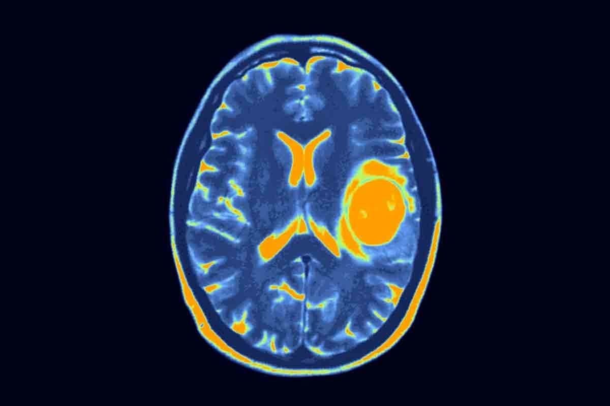
A PET scan can spot FDG uptake in lymph nodes, which means cancer might be there. This is very important for knowing how far the cancer has spread.
Key Takeaways
- PET scans can detect cancerous activity in lymph nodes.
- FDG uptake in lymph nodes is a key indicator of cancerous cells.
- Accurate diagnosis using PET scans is vital for cancer treatment.
- PET detection of lymph node metastasis helps determine cancer stage.
- PET scan results help identify cancerous lymph nodes with greater accuracy.
Understanding PET Scans and Their Role in Cancer Detection
PET scans are vital for finding and managing cancer, like in lymph nodes. They are a key tool in cancer diagnosis and treatment planning.
What is a PET Scan?
A PET scan uses a special sugar molecule to find cancer and other diseases. It injects a small amount of radioactive material, like Fluorodeoxyglucose (FDG), into your blood. Cancer cells, which use more sugar, show up more on the scan.
How PET Scans Work to Detect Cancer
PET scans highlight areas with high activity in the body. Cancer cells, being more active, take up more FDG. This makes them appear as “hot spots” on the scan.
“The ability of PET scans to detect metabolic changes before they result in anatomical changes makes them invaluable in early cancer detection and monitoring,” notes a leading oncologist.
FDG Uptake and Metabolic Activity
The amount of FDG a cell takes up shows its metabolic activity. Cancer cells, being more active, take up more FDG. This helps doctors spot cancerous lymph nodes and understand how aggressive the cancer is.
By understanding PET scans and FDG uptake, doctors can make better decisions about cancer diagnosis and treatment. The Significance of Lymph Nodes in Cancer Diagnosis
Lymph nodes are key to our immune system. They help in cancer diagnosis and staging. These nodes filter out pathogens, including cancer cells, and start the immune response.
Normal Lymph Node Function
Lymph nodes are small, bean-shaped structures found in the neck, armpits, and groin. They filter lymphatic fluid, catching bacteria, viruses, and cancer cells. Normally, you can’t feel or see them on most scans.
When they work right, lymph nodes:
- Filter out pathogens and foreign substances
- Activate immune cells, such as lymphocytes
- Produce antibodies to fight infection
How Cancer Affects Lymph Nodes
Cancer can spread to lymph nodes through the lymphatic system. This is called lymphatic metastasis. When cancer cells get stuck in a node, it grows. Cancer in lymph nodes is key for staging and prognosis.
Cancer can change lymph nodes in several ways:
- Cancer cells can metastasize to lymph nodes, altering their structure and function.
- Lymph nodes can become enlarged due to cancer cell infiltration.
- The involvement of lymph nodes can indicate the spread of cancer beyond its original site.
Why Lymph Node Assessment Matters in Cancer Staging
Checking lymph nodes is vital for accurate cancer staging. It affects treatment decisions and outcomes. Lymph node status shows how far cancer has spread, guiding treatment.
There are several ways to assess lymph nodes:
- Physical examination to detect palpable nodes
- Imaging studies, such as CT scans, MRI, or PET scans
- Biopsy or fine-needle aspiration to confirm cancer presence
Understanding lymph node involvement is key for effective treatment. Accurate assessment is essential for managing cancer, affecting both treatment and prognosis.
Cancerous Lymph Nodes PET Scan: Detection Capabilities

PET scans are key in finding cancer in lymph nodes. They use FDG (fluorodeoxyglucose) to spot areas where cells are very active. This is common in cancerous cells.
Sensitivity Rates for Different Cancer Types
PET scans work differently for each cancer type. For example:
- Lymphoma: PET scans are very good at finding lymphoma, showing high FDG uptake.
- Lung Cancer: They are important for checking lymph nodes in the chest.
- Breast Cancer: PET scans can find cancer in lymph nodes in the armpit, but their success can vary.
Factors Affecting Detection Success
Several things can affect how well PET scans work:
- Size of the lymph node: Smaller nodes are harder to spot.
- Metabolic activity: Nodes that are more active are easier to find.
- Type of cancer: Different cancers take up FDG at different levels.
PET “Hot Spots” in Lymph Nodes: What They Mean
A “hot spot” on a PET scan means high FDG uptake, showing active cells. In lymph nodes, it usually means cancer. But, not all hot spots are cancer; inflammation or infection can also cause them.
In summary, PET scans are very useful in finding cancer in lymph nodes. Their success depends on the cancer type and other factors. Knowing these details is key for accurate diagnosis and treatment.
Size Matters: Detection Limits for Lymph Node Metastases
Finding cancer in lymph nodes is key for cancer staging and treatment plans. The size of these metastases affects how well PET scans can spot them.
Minimum Size Requirements for Detection
PET scans can’t always find very small lymph node metastases. Studies say PET scans can spot metastases around 5-8 mm in size. But, new PET tech has made detection better, and size is very important.
Micrometastasis and PET Scan Limitations
Micrometastasis means tiny cancer cells in lymph nodes, smaller than 2 mm. PET scans have trouble finding these tiny cancer spots because they’re not very detailed or sensitive. This can mean cancer is not fully found, leading to wrong cancer stages.
“The detection of micrometastasis remains a significant challenge in cancer diagnosis, and PET scans are not always able to identify these small cancer deposits.”
Normal-Sized Nodes with Malignancy: Detection Challenges
Normal-sized lymph nodes with cancer are hard to find. Even if a node looks normal, it can have cancer cells. PET scans look for cancer by checking for high activity, but sometimes normal nodes don’t show enough activity.
It’s important to know these limits when looking at PET scan results. New imaging tech is being worked on to better find cancer in lymph nodes, even in normal-sized ones or when there are tiny cancer spots.
PET Scan vs. CT Scan for Lymph Node Cancer Detection

Finding cancer in lymph nodes is key in diagnosing cancer. PET scans and CT scans are used for this purpose. Knowing what each can do helps doctors diagnose and plan treatment better.
Anatomical vs. Metabolic Imaging
CT scans show the body’s inside structures well. They help find where lymph nodes are and how big they are. But, they might not tell if a lymph node is cancerous or not.
PET scans, though, look at how cells work. They find cancer cells because they use more energy than normal cells. This makes PET scans great at spotting cancer in lymph nodes, even if they’re not big.
Comparative Detection Rates
PET scans are better at finding cancer in lymph nodes than CT scans, studies say. This is true for cancers like lymphoma. But, how well each works can change based on the cancer type and where the lymph nodes are.
A study might show PET scans find more cancer in some cases. This is because they show where cells are using a lot of sugar. This is important for cancers that use a lot of sugar.
Benefits of Integrated PET/CT Imaging
PET/CT scans use both PET and CT imaging. They show how cells work and the body’s structure. This helps doctors find cancer in lymph nodes more accurately.
PET/CT scans are key in planning cancer treatment. They help see how far cancer has spread. This helps doctors decide on treatments like surgery or chemo.
Using PET and CT scans together helps doctors make better choices for patients. This leads to better cancer care.
PET Scan Accuracy in Lymph Node Cancer Diagnosis
PET scans are key in finding lymph node cancer. They show how active tissues are, like lymph nodes. This helps doctors plan treatments.
Sensitivity and Specificity Rates
Sensitivity and specificity are important for PET scans. Sensitivity is how well the scan finds cancer in lymph nodes. Specificity is how well it misses cancer in lymph nodes.
PET scans are good at finding cancer in lymph nodes, like in lymphoma. But, how well they work depends on the cancer type, lymph node size, and cancer cell activity.
False Positive Results: Causes and Frequency
False positives happen when a scan shows cancer where there isn’t any. Inflammation, infection, and granulomatous diseases can look like cancer. They make lymph nodes seem active like cancer does.
How often false positives happen can vary. Doctors must use other tests and information to make sure the scan results are right.
False Negative Results: When Cancer Goes Undetected
False negatives occur when a scan misses cancer in a lymph node. This can happen if the cancer cells don’t show up well or if the node is small.
Micrometastases, or tiny cancer spots, are hard for PET scans to find. This shows why doctors need to use many tests to find cancer.
Cancer-Specific Considerations in Lymph Node PET Imaging
Lymph node PET imaging is key in diagnosing and staging cancers. It helps plan treatments and improves patient outcomes. Each cancer type has its own needs for PET imaging.
Breast Cancer Nodal Metastasis Detection
PET scans help find cancer in lymph nodes for breast cancer. They are very good at spotting big cancer cells but not always small ones. Studies show PET/CT’s accuracy varies, depending on the cancer and how it’s checked.
Table: Sensitivity of PET/CT in Detecting Axillary Lymph Node Metastases in Breast Cancer
| Tumor Size | Sensitivity (%) | Specificity (%) |
| T1 (<2 cm) | 40-60 | 90-95 |
| T2 (2-5 cm) | 60-80 | 85-90 |
| T3 (>5 cm) | 80-90 | 80-85 |
Lung Cancer Mediastinal Lymph Node Assessment
PET scans are vital for lung cancer to check lymph nodes in the chest. They are more accurate than other methods, spotting cancer in 80% to 90% of cases.
Head and Neck Cancer Lymph Node Evaluation
PET/CT is essential for head and neck cancers to find cancer in neck lymph nodes. The area’s complex anatomy makes it hard to stage accurately. PET/CT can spot cancer that other scans miss.
PET imaging is vital for each cancer type. Knowing its strengths and weaknesses helps doctors give the best care.
Lymphoma Staging with PET Scans
PET scans have changed how we fight cancer, like lymphoma. They show how far the disease has spread. Knowing this is key to picking the right treatment.
Detection Differences Between Hodgkin and Non-Hodgkin Lymphoma
Hodgkin lymphoma (HL) and non-Hodgkin lymphoma (NHL) are two cancers that start in the lymph system. They need different treatments. PET scans help tell them apart and see how far they’ve spread.
HL spreads in a more set way, but NHL can be unpredictable. This makes PET scans very important for both types.
PET scans are very good at finding HL because it shows up well on scans. NHL can be harder to spot, depending on its type. Some NHL types show up well, while others don’t.
Deauville Criteria for Response Assessment
The Deauville criteria help doctors check if lymphoma treatment is working. They look at how active the tumor is after treatment. Scores range from 1 to 5, with 1-3 meaning the treatment is working well.
- A score of 1 means no extra activity seen.
- A score of 2 shows activity less than the blood pool in the chest.
- A score of 3 means activity is more than the blood pool but less than the liver.
- A score of 4 shows activity more than the liver but not too high.
- A score of 5 means activity is much higher than the liver.
Impact on Treatment Planning
PET scans help plan treatment for lymphoma. Knowing how far the disease has spread helps choose the best treatment. This could be chemotherapy, radiation, or both.
In summary, PET scans are key in lymphoma staging. They give insights into the disease’s spread and activity. The Deauville criteria help assess treatment success. This information is vital for planning treatment.
Differentiating Between Inflammation and Cancer in Lymph Nodes
PET scans are good at finding cancer, but telling the difference between inflammation and cancer in lymph nodes is hard. Inflammation can make lymph nodes look like cancer on PET scans.
Inflammatory Conditions That Mimic Cancer
Many inflammatory conditions can make PET scans look like cancer. These include:
- Infections such as tuberculosis or sarcoidosis
- Autoimmune diseases like rheumatoid arthritis
- Granulomatous diseases
- Post-surgical or post-radiation inflammation
It’s important to know about these conditions to understand PET scan results. For example, a study found that sarcoidosis and lymphoma can look similar on PET scans. This shows we need to look at the whole picture, not just the scan.
| Condition | PET Scan Characteristics | Clinical Correlation |
| Sarcoidosis | Symmetrical uptake in hilar and mediastinal lymph nodes | Clinical history, biopsy |
| Lymphoma | Asymmetrical uptake, often with higher SUV values | Biopsy, clinical presentation |
| Rheumatoid Arthritis | Symmetrical uptake in lymph nodes, often with joint involvement | Clinical history, serological tests |
Advanced Techniques to Improve Specificity
To make PET scans better at telling the difference, new methods are being tried. These include:
- Dual-time point PET scanning: This scans the patient twice to see how uptake changes. Cancer usually shows more uptake over time, while inflammation might not.
- PET/CT with contrast: Contrast agents help see structures better and can tell more about lesions.
- New tracers: Scientists are looking for PET tracers that are more specific to cancer. These might be better at finding cancer without false positives.
Using these new methods and looking at the whole patient picture can make PET scans more accurate. This helps tell the difference between inflammation and cancer in lymph nodes.
PET Scan Results and Biopsy Correlation
PET scan results and biopsy correlation are key in cancer diagnosis and staging. They help understand how accurate PET scans are in finding cancer in lymph nodes. This information guides treatment plans.
When Biopsy Is Needed After PET Findings
A biopsy is often needed after a PET scan to confirm cancer in lymph nodes. The need for a biopsy depends on the PET scan results, patient history, and symptoms.
If a PET scan shows abnormal activity in a lymph node, a biopsy is needed. This is to check if the activity is from cancer or another issue.
Concordance Rates Between PET and Pathology
The match between PET scan results and pathology findings varies by cancer type and lymph node. PET scans are very good at spotting cancer in lymph nodes for some cancers.
| Cancer Type | PET Sensitivity | PET Specificity |
| Lymphoma | 85% | 90% |
| Breast Cancer | 80% | 85% |
| Lung Cancer | 90% | 95% |
PET-Guided Biopsy Approaches
PET-guided biopsy is a valuable tool for better biopsy results. It uses PET scans to guide the biopsy needle. This helps target areas of abnormal activity more precisely.
This method is great for hard-to-reach lymph nodes or those with complex anatomy.
Clinical Impact: How PET Findings Influence Treatment Decisions
PET scans are key in finding the best treatment for cancer patients. They show how far cancer has spread. This helps decide on different treatments.
Surgical Planning Based on PET Results
PET scans change how surgeons plan operations. PET scans show the main tumor and any cancer spread. This helps surgeons plan the best surgery.
If cancer has spread to lymph nodes, surgeons might need to remove these nodes. PET scans help decide this. They also help plan less invasive surgeries when cancer hasn’t spread far.
Radiation Therapy Field Determination
PET scans are vital for planning radiation therapy. They give detailed info on tumor size better than other images. This means radiation targets the tumor well, protecting healthy tissue.
PET scans show how far cancer has spread and where it’s most active. This lets radiation doctors make treatment plans that fit each patient. It makes radiation therapy more effective and reduces side effects.
Systemic Therapy Selection and Monitoring
Choosing systemic therapy, like chemotherapy, depends on PET scans. PET scans help find who needs systemic therapy by showing widespread disease.
PET scans also check how well systemic therapy works. They look at changes in tumor activity. This helps doctors adjust treatments if needed.
Limitations of PET Scans in Lymph Node Cancer Diagnosis
PET scans are a key tool in finding cancer, but they have their limits. It’s important for doctors and patients to know these limits. This helps in making the right choices for cancer treatment.
Technical and Physical Limitations
PET scans work by showing how active cells are, using FDG. But, this method can miss some cancers. This is because some cancer cells don’t take up much FDG.
Also, PET scans can’t always spot small tumors in lymph nodes. This is because their resolution isn’t perfect.
“The spatial resolution of PET scans is limited, which can make it challenging to detect small tumor deposits in lymph nodes,” notes a study on PET scan limitations. This shows why using PET scans alone isn’t enough for cancer staging.
Cost and Accessibility Considerations
PET scans are also pricey and not easy to get. They cost more than CT scans or MRI. This makes them hard for some to access.
- High cost of PET scans compared to other imaging techniques
- Limited availability in rural or under-resourced areas
- Insurance coverage variability for PET scans
Even with these issues, PET scans are very useful in finding and staging lymph node cancer. Knowing their limits helps doctors make better treatment plans.
Conclusion: The Role of PET Scans in the Future of Lymph Node Cancer Detection
PET scans have changed how we find cancer in lymph nodes. They show how tissues work, helping doctors plan treatments better.
The future looks bright for PET scans in finding lymph node cancer. New tech and methods will make them even better at spotting cancer.
PET scans will keep being key in making treatments fit each patient. They will work with CT and MRI scans to help doctors more. This will make cancer care even better.
Keeping PET scans up to date is vital. It will help patients and move cancer research forward.
FAQ
What is a PET scan, and how does it detect cancer in lymph nodes?
A PET scan is a test that uses a special tracer to see how active cells are in the body. It finds cancer in lymph nodes by spotting areas that take up more glucose, which means cancer cells are there.
How accurate are PET scans in detecting cancerous lymph nodes?
PET scans are very good at finding cancer in lymph nodes, but they’re not 100% accurate. They can sometimes say a node has cancer when it doesn’t, or miss cancer when it’s there.
What are the limitations of PET scans in detecting lymph node metastases?
PET scans can miss small cancer spots in lymph nodes, and they might not work well on nodes that are the normal size. They can also have trouble telling the difference between inflammation and cancer.
How do PET scans compare to CT scans in detecting lymph node cancer?
PET scans show how active cells are, while CT scans show the body’s structure. Using both together, called PET/CT imaging, can make finding cancer more accurate.
Can PET scans detect lymphoma, and how are they used in lymphoma staging?
Yes, PET scans are great at finding lymphoma. They help doctors figure out how far the cancer has spread, which is important for treatment planning.
How do PET scan results influence treatment planning for cancer patients?
PET scan results can change how doctors plan treatment. They help decide if surgery or radiation is needed, and what kind of treatment to use.
What are the causes of false positive results on PET scans for lymph node cancer?
False positives on PET scans can happen when inflammation, infection, or other non-cancerous things make lymph nodes take up more glucose.
When is biopsy necessary after PET scan findings indicate possible lymph node cancer?
Biopsy is often needed to confirm cancer in lymph nodes. This is true when PET scan results are unclear or when treatment plans depend on knowing for sure if cancer is there.
What are the technical and physical limitations of PET scans in lymph node cancer diagnosis?
PET scans have some technical and physical limits. Things like how clear the image is, how well the tracer works, and how the patient is prepared can affect how well the scan works.
How do PET scans contribute to the management of different types of cancer, such as breast, lung, and head and neck cancers?
PET scans are very important in managing many cancers. They help doctors see if cancer has spread to lymph nodes, which helps make treatment plans and improve patient outcomes.
References
- Khan, I. S., et al. (2025). Diagnostic accuracy of FDG PET-CT in lymph nodal staging of lung cancer: A correlation with histopathology. Journal of Nuclear Medicine and Molecular Imaging, 5(1), 25-35. https://pmc.ncbi.nlm.nih.gov/articles/PMC11846659/


