Last Updated on November 25, 2025 by Ugurkan Demir

Understanding the cardiovascular electrical system is key to knowing how your heart works. It’s what keeps your heart beating in rhythm. At Liv Hospital, we give you the latest and most reliable info on your heart’s electrical activity.
The sinoatrial node, or sinus node, is like the heart’s natural clock. It sends electrical impulses that start the heartbeat. This important process helps keep blood flowing well all over your body.
We’ll show you the main facts about the cardiovascular electrical system. You’ll learn how it helps control your heart’s rhythm.

The heart’s electrical system is key to its function. It sends signals to the heart’s upper and lower chambers. This makes the heart beat in a steady rhythm.Learn 7 key facts about the cardiovascular electrical system and how it powers heart function.
The sinoatrial node, or SA node, acts as the heart’s natural pacemaker. It starts the electrical impulses that control the heartbeat. These impulses then move through the heart, making sure the chambers contract and relax together.
Electrical impulses are vital for a normal heart rhythm. The SA node sets the heart rate, which changes with the body’s needs. This can happen during exercise or when resting.
The electrical system’s proper function is essential for:
Effective blood circulation depends on the heart’s electrical signals. When these signals work well, the heart’s chambers beat in sync. This ensures blood flows efficiently.
Key aspects of electrical coordination include:
The heart’s electrical system is vital for good blood flow and heart health. Understanding how electrical signals control the heart gives us insights into life’s complex processes.

The sinoatrial node is a small but key part of our heart. It’s found in the right atrium’s upper part. It’s about 15 millimeters long and 4 mm wide. This tiny node is the heart’s natural pacemaker, starting the electrical impulses that control our heartbeat.
The sinoatrial (SA) node is at the junction of the superior vena cava and the right atrium’s lateral wall. Its location is perfect for starting the electrical impulses that move through the heart. The SA node’s special cells and structure make it the heart’s primary pacemaker.
The SA node’s specialized cells can start electrical impulses on their own. This is called automaticity.
Automaticity lets certain heart cells depolarize on their own. In the SA node, this happens through ionic movements across cell membranes. The SA node’s cells have lots of hyperpolarization-activated cyclic nucleotide-gated channels. These help them act as pacemakers.
The sinoatrial node’s ability to start impulses is key for a regular heartbeat. Its automaticity keeps our heart beating rhythmically, even without outside help.
In summary, the sinoatrial node is a vital part of our heart. Its unique structure and location let it start the electrical impulses that control our heartbeat. This ensures our heart works well together.
The heart’s electrical impulses travel through a specific conduction pathway. This pathway is key for the heart’s chambers to contract together well and efficiently.
The journey starts at the sinoatrial (SA) node. From there, the impulse goes through the internodal pathways to the atrioventricular (AV) node. These pathways are special for fast electrical signal transmission.
When the impulse reaches the AV node, it gets delayed. This pause is important. It lets the atria fully contract before the ventricles start, ensuring good blood flow.
After the delay, the impulse goes through the Bundle of His. It then splits into the left and right bundle branches. These branches further split into Purkinje fibers. These fibers send the impulse to the ventricular muscle cells, making the ventricles contract.
The heart’s chambers working together shows the complex and specialized nature of the cardiovascular electrical system’s conduction pathway.
The heart’s electrical system makes sure it beats in the right order. It starts with the atria and then moves to the ventricles. This order is key for pumping blood well.
The heart’s electrical system makes the atria contract before the ventricles. This is thanks to a precise sequence of electrical signals. The sinoatrial (SA) node, the heart’s natural pacemaker, starts this process.
The signal then goes through the internodal pathways to the atrioventricular (AV) node. There, it is slowed down. This pause is important. It lets the atria fully contract before the ventricles start.
The cardiac cycle has two main parts: diastole and systole. In diastole, the heart relaxes and fills with blood. During systole, the heart contracts and pumps blood out.
The timing between the SA node, AV node, and ventricles is vital. The delay between atrial and ventricular contractions ensures the ventricles are full before they contract. This boosts the heart’s efficiency.
Knowing these timing relationships helps us understand the heart’s rhythm. Any problem in this rhythm can cause heart issues or arrhythmias.
Seeing how electricity moves through the heart is key for diagnosing and treating heart issues. The heart’s electrical system is complex. It needs detailed views to fully grasp its role.
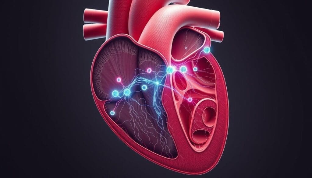
We use accurate diagrams to show how electrical signals move through the heart. These diagrams help teach and guide doctors in understanding the heart’s electrical activity.
These diagrams show the exact path of electrical signals through the heart. They highlight the SA node, AV node, Bundle of His, and the ventricular system.
They are essential for seeing how the heart’s electrical system works. They also show how heart conditions can affect it.
| Component | Function |
| SA Node | Initiates the heartbeat |
| AV Node | Delays the impulse before it reaches the ventricles |
| Bundle of His | Transmits the impulse to the ventricles |
| Bundle Branches | Distributes the impulse to the ventricular muscle |
Today’s imaging lets us see the heart’s electrical activity in detail. Tools like electrocardiographic imaging (ECGI) and cardiac MRI give deep insights into the heart’s electrical system.
These methods help doctors map the heart’s electrical activity. This helps in diagnosing heart rhythm problems and other electrical issues. Knowing the heart’s electrical flow helps doctors create better treatment plans.
Electrical currents in the heart drive each heartbeat. They make sure the heart contracts and relaxes in sync. These currents are made when ions move across cell membranes, key to the heart’s pumping action.
The heart’s electrical currents are complex and vital. They help keep the heart’s rhythm steady. Knowing how they work helps us understand how the heart responds to needs and sickness.
Ions like sodium, potassium, calcium, and chloride move across heart cells. Ion channels and pumps help control this movement. This creates electrical gradients that spark action potentials.
The action potentials are quick changes in heart cell voltage. They happen when a cell is excited. This is due to ion channels opening and closing, causing a fast depolarization and then repolarization.
Cardiac tissues have unique action potentials. For example, the SA node and AV node have different profiles than atrial and ventricular cells. The SA node, the heart’s pacemaker, has a special profile for starting electrical activity.
Knowing these differences helps us see how electrical signals move through the heart. This ensures the heart beats in sync and pumps blood well.
The heart’s electrical system is complex and sophisticated. This complexity lets the heart adjust to different situations and stay efficient.
An electrocardiogram (ECG) is a key tool that lets us see the heart’s electrical system. It records the heart’s electrical activity. This helps doctors diagnose and manage heart conditions well.
The ECG is more than a tool; it’s a way to understand the heart’s electrical system. By looking at the ECG, we learn how electrical impulses move through the heart. This is important for keeping the heart rhythm normal.
The ECG waveform shows the heart’s electrical activity. It has parts like the P wave, QRS complex, and T wave. Each part shows a different electrical event in the heart.
The P wave shows when the atria depolarize, the QRS complex shows ventricular depolarization, and the T wave shows ventricular repolarization. By linking these waves with heart events, we see how electrical impulses move through the heart. For example, a normal P wave followed by a QRS complex means the heart is working right. But any problem in this sequence can cause heart issues like arrhythmias or conduction blocks.
Understanding ECG patterns is key for doctors. A normal ECG means the heart’s electrical system is working well. But, abnormal patterns can show heart problems.
For example, a long QRS complex might mean a bundle branch block. An irregular rhythm could suggest atrial fibrillation. By knowing how to read these patterns, doctors can better diagnose and treat heart conditions. This helps improve patient care.
In summary, the electrocardiogram is a powerful tool for understanding the heart’s electrical function. By knowing how to link ECG waves with heart events and understand normal and abnormal patterns, we can better diagnose and treat heart conditions.
The autonomic nervous system is key in controlling the heart’s electrical activity. It makes sure our heart rate and rhythm change as needed. This is true during exercise or when we’re stressed.
We’ll look at how the autonomic nervous system affects the heart’s electrical system. We’ll focus on the sympathetic and parasympathetic branches. We’ll also talk about how neurohumoral modulation impacts cardiac conduction.
The autonomic nervous system has two main parts: the sympathetic and parasympathetic systems. The sympathetic nervous system is known as the “fight or flight” response. It makes the heart beat faster and stronger.
The parasympathetic nervous system helps us relax and conserve energy. It slows down the heart rate. This is the “rest and digest” state.
The sympathetic influences on the heart are controlled by neurotransmitters like norepinephrine. This increases the heart rate by making the SA node work faster. On the other hand, parasympathetic influences slow down the heart rate. This is done by the vagus nerve releasing acetylcholine, which decreases the SA node’s automaticity.
The heart’s electrical activity is also influenced by neurohumoral factors. Hormones like adrenaline and thyroxine can change heart rate and conduction. For example, adrenaline increases heart rate and contractility during stress.
The balance between the autonomic nervous system and neurohumoral factors makes the heart’s electrical system very adaptable. This complex regulation helps the heart respond well to different situations. It goes from rest to intense activity smoothly.
Problems with the heart’s electrical system can really affect how well it works. This can cause arrhythmias and conduction blocks. These issues can lead to irregular heartbeats and lower heart efficiency. They can even be life-threatening.
Arrhythmias happen when the heart’s electrical signals get mixed up. This can cause heartbeats to become irregular. The reasons for arrhythmias vary, including:
Symptoms of arrhythmias can be mild or severe. They can range from feeling your heart skip a beat to chest pain and trouble breathing. Doctors use electrocardiogram (ECG) tests to diagnose arrhythmias.
Conduction blocks happen when electrical signals can’t move through the heart properly. This can slow down or block heartbeats. Depending on where and how severe the block is, it might need a pacemaker to fix.
The effects of conduction blocks can be serious. They can lead to:
Today, there are many ways to treat heart electrical problems. These include:
A leading cardiologist says, “The treatment of heart rhythm disorders has greatly improved. Now, patients have more effective and personalized treatments than ever.”
“The key to successful treatment lies in accurate diagnosis and a tailored approach to managing each patient’s unique condition.”
Understanding the heart’s electrical system and its disorders helps doctors provide better care. This leads to better patient outcomes and a better quality of life.
Knowing how the heart’s electrical system works is key. It helps us understand how the heart beats and pumps blood. This knowledge is vital for keeping our heart healthy.
Healthcare experts use this knowledge to spot and treat heart problems. They learn about the heart’s electrical activity. This helps them give better care to their patients.
Managing heart conditions well needs a deep understanding of the heart’s electrical system. We must look at the sinoatrial node, the conduction pathway, and how the heart’s activity is controlled. This way, we can help our patients stay healthy and do better.
In healthcare, knowing about the heart’s electrical system is very important. It helps us create better treatments for heart problems. This leads to better care for our patients.
The sinoatrial node, or sinus node, is the heart’s main pacemaker. It starts the electrical impulses that control heart function.
The electrical impulse begins in the sinoatrial node. It’s located in the right atrium.
The heart’s electrical signal follows a specific path. It starts in the sinoatrial node, then moves through the internodal pathways. Next, it goes through the atrioventricular node, the Bundle of His, and the bundle branches.
Electrical signals control the heart’s rhythm. They make sure the heart chambers contract and relax properly. This ensures blood flows well.
The atrioventricular node is key in the heart’s electrical system. It delays the signal. This lets the atria fully contract before the ventricles do.
We can see the heart’s electrical flow through diagrams and imaging. Techniques like cardiac electrical mapping help us understand it better.
Cardiac electrical activity comes from ion movement. Ions like sodium, potassium, and calcium move across cell membranes. This creates action potentials that control the heart’s activity.
The autonomic nervous system controls the heart’s electrical activity. It uses sympathetic and parasympathetic influences. It also uses neurohumoral modulation. This helps the heart adapt to different situations.
Common heart electrical system disorders include arrhythmias and conduction blocks. These conditions disrupt normal heart rhythm.
The electrocardiogram helps us understand the heart’s electrical function. It shows how ECG waves relate to heart conduction. This lets doctors diagnose and manage heart conditions effectively.
Subscribe to our e-newsletter to stay informed about the latest innovations in the world of health and exclusive offers!