Getting a correct diagnosis for brain tumors is key to good treatment. At Liv Hospital, we use top-notch imaging to diagnose and treat many conditions. We mainly use CT scans and MRI, and many patients often ask about the difference between CAT scan vs MRI brain tumors. Understanding how each imaging method works helps determine the most accurate approach for detecting and managing brain tumors.
CT scans are quicker because they use X-rays. On the other hand, MRI uses magnetic fields and radio waves for better soft tissue detail. Knowing the differences between these methods helps pick the best one for diagnosis.
We use the latest technology and expert care to give our patients the best diagnosis and treatment plan.
Key Takeaways
- CT scans are faster and use X-ray technology.
- MRI provides superior detail of soft tissues.
- Choosing the right imaging technique is key to accurate diagnosis.
- Liv Hospital offers advanced technology and expert care.
- Understanding the differences between CT scans and MRI is vital for effective treatment.
Understanding Medical Imaging for Brain Tumors

Getting an accurate diagnosis is key to treating brain tumors well. Medical imaging is vital in this process. It uses advanced tech to give detailed views of brain tumors. This helps doctors plan the best treatment.
The Importance of Accurate Diagnosis
Knowing exactly what a brain tumor is helps doctors choose the right treatment. Medical imaging technologies like CT scans and MRI help tell different tumors apart. They also show how serious the tumor is. This info is vital for making treatment plans that work.
Brain anatomy is complex, and tumors come in many types. But advanced imaging helps us understand tumors better. We can see where they are, how big they are, and how they might affect the brain around them.
Overview of Neuroimaging Technologies
Neuroimaging has changed how we deal with brain and cancer issues. CT scans and MRI are the main tools for diagnosing brain tumors. Each has its own strengths and is used in different situations.
CT scans are great in emergencies because they’re fast and can spot bleeding right away. MRI, on the other hand, shows soft tissues better. This makes MRI perfect for looking at tumors and how they relate to other brain parts. Knowing what each can do helps pick the best imaging for each patient.
Using these imaging tools helps us understand brain tumors better. This leads to better treatments. The choice between CT and MRI depends on the patient’s situation and what the doctor needs to see.
What is a CT Scan (Cat Scan)?

CT scans are key in medical imaging, mainly for finding brain tumors. They help us see the brain in detail. This is important for diagnosing and treating many conditions.
How CT Technology Works
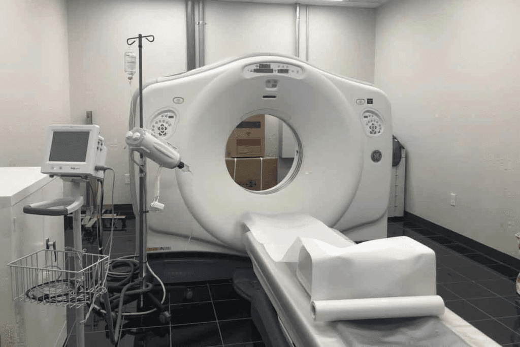
CT scans use X-rays to show the brain. They work by moving an X-ray source and detectors around the patient. This captures data from many angles.
Then, a computer turns this data into clear images. This is done using advanced algorithms.
Key components of CT technology include:
- X-ray tube: Produces X-rays that penetrate the body.
- Detectors: Capture X-rays that have passed through the body.
- Computer system: Reconstructs images from the captured data.
Types of CT Scans for Brain Imaging
There are many CT scans for brain imaging, each for different uses.
Common types include:
- Non-contrast CT: Used for detecting acute hemorrhage, fractures, and calcifications.
- Contrast-enhanced CT: Uses a contrast agent to highlight certain areas or structures.
- CT Angiography: Shows blood vessels and is good for vascular abnormalities.
In summary, CT scans are essential for brain imaging. Knowing how they work and the different types helps doctors make better decisions. This is true when comparing cat scan vs an MRI of the brain for diagnosis.
What is an MRI?
MRI is a non-invasive imaging technique. It uses strong magnetic fields and radio waves to create detailed images of the brain.
We use MRI technology to get high-resolution images of soft tissues. This is key for diagnosing and monitoring brain tumors. MRI doesn’t use ionizing radiation, making it safer for patients needing repeated scans.
How MRI Technology Works
MRI technology aligns the hydrogen atoms in the body with a strong magnetic field. Radio waves disturb these atoms, creating signals. These signals are used by the MRI machine to form detailed images.
The process involves several key components. These include the main magnetic field, gradient coils, and radiofrequency coils. Together, they generate high-quality images of the brain’s anatomy.
Types of MRI Sequences for Brain Imaging
There are several MRI sequences used in brain imaging. Each provides different information about the brain’s structures.
The most common sequences are T1-weighted, T2-weighted, FLAIR (Fluid Attenuated Inversion Recovery), and DWI (Diffusion-Weighted Imaging). Each sequence has its specific uses in diagnosing brain conditions, including tumors.
| MRI Sequence | Application |
| T1-weighted | Provides detailed anatomy, useful for tumor localization |
| T2-weighted | Highlights differences in water content, useful for detecting edema |
| FLAIR | Suppresses signal from fluids, useful for detecting lesions |
| DWI | Sensitive to early changes in brain ischemia, useful in stroke diagnosis |
Understanding MRI technology and the different sequences helps healthcare professionals. They can make better decisions about the best imaging approach for diagnosing and managing brain tumors.
Cat Scan vs MRI Brain Tumors: 7 Key Differences
It’s important to know the differences between CT scans and MRI for diagnosing brain tumors. Both CT scans and MRI are key in medical imaging for brain tumors. But they have their own strengths and weaknesses.
Difference 1: Imaging Technology
CT scans and MRI use different technologies. CT scans use X-rays to create detailed images of the body, including the brain. MRI, on the other hand, uses a strong magnetic field and radio waves to show brain structures.
CT scans are great at showing bones and detecting calcifications in tumors. MRI is better at showing soft tissues. This makes MRI perfect for seeing how far a tumor has spread into the brain.
Difference 2: Radiation Exposure
CT scans expose patients to radiation, which is a concern for repeated scans. MRI, on the other hand, is safer because it doesn’t use radiation. This makes MRI a better choice for long-term monitoring and for pregnant women or those sensitive to radiation.
Difference 3: Tissue Contrast Resolution
MRI can better distinguish between different soft tissues than CT scans. This is important for identifying tumor types and distinguishing tumors from surrounding swelling.
Difference 4: Spatial Resolution
Both CT and MRI can produce high-resolution images. But MRI is better at showing details in soft tissues. This is key for planning surgeries and checking tumor edges.
| Feature | CT Scan | MRI |
| Imaging Technology | Uses X-rays | Uses magnetic field and radio waves |
| Radiation Exposure | Involves ionizing radiation | No ionizing radiation |
| Tissue Contrast Resolution | Lower contrast resolution | Superior contrast resolution |
| Spatial Resolution for Soft Tissues | Lower spatial resolution for soft tissues | Higher spatial resolution for soft tissues |
Advantages of CT Scans for Brain Tumor Detection
CT scans are great for finding brain tumors. They are fast, show bones well, and spot calcifications and hemorrhages. This makes them key in neuroimaging, even in emergencies.
Speed and Emergency Situations
CT scans are quick. In emergencies like head trauma or bleeding, they are lifesaving. Quick diagnosis means timely treatment, which is vital for serious conditions.
In emergencies, time is everything. CT scans are quicker than MRI scans. They don’t lose image quality, helping doctors make accurate diagnoses.
Bone Structure Visualization
CT scans are top-notch at showing bone structures. This is key to seeing how far a brain tumor has spread. Detailed bone imaging helps plan surgeries and understand the tumor’s effect on bones.
For brain tumor patients, knowing how the tumor affects bones is essential. CT scans give clear images, helping neurosurgeons plan better.
Detecting Calcifications and Hemorrhage
CT scans are great at finding calcifications in brain tumors and spotting bleeding. Spotting calcifications is key to diagnosing some tumors, like oligodendrogliomas.
They also excel at finding bleeding. For suspected bleeding in the brain or inside tumors, CT scans quickly show where and how much. This helps doctors decide on treatment right away.
In summary, CT scans are a big help in finding brain tumors. They are fast, show bones well, and spot calcifications and bleeding. These benefits make CT scans a top choice for both urgent and non-urgent cases.
Advantages of MRI for Brain Tumor Detection
MRI technology is key in finding brain tumors. It gives us detailed images that help doctors plan treatments. These images are vital for accurate diagnosis.
Superior Soft Tissue Contrast
MRI shines in showing soft tissues in the brain. It can tell different tissues apart, unlike CT scans. This helps doctors spot tumors and know how far they’ve spread.
Tumor Characterization Capabilities
MRI lets us learn more about brain tumors. It shows the tumor’s makeup, blood flow, and how it works. This info helps doctors figure out how aggressive the tumor is and what treatment to use.
Multiplanar Imaging Advantages
MRI can show images from different angles. This is a big plus compared to CT scans. It helps doctors see how tumors relate to other parts of the brain, which is key for surgery and radiation therapy.
To wrap it up, MRI is a big help in finding and treating brain tumors. It offers clear images, helps understand tumors better, and shows them from different angles. These benefits make MRI a must-have for brain tumor care.
Limitations of Each Imaging Modality
It’s important to know the limits of CT scans and MRI for brain tumor diagnosis. Both have changed neuro-oncology a lot. But, they each have their own problems.
CT Scan Limitations for Brain Tumors
CT scans are fast and easy to get, but they have some big downsides. Here are a few:
- Radiation Exposure: CT scans use radiation, which is a big worry for patients, even more so for those needing many scans.
- Limited Soft Tissue Contrast: CT scans don’t show soft tissues well, making it hard to spot some tumor details.
- Beam Hardening Artifacts: CT scans can get blurry in areas with lots of bone or metal, hiding important details.
MRI Limitations for Brain Tumors
MRI is great for soft tissue, but it has its own issues. Here are a few:
- Claustrophobia and Patient Comfort: MRI scans can be long and tight, making some patients uncomfortable or scared.
- Contraindications for Certain Patients: Some patients with metal implants or pacemakers can’t have MRI scans for safety reasons.
- Cost and Availability: MRI scans cost more and might not be available everywhere, making them less accessible.
Knowing these limits helps doctors choose the right scan for brain tumors. This can lead to better care for patients.
Clinical Decision Making: When to Use CT vs MRI
Choosing between CT scans and MRI for brain tumors involves many factors. We need to look at the good and bad of each to pick the best for each situation.
Emergency Scenarios
In emergencies like head trauma or bleeding, CT scans are often the first choice. They’re quick and good at finding bleeding. For example, in brain injuries, CT scans can spot bleeding that needs surgery fast.
But, for thinking stroke might be happening, MRI is better. It shows early signs of stroke well with special sequences.
Initial Diagnosis Considerations
Choosing between CT and MRI for the first diagnosis depends on the case. For brain tumors, MRI is usually better. It shows soft tissues well and helps figure out tumor type and size.
Follow-up Imaging Protocols
For checking up on tumors, the choice between CT and MRI depends on the initial diagnosis and treatment. MRI is best for tumor follow-ups because it’s detailed and doesn’t use radiation. But, if MRI can’t be used, CT scans can help, even if they’re not as detailed.
In the end, picking between CT and MRI for brain tumor imaging should be based on each patient’s needs and situation.
Brain Tumor Types and Optimal Imaging Approaches
Diagnosing brain tumors needs a deep understanding of the tumor type. This helps choose the best imaging method. Brain tumors are divided into primary and metastatic types, each with unique features.
Primary Brain Tumors
Primary brain tumors start in the brain and can be benign or malignant. Common types include gliomas, meningiomas, and acoustic neuromas. Gliomas are a wide range of tumors, from low-grade to high-grade glioblastomas.
MRI is the top choice for primary brain tumors. It offers better soft tissue contrast. This helps see the tumor’s extent and type clearly.
For tumors with calcifications or hemorrhage, CT scans are sometimes used, too. But MRI’s multiplanar imaging and sensitivity to tumor vascularity make it key for diagnosis and management.
Metastatic Brain Tumors
Metastatic brain tumors come from cancers outside the brain. Common sources are the lung, breast, and melanoma. These tumors are usually well-defined and often multiple.
Both CT and MRI can spot metastatic brain tumors. But MRI is more sensitive, great for small or hard-to-find lesions.
Contrast agents like gadolinium in MRI make tumors more visible. MRI’s high contrast resolution helps tell tumor types apart and see how far the disease has spread. This is vital for planning treatment.
In summary, knowing the brain tumor type is key to picking the right imaging method. MRI is usually the best choice for both primary and metastatic tumors because of its superior soft tissue contrast and multiplanar imaging. But CT scans are useful in emergencies or when looking for calcifications.
Patient Experience: What to Expect
When dealing with brain tumors, knowing what to expect from CT scans and MRI is key. These tests are vital for diagnosis but offer different experiences. Let’s look at what patients can expect from each.
Preparing for a CT Scan
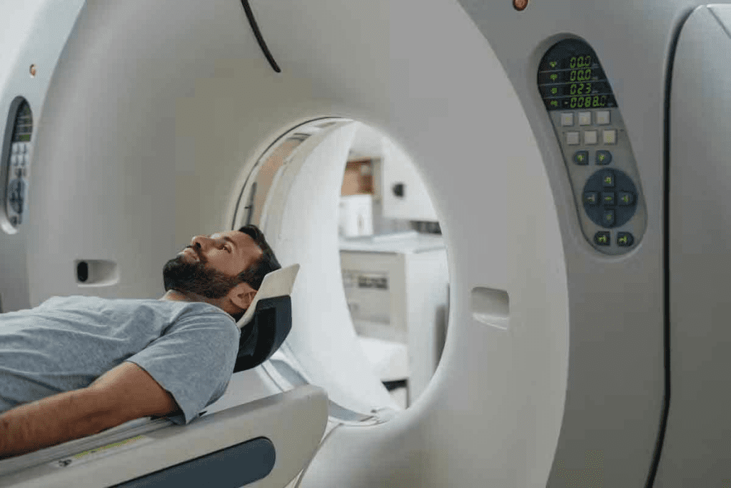
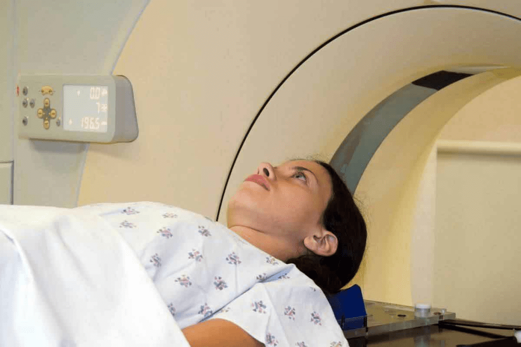
Getting ready for a CT scan is easy. You’ll need to take off any metal items like jewelry or glasses. You might also wear a hospital gown. Sometimes, a contrast dye is used to make images clearer.
Tell your doctor about any allergies or sensitivities to contrast dye. If you’re pregnant or think you might be, let your doctor know. This can help you choose the right test for you.
Preparing for an MRI
Preparation for an MRI is similar to a CT scan, but with extra steps. You’ll need to remove all metal items, including jewelry and glasses. Clothes with metal fasteners are also a no-go. If you have metal implants, like pacemakers, tell your doctor.
You’ll lie on a table that slides into the MRI machine. The scan can last from 15 to 90 minutes. Try relaxation techniques, like deep breathing, to ease any anxiety.
Contrast Agents and Their Use
Both CT scans and MRI might use contrast agents to make images clearer. CT scans use iodine-based agents, while MRI uses gadolinium-based agents.
These agents are usually safe but can cause side effects like nausea or dizziness. Rarely, serious allergic reactions can happen. Always tell your doctor about any allergies or past reactions.
| Imaging Modality | Contrast Agent | Common Side Effects |
| CT Scan | Iodine-based | Nausea, headache, dizziness |
| MRI | Gadolinium-based | Headache, dizziness, coldness at the injection site |
The table shows that CT scans and MRI use different contrast agents. Knowing this can help you prepare better for your test.
Conclusion: The Future of Brain Tumor Imaging
The choice between CT scans and MRI for brain tumors is key for diagnosis and treatment. Both have their strengths. CT scans are fast and good for emergencies. MRI gives better detail and helps see soft tissues.
New technologies are making brain tumor imaging better. Advances like artificial intelligence and new imaging methods will help. These changes will make treatments more precise and tailored to each patient.
Knowing the differences between CT and MRI scans is vital for doctors and patients. As these technologies get better, diagnosing brain tumors will become more accurate. This will lead to better care for patients with brain tumors.
FAQ
What is the main difference between a CT scan and an MRI for brain tumor diagnosis?
CT scans use X-rays, while MRI uses a strong magnetic field and radio waves. This means MRI can show soft tissues better than CT scans.
Which is better for detecting brain tumors, a CT scan or an MRI?
MRI is better for finding and understanding brain tumors. It shows soft tissues more clearly, giving detailed images of tumors and brain tissue.
Are CT scans or MRI scans faster for brain imaging?
CT scans are quicker than MRI scans. This makes them better for emergency situations where fast decisions are needed.
Do CT scans or MRI scans use radiation?
CT scans use X-rays, which involve radiation. MRI scans do not use ionizing radiation. This makes MRI safer for patients needing repeated scans.
Can both CT scans and MRI detect calcifications in brain tumors?
Yes, CT scans are great at finding calcifications in brain tumors. MRI can also find calcifications, but not as well as CT scans.
How do I prepare for a CT scan or MRI for a brain tumor diagnosis?
You might need to remove metal objects and avoid certain foods or medications. You might also use contrast agents. Your healthcare provider will give you specific instructions.
What are the limitations of using CT scans for brain tumor detection?
CT scans have trouble showing soft tissues clearly compared to MRI. This can make it hard to tell apart different types of tumors or tumors from swelling.
What are the advantages of MRI over CT scans for brain tumor imaging?
MRI shows soft tissues better, helps understand tumors more, and can be viewed from different angles. These are big advantages for planning surgery and seeing how far tumors have spread.
Are there specific brain tumor types better diagnosed with CT scans or MRI?
Some tumors are better seen with one method over the other. MRI is usually best for most tumors because of its detailed soft tissue images. CT scans might be used for specific cases, like finding calcifications.
Can I undergo an MRI if I have certain metal implants or devices?
It depends on the metal implant or device. Some are safe for MRI, while others are not. Always tell your healthcare provider about any implants or devices before an MRI.
How do emerging technologies impact brain tumor imaging?
New imaging technologies, like better MRI sequences and contrast agents, are helping diagnose and manage brain tumors better. This could lead to better patient care and outcomes.
References
- Hope, T. A., et al. (2019). Meta-analysis of 68Ga-PSMA-11 PET accuracy for the detection of prostate cancer. Journal of Nuclear Medicine, 60(2), 197“201. https://pmc.ncbi.nlm.nih.gov/articles/PMC6581235/
- Jochumsen, M. R., et al. (2024). PSMA PET/CT for primary staging of prostate cancer: A systematic review and meta-analysis. European Urology, 85(3), 245“256. https://www.sciencedirect.com/science/article/pii/S0001299823000557


