Last Updated on November 26, 2025 by Bilal Hasdemir

Discover how a cholelithiasis CT scan compares to ultrasound for accurate gallstone detection. Getting gallstones diagnosed right is key to good treatment. At Liv Hospital, we focus on what’s best for each patient. Imaging is very important in finding gallstones. The choice between CT scans and ultrasound is a big part of our decisions.
CT scans and ultrasound are both used to find gallstones. Each has its own benefits. Ultrasound is usually the first choice, but CT scans offer more details in some cases.
Cholelithiasis CT Scan vs Ultrasound: 7 Insights
It’s key to understand how gallstones form to treat cholelithiasis well. Gallstones are solid lumps in the gallbladder, made mostly of cholesterol or bilirubin.
Gallstones form when bile’s mix is off balance. Bile has water, cholesterol, bile salts, and bilirubin. Cholesterol gallstones are common, linked to too much cholesterol in bile. Pigment stones are made of bilirubin, often in people with hemolytic disorders.
Cholelithiasis is a big health problem globally, with different rates in different groups. Risk factors include obesity, female gender, age over 40, and certain ethnicities. Other risks are metabolic syndrome, quick weight loss, and some medicines.
Gallstones can show up in many ways. Some people don’t have symptoms, while others get biliary colic. This is pain in the right upper abdomen that comes and goes. Serious problems like cholecystitis or choledocholithiasis need quick doctor visits.
Advanced medical imaging greatly helps in diagnosing cholelithiasis. It detects gallstones, their type, and any complications. This is key for treating the condition effectively.
Getting the diagnosis right is vital for treating cholelithiasis well. It helps tell apart gallstones that cause symptoms from those that don’t. Imaging modalities give important details about gallstones and gallbladder issues.
Accurate diagnosis is very important. It affects how well a patient does and how much healthcare costs. If a diagnosis is wrong or late, it can lead to serious problems like cholecystitis or pancreatitis.
There are many ways to diagnose cholelithiasis, each with its own benefits and drawbacks. The main ones are:
| Imaging Modality | Sensitivity for Gallstones | Advantages | Limitations |
| Ultrasound | High (95%) | Non-invasive, no radiation, cost-effective | Operator-dependent, limited in obese patients |
| CT Scan | Moderate (70-80%) | Detects complications, evaluates surrounding structures | Involves radiation, less sensitive for small stones |
| MRI/MRCP | High for bile duct stones | Detailed biliary tract imaging, no radiation | Expensive, limited availability |
Choosing the right imaging test for cholelithiasis depends on several things. These include the patient’s symptoms, how they’re feeling, and if there are any complications. Ultrasound is usually the first choice because it’s safe and works well.
If there’s a chance of complications or if ultrasound results are unclear, CT scans might be used. The right imaging test depends on the patient’s needs and the risks and benefits of each test.

Ultrasound is the top choice for checking gallbladder health. It’s safe, doesn’t use harmful radiation, and works well. This makes it a go-to for finding gallstones.
Ultrasound uses echo-location to see inside the body. It sends sound waves and catches the echoes to make pictures. Gallstones show up well because they bounce sound waves.
The ultrasound test for gallstones is simple. Patients usually fast before to make the gallbladder easier to see. A gel is applied to the belly, and a transducer takes pictures of the gallbladder.
Ultrasound is very good at finding gallstones. It has a high sensitivity and specificity. Studies show it can spot gallstones up to 87% of the time.
Ultrasound is also cost-effective and easy to find. It’s cheaper and more available than other tests like CT scans. This makes it a great first choice for gallstone checks.
Gallstones show up in a specific way on ultrasound. This helps doctors spot and understand them.
Gallstones look like echogenic foci with posterior acoustic shadowing on ultrasound. This is because the stone’s surface reflects sound waves well. The sound waves then get blocked by the stone, creating a shadow.
The shadow is key for doctors to tell gallstones apart from other things. It happens because the ultrasound waves can’t pass through the stone, making an area behind it dark.
Ultrasound can also show if a gallstone is moving or stuck. Mobile stones change position when you move, while impacted stones stay put. Knowing this is important because stuck stones can cause problems like cholecystitis.
The Wall Echo Shadow (WES) sign is a special ultrasound finding for gallstones. It happens when the gallbladder is full of stones. You see the gallbladder wall, the stone, and the shadow all together.
The WES sign is a good sign for gallstones. It’s very helpful when the gallbladder is hard to see.
It’s important to tell gallstones apart from other things seen on ultrasound. Some things to keep in mind are:
By paying close attention to these details, doctors can accurately find and identify gallstones.
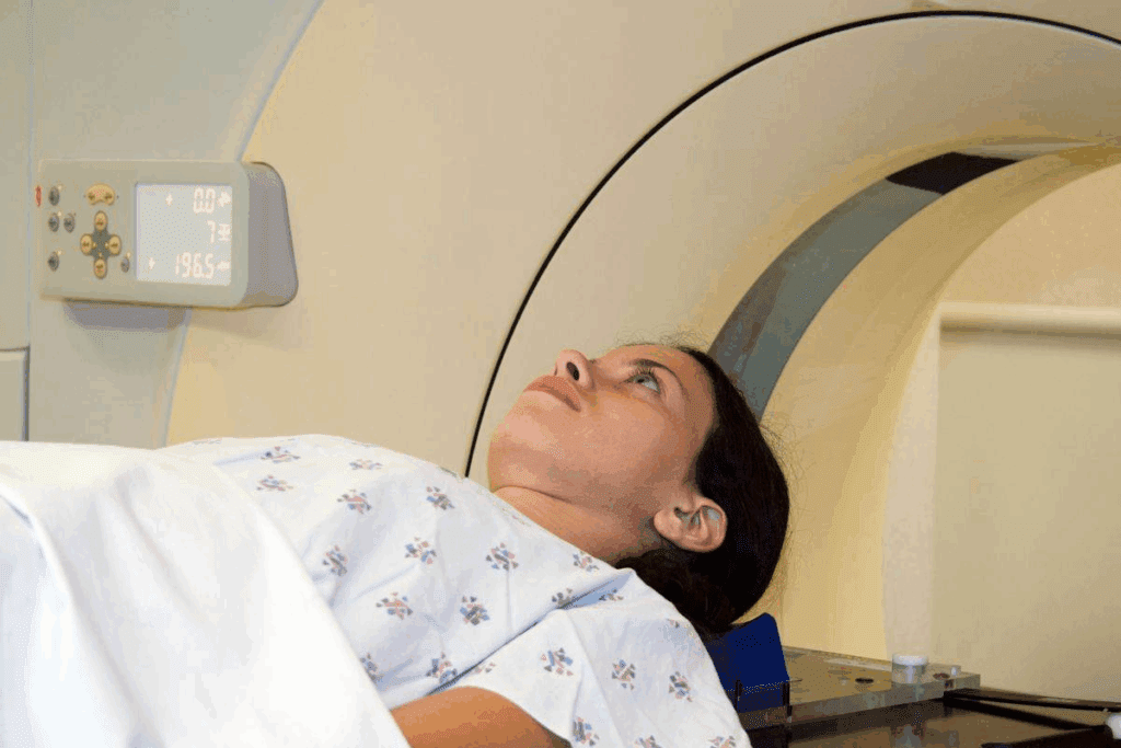
Diagnosing cholelithiasis with CT scans involves several technical factors. These include the use of contrast and scanning parameters.
Non-contrast CT scans are often enough for finding gallstones, mainly for calcified ones. But, contrast-enhanced CT is needed to check for complications. It also helps see the gallbladder and nearby tissues for inflammation or other issues.
The quality of a CT scan for cholelithiasis depends on the right scanning parameters. These include:
Proper patient preparation is key for high-quality CT images. Patients are usually asked to:
CT scans involve radiation, so it’s important to keep the dose low while keeping image quality high. Using dose modulation and the lowest necessary mAs can help reduce radiation.
Gallstones are hard to spot on CT scans because most aren’t calcified. Their look on CT images can change a lot. This makes finding them tricky sometimes.
Gallstones are split into two types based on their CT scan look. Radiolucent stones are hard to see because they blend in with bile. They’re mostly made of cholesterol. Radiopaque stones, on the other hand, are calcified and show up clearer on CT scans.
Most gallstones are cholesterol stones, which are hard to spot on CT scans. Spotting these stones is key for correct diagnosis and treatment.
Several things can make gallstones hard to see on CT scans. These include:
Knowing these factors helps make CT scans better for finding gallstones.
Reading CT scans for gallstones can be tricky. There are a few common mistakes. These include:
Radiologists need to watch out for these mistakes to give accurate diagnoses.
New CT methods can help find gallstones better. These include:
These advanced methods can help solve some of the challenges in spotting gallstones on CT scans.
Ultrasound and CT scans are used to find gallstones. They help doctors make important decisions. The choice between them depends on the patient’s health and what complications might be present.
Ultrasound is often the first choice for finding gallstones. It’s very good at it, with an 87% sensitivity. This is because it can spot small stones and show what’s happening in real-time.
Ultrasound is also safe because it doesn’t use radiation. This makes it a great option for patients.
CT scans, on the other hand, are not as good at finding simple gallstones. They’re used more when there’s a chance of complications or when the diagnosis is tricky. CT scans might miss some stones because they’re not visible on the scan.
But, CT scans are useful in some situations. They help check for other problems in the belly.
The difference in how well ultrasound and CT scans work is important. For simple gallstone cases, ultrasound is the best first choice. It’s safe and very accurate.
When there’s a chance of serious problems, like inflammation or pancreatitis, CT scans are used. They help doctors understand how bad the situation is.
Knowing how well different tests work is key for making the right decisions. It helps doctors take the best care of their patients.
CT imaging is top-notch for complicated gallstone disease. It can spot complications like acute cholecystitis, gangrene, or perforation. These need quick and correct diagnosis.
CT scans are 92% sensitive in spotting acute cholecystitis. This is key for fast action in treatment.
A study in the Journal of Radiology showed CT scans do more than just find gallstones. They also show how severe the inflammation is and any complications. This makes CT a valuable tool in managing complicated gallstone disease.
“CT is very useful in suspected complicated cholecystitis. It can spot gangrene or perforation, helping guide surgery.”
CT scans are great at finding complications from gallstone disease. These can include:
Finding these complications early is vital for patient care. CT scans give detailed images for quick identification.
CT scans also check surrounding areas, which is key for a full patient check-up. They look at bile ducts, liver, and nearby tissues for any problems.
| Imaging Modality | Sensitivity for Acute Cholecystitis | Ability to Detect Complications |
| CT Scan | 92% | High |
| Ultrasound | 87% | Moderate |
CT scan info is key for planning treatments. It helps doctors see the gallbladder and nearby areas clearly. This leads to better treatment plans.
There are more ways to find gallstones than just ultrasound and CT scans. Ultrasound is usually the first choice, but other methods help in certain cases. They are useful when ultrasound results are not clear.
Abdominal X-rays are not very good at finding gallstones. They can spot only 10% to 20% of them. This is because most gallstones don’t show up on X-rays because they don’t have enough calcium.
But, X-rays can help in some cases. They can show calcified stones and help rule out other pain causes.
Radiopaque stones, which have calcium, can be seen on X-rays. But these are rare. X-rays are not the main tool for finding gallstones.
MRI and MRCP give detailed views of the biliary tract. They are great for checking gallstone complications or bile ducts. MRCP is special because it shows the biliary and pancreatic ducts without needing contrast agents.
MRCP’s advantages include spotting bile duct stones well. It also shows detailed anatomy. This helps plan surgeries or endoscopic treatments.
Nuclear medicine, like HIDA scans, shows how the gallbladder and biliary system work. These scans are good for finding acute cholecystitis. They show if the gallbladder is working right and if there are blockages.
HIDA scans use a radioactive tracer that the liver and bile take up. The scan checks if the gallbladder is working and if there are blockages in the bile ducts.
In summary, while ultrasound and CT scans are key, MRI, MRCP, and nuclear medicine studies also help. They provide important information in certain situations.
Ultrasound is usually the first choice for finding gallstones. But, CT scans have their own benefits in certain situations. The choice between CT and ultrasound depends on the case’s complexity and the need for detailed images.
When ultrasound results are unclear, CT scans offer a more detailed look. This is helpful when ultrasound can’t see the gallbladder or gallstones well.
For suspected complications like acute cholecystitis or gangrene, CT scans are key. They show the gallbladder and its surroundings well. This helps spot complications ultrasound might miss.
Some patients face challenges with ultrasound, like obesity or bowel gas. In these cases, CT scans are a better choice.
Knowing when to choose CT over ultrasound helps doctors make better decisions for their patients.
Managing gallstones better comes from a detailed diagnostic method. This method uses different imaging and lab tests to fully understand a patient’s situation.
Using different imaging methods in order helps diagnose and treat gallstones well. Ultrasound is often the first choice because it’s non-invasive and good at finding gallstones. But if ultrasound isn’t clear or if there’s a complication, CT scans offer more details.
“The choice of imaging modality should be guided by the clinical presentation and initial findings,” as recent guidelines say. This step-by-step method makes sure patients get the right tests, saving resources and improving care.
Linking imaging results with lab tests is key for accurate gallstone diagnosis and treatment. Tests like liver function and blood counts give clues about the patient’s health. For example, high liver enzymes might mean gallstones in the bile ducts, while high white blood cells could point to inflammation of the gallbladder.
A study in a top medical journal found that combining imaging and lab results boosts accuracy in diagnosing gallstone complications. This approach helps doctors make better decisions for patient care.
When picking diagnostic methods for gallstones, cost matters. CT scans give detailed views but cost more and use radiation. A cost analysis helps decide the best first imaging test based on the patient’s risk and symptoms.
Research shows that starting with ultrasound is usually the most affordable for simple gallstone cases. But for high-risk patients or those with possible complications, starting with CT might be worth the extra cost for its detailed insights.
Choosing the right imaging is key for accurate gallstone diagnosis. The best imaging method depends on the patient’s symptoms and the need for clear diagnosis.
Ultrasound is often the first choice for simple gallstone cases. It’s very sensitive, affordable, and easy to get. But, CT scans are important for complex gallstone cases and finding related problems.
Using a mix of clinical checks, lab tests, and imaging is best for managing gallstones. This approach helps doctors make accurate diagnoses. It also saves money and improves patient care.
In short, knowing the strengths and weaknesses of different imaging tests is vital. It helps doctors make the best choices for diagnosing gallstones. This leads to better care for patients.
Ultrasound is the top choice for finding gallstones. It’s very sensitive, affordable, and easy to get.
On ultrasound, gallstones show up as bright spots with a shadow behind. They can move or get stuck.
The WES sign is a special ultrasound feature. It shows the gallbladder wall, a bright stone, and a shadow. This means there are gallstones.
Yes, CT scans can spot gallstones. But they’re not as good as ultrasound for simple gallstone cases, mainly for stones that don’t show up well.
CT scans are better at finding serious gallstone problems and acute cholecystitis. They also check other areas around the gallbladder.
Radiopaque stones show up on CT scans because they have calcium. But radiolucent stones are harder to see. You might need special CT methods to find them.
Yes, other options include MRI, MRCP, and nuclear medicine studies. They’re useful for certain situations, like checking the biliary tract or gallbladder function.
CT is better when ultrasound results are unclear, there’s a chance of serious gallstone issues, or ultrasound can’t be used well, like in obesity or gas in the bowel.
Sequential imaging uses different imaging methods step by step. It matches findings with lab results to give the best care for patients.
X-rays aren’t very good at finding gallstones. They only work for about 10-20% of cases because most stones don’t show up well.
Subscribe to our e-newsletter to stay informed about the latest innovations in the world of health and exclusive offers!