Last Updated on November 27, 2025 by Bilal Hasdemir

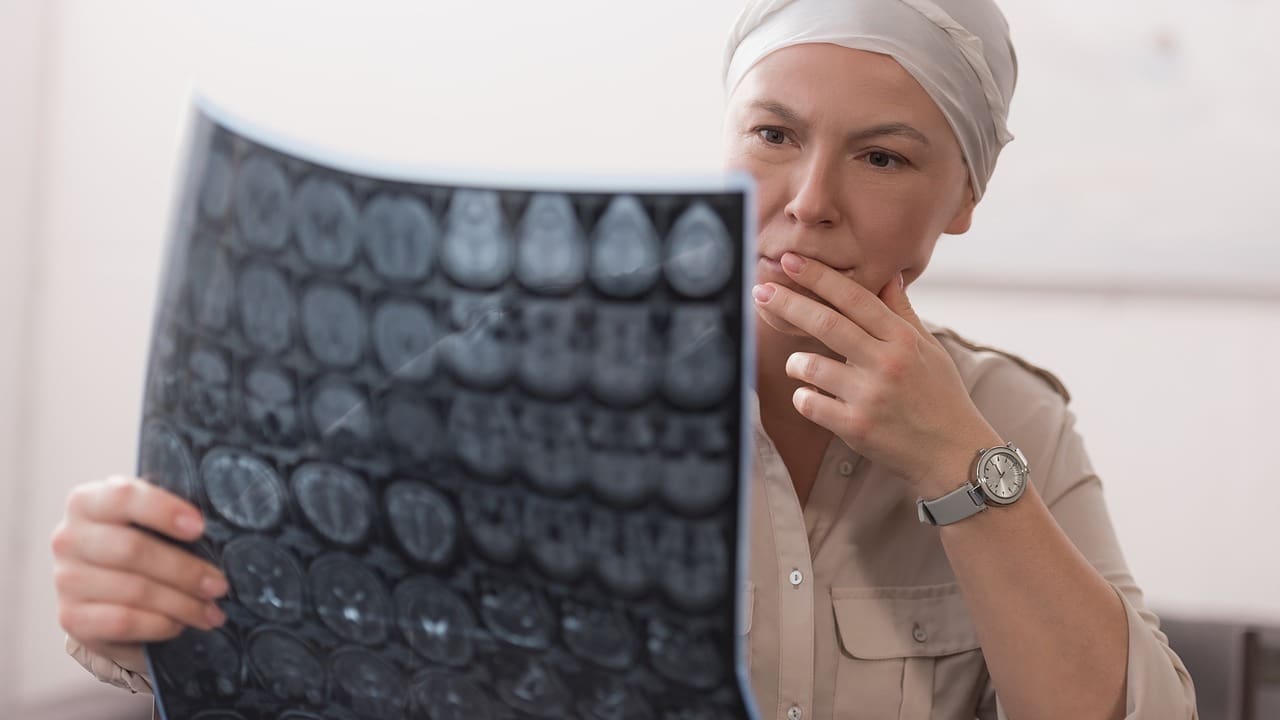
Diagnosing complex neck and soft tissue issues often requires a CT neck with contrast. At Liv Hospital, we use advanced imaging techniques like CT neck with contrast to ensure accurate and detailed diagnoses for effective treatment planning.
A CT neck with contrast helps spot infections, tumors, and vascular issues. The contrast makes these problems easier to see. This helps our team make better diagnoses and treatment plans.
Recent studies show contrast in CT scans boosts accuracy. For example, a study on CT neck in emergent settings shows contrast’s value in finding and identifying abscesses and masses.
Key Takeaways
- CT neck with contrast is used to assess various neck conditions, including infections and tumors.
- Contrast material enhances visualization, improving diagnostic accuracy.
- Advanced imaging protocols ensure clear and accurate diagnoses.
- CT scans are a key tool for diagnosing complex neck conditions.
- Liv Hospital’s medical team develops effective treatment plans based on accurate diagnoses.
Understanding CT Neck Imaging Fundamentals

CT neck imaging is key for doctors to diagnose and treat many health issues. It helps us see the neck’s detailed anatomy.
Basic Principles of CT Scanning
CT scans use X-rays to make detailed images of the body. A CT scanner moves around the patient, taking pictures from different angles. These images are then put together to form a complete picture.
Key components of a CT scanner include:
- X-ray tube
- Detectors
- Computer system
The X-ray tube sends X-rays through the patient’s body. The detectors catch the X-rays that pass through, sending this info to the computer. The computer then makes the images.
Anatomical Structures Visualized in Neck CT
A CT scan of the neck shows many structures, including:
| Anatomical Structure | Description |
| Vertebral column | The series of vertebrae that make up the neck |
| Soft tissues | Muscles, lymph nodes, and other non-bony structures |
| Vascular structures | Arteries and veins that supply the neck and head |
These structures are important for diagnosing many conditions, from injuries to vascular diseases.
The Role of Contrast Media in Medical Imaging
Contrast media make certain tissues or structures more visible during a CT scan. In neck imaging, they help show soft tissues and blood vessels better.
Contrast agents highlight important areas, making CT scans more accurate. This helps doctors make better decisions for patient care.
What Is CT Neck with Contrast and How Does It Work?

CT neck with contrast is a detailed imaging method. It makes blood vessels and some tissues in the neck stand out. This tool is key for spotting and checking many neck health issues.
Definition and Technical Aspects
A CT scan of the neck with contrast uses X-rays and a contrast agent. This combo creates detailed pictures of the neck’s inside. The contrast agent, given through an IV, lights up blood vessels and tissues, making them easier to see.
The technical process involves a CT scanner moving around the patient. It takes many X-ray images. Then, these images are turned into clear 2D and 3D pictures of the neck’s layout.
Types of Contrast Agents Used
Iodinated contrast media are the main contrast agents for CT neck scans. Iodine is great at blocking X-rays. This makes it perfect for making body structures or fluids show up better in scans.
“The development of iodinated contrast agents has significantly improved the diagnostic capability of CT scans, allowing for better visualization of vascular structures and certain pathologies.”
Administration Methods and Protocols
The contrast agent goes into a vein through an IV line in the arm. The exact way it’s given can change based on the scan’s needs and the patient’s health.
Key considerations for giving the contrast include how much and how fast to inject it. These decisions depend on the scan type, the patient’s weight, and kidney health.
Knowing how CT neck with contrast works and the contrast agents used helps doctors diagnose and treat neck problems better.
Key Clinical Indications for CT Neck with Contrast
Using contrast in CT neck scans makes it easier to diagnose several important conditions. Doctors use CT neck with contrast to see the soft tissues, blood vessels, and other neck structures clearly. This helps them understand what’s going on in the neck.
Suspected Infections and Inflammatory Conditions
CT neck with contrast is great for spotting infections and inflammation in the neck. The contrast agent shows how big an infection is, if there are abscesses, and if other areas are affected. This info is key for deciding how to treat the infection.
Tumor Detection and Characterization
For people with neck tumors, CT neck with contrast is a top choice for diagnosis. It shows where the tumor is, how big it is, and how it relates to nearby tissues. This info is vital for figuring out the cancer’s stage and choosing the right treatment.
Evaluation of Lymphadenopathy
Lymph nodes getting bigger can mean many things, like infections, autoimmune diseases, or cancer. CT neck with contrast helps figure out why lymph nodes are swollen. It shows how big and shaped the nodes are, and how they react to the contrast. This helps doctors narrow down what might be wrong and how to treat it.
Assessment of Vascular Anomalies
Vascular issues like aneurysms, stenoses, or blood clots can be seen clearly with CT neck with contrast. The contrast makes blood vessels stand out, so doctors can check their shape and any problems. This is super important for planning surgeries or other treatments for blood vessel issues.
| Clinical Indication | Diagnostic Benefit of Contrast | Impact on Patient Management |
| Suspected Infections | Delineates the extent of infection, identifies abscesses | Guides drainage or surgical intervention |
| Tumor Detection | Identifies tumor location, size, and relation to adjacent structures | Aids in cancer staging and treatment planning |
| Lymphadenopathy | Characterizes lymph node enlargement | Narrows differential diagnosis and guides further management |
| Vascular Anomalies | Enhances visibility of blood vessels and their pathology | Assists in planning interventions or surgeries |
Diagnostic Advantages of Using Contrast in Neck CT
Using contrast media in neck CT scans makes them much better. It helps us see more clearly, leading to better diagnoses. This is good for patients.
Enhanced Tissue Differentiation
Contrast agents make it easier to see different tissues in CT scans. They help spot problems by showing what’s different. This is really helpful in tricky areas like the neck.
Improved Visualization of Vascular Structures
Contrast makes blood vessels stand out in neck CT scans. This is key for finding and fixing blood vessel issues. It helps doctors see problems like blockages or weak spots.
Superior Detection of Pathological Processes
Contrast makes it easier to find diseases like tumors and infections in neck CT scans. This is thanks to the contrast media. For more on how to use CT scans, check out medical guidelines.
Here’s what contrast in neck CT scans does well:
| Diagnostic Advantage | Description | Clinical Benefit |
| Enhanced Tissue Differentiation | Contrast highlights differences between tissues | Better identification of abnormalities |
| Improved Visualization of Vascular Structures | Clear visualization of blood vessels | Accurate diagnosis of vascular conditions |
| Superior Detection of Pathological Processes | Effective identification of tumors and infections | Enhanced patient outcomes through timely diagnosis |
In short, contrast in CT neck scans is very helpful. It makes it easier to see tissues, blood vessels, and diseases. This leads to more accurate diagnoses and better care for patients.
CT Neck with Contrast in Oncological Assessment
CT neck with contrast is key in cancer assessments. It helps us see the extent of cancer spread. This imaging modality gives us detailed views of the neck’s soft tissues. It’s vital for accurate diagnosis and treatment planning.
Tumor-Node-Metastasis (TNM) Staging
CT neck with contrast is mainly used for TNM staging. This system checks the tumor’s size and spread. It also looks at nearby lymph nodes and metastasis.
With contrast, we can see the tumor’s edges and its relation to other tissues. This is key for accurate staging.
The staging info from CT neck with contrast guides treatment choices. It also helps predict patient outcomes.
Pre-Treatment Planning
CT neck with contrast is also vital for pre-treatment planning. It gives us detailed images of the tumor’s size and location. This info is essential for planning surgeries or radiation therapy.
Knowing the tumor’s extent helps us tailor a treatment plan. It’s made for each patient’s specific needs.
Post-Treatment Surveillance
After treatment, post-treatment surveillance is key. It helps monitor the patient’s response and catch complications early. CT neck with contrast lets us check the treated area for any signs of disease.
Regular checks of CT neck with contrast help us act fast. This improves patient outcomes.
Recurrence Detection
Detecting recurrence early is vital. CT neck with contrast is very good at spotting tumors that come back. By watching patients closely, we can catch recurrence early.
Using contrast makes tumors more visible. This allows for timely treatment.
When CT Neck Without Contrast Is Preferred
Non-contrast CT scans of the neck are sometimes chosen over contrast-enhanced ones. This choice depends on the patient’s symptoms and what the doctor needs to diagnose.
Trauma Evaluation
For acute trauma, a CT neck without contrast is often the first choice. It quickly checks for bone injuries and finds fractures or dislocations.
Detection of Calcifications
Non-contrast CT scans are better at finding calcifications in the neck. This is key for diagnosing issues like parathyroid adenomas or calcified lymph nodes.
Foreign Body Identification
If a foreign body is suspected in the neck, a non-contrast CT can pinpoint its location and type. This is done without the need for contrast.
Contraindications to Contrast Use
Some patients can’t use contrast media due to kidney problems or allergies. For them, a non-contrast CT neck is safer.
| Clinical Scenario | Preferred Imaging | Rationale |
| Acute Trauma | CT Neck without Contrast | Quick assessment of bony structures and detection of fractures |
| Suspected Calcifications | CT Neck without Contrast | Superior detection of calcifications |
| Foreign Body Suspected | CT Neck without Contrast | Identification of foreign body without contrast |
| Contraindications to Contrast | CT Neck without Contrast | Safer alternative for patients with renal impairment or contrast allergy |
In summary, while contrast CT scans are useful, there are times when a non-contrast CT is better. Knowing when to use each is key for the best patient care.
Combined Protocols: CT Neck With and Without Contrast
For complex neck conditions, doctors use a special CT scan method. This method scans the neck with and without contrast. It gives a detailed look at the anatomy and any problems.
Clinical Scenarios Requiring Dual-Phase Imaging
Some cases need both contrast and non-contrast CT scans of the neck. These include:
- Evaluating complex tumors or masses
- Assessing vascular anomalies or diseases
- Investigating infections or inflammatory conditions
In these situations, the two-phase scan helps in characterizing lesions. It shows how these lesions relate to the surrounding tissues.
Benefits of Comparative Assessment
Looking at images with and without contrast has many advantages. It helps in:
- Better differentiation between various tissue types
- Enhanced detection of pathological processes
- More accurate assessment of the extent of disease
By comparing these images, doctors can gain a more nuanced understanding of the patient’s condition. This leads to more accurate diagnoses.
Radiation Dose Considerations
One important thing to think about with combined CT neck protocols is the higher radiation dose. We must balance the benefits of this imaging against the risks of more radiation.
To reduce these risks, we can:
- Optimize scanning protocols to lower the dose
- Use technologies that reduce radiation
- Pick patients who will most benefit from the combined protocol
By being careful, we can make sure the benefits of CT neck with and without contrast are worth the risks.
Soft Tissue Neck CT with Contrast: Special Considerations
Contrast in soft tissue neck CT scans makes it easier to see structures and problems. CT scans with contrast are key for looking at soft tissue in the neck. They give detailed views of different diseases.
Optimizing Protocols for Soft Tissue Evaluation
To get the most from soft tissue neck CT with contrast, we need to fine-tune the imaging. This means picking the right contrast agent, how fast to inject it, and when to take the scan. Contrast agents with iodine are often used because they make blood vessels and some diseases stand out.
We also have to think about the patient’s health history. This includes any allergies or kidney issues to avoid risks from the contrast.
Common Pathologies Better Visualized with Contrast
Soft tissue neck CT with contrast is great for spotting certain diseases. These include abscesses, tumors, and swollen lymph nodes. The contrast makes it easier to see these problems and plan treatment.
- Abscesses: Contrast helps find the abscess and the inflammation around it.
- Tumors: It shows how blood vessels are in the tumor, helping tell if it’s cancerous.
- Lymphadenopathy: It lets us see how lymph nodes look and if there’s a problem.
Case Examples Demonstrating Clinical Value
There are many examples showing how useful soft tissue neck CT with contrast is. For example, it’s great for finding infections in the deep neck. It helps doctors know what treatment to use.
In cancer cases, it helps figure out how big the tumor is and if treatment is working. These examples show how important contrast-enhanced CT is for accurate diagnosis.
Potential Risks and Contraindications of CT Neck with Contrast
CT Neck with Contrast is a key tool for doctors. But, it’s important to know its risks and when not to use it. This helps keep patients safe and gets the best results from tests.
Allergic Reactions and Hypersensitivity
One big risk of CT Neck with Contrast is allergic reactions to the contrast. These can be mild, like itching, or very serious, like anaphylaxis. We check patients for allergies before the test.
- Premedication may be necessary for patients with a history of allergic reactions.
- Emergency protocols should be in place in case of severe reactions.
- Patients should be monitored closely during and after the procedure.
Renal Function Concerns
Using contrast in CT Neck with Contrast can harm kidneys, mainly in those with diabetes or kidney problems. We check kidney health before using contrast to spot risks.
- Hydration protocols can help reduce the risk of CIN.
- Alternative imaging methods should be considered for high-risk patients.
- Monitoring renal function post-procedure is advisable for at-risk patients.
Thyroid-Related Considerations
Patients getting CT Neck with Contrast need to be careful about thyroid issues. The contrast has iodine, which can mess with thyroid function. We’re careful with patients who have thyroid problems.
We also tell patients that their thyroid tests might change after the test.
Patient Preparation and Screening
Preparation and screening are key to avoid risks with CT Neck with Contrast. We check for allergies, kidney health, and thyroid status. We also make sure patients know what to expect and the risks involved.
By carefully checking patients and taking steps to prevent problems, we make sure the test is safe and effective.
Conclusion: The Future of CT Neck with Contrast Imaging
CT neck with contrast has changed how we look at neck problems. Every year, about 70 million CT scans are done in the U.S. This shows how vital it is to know about contrast agents in medical imaging. Nonionic contrast agents are now the go-to because they cause fewer side effects.
For more on contrast agents and their uses, check out the American Academy of Family Physicians.
The future of CT neck with contrast looks bright. New tech and methods are coming to make diagnoses better and safer. We’ll see clearer images of soft tissues and blood vessels.
This will help a lot in cancer care, where knowing the cancer’s stage and how it’s responding to treatment is key.
As the technology and agents keep getting better, we’ll see more accurate imaging. This is good news for everyone involved in healthcare. It means we can give our patients the best care possible.
FAQ
What is a CT neck with contrast, and how does it differ from a standard CT scan?
A CT neck with contrast uses a special dye to show more details in the neck. This makes it better than a regular CT scan for seeing certain things.
Why is contrast used in CT neck imaging?
Contrast helps doctors see different parts of the neck more clearly. It’s key for spotting problems like tumors or blood vessel issues.
What are the common types of contrast agents used for CT neck imaging?
Iodine-based agents are the most used for CT neck scans. They are given through an IV to make blood vessels and other areas stand out.
What are the clinical indications for CT neck with contrast?
Doctors use CT neck with contrast to check for infections, find tumors, and look at blood vessels. It’s also used for other health issues.
How does CT neck with contrast aid in oncological assessment?
It helps a lot in cancer care. It’s used for planning treatments, checking how well treatments work, and finding cancer again.
Are there any scenarios where CT neck without contrast is preferred?
Yes, without contrast is better for some things. It’s used for trauma, finding old injuries, and when dye can’t be used because of health reasons.
What are the possible risks and things you shouldn’t do before a CT neck with contrast?
There are risks like allergic reactions and problems with the kidneys or thyroid. It’s important to be ready and checked before getting the dye.
How does combined CT neck protocol (with and without contrast) benefit diagnostic assessment?
Using both types together can help doctors make better diagnoses. But, they have to think about how much radiation it uses.
What are the special considerations for soft tissue neck CT with contrast?
For soft tissue, the right settings are key. Knowing what the dye shows and seeing examples helps doctors use it well.
How is CT neck with contrast used in evaluating lymphadenopathy and vascular anomalies?
It’s great for looking at lymph nodes and blood vessels. It helps doctors see these areas clearly, which is important for diagnosis.
What is the role of patient preparation and screening before undergoing CT neck with contrast?
Getting ready and checked before is very important. It helps avoid problems, like allergic reactions or kidney issues, when the dye is given.
References
- RadiologyInfo.org. (2024). Patient Safety – Contrast Material. https://www.radiologyinfo.org/en/info/safety-contrast


